THE JOURNAL OF BIOLOGICAL CHEMISTRY VOL. 280, NO. 41, pp. 34521–34529, October 14, 2005 © 2005 by The American Society for Biochemistry and Molecular Biology, Inc. Printed in the U.S.A.
The Switch in Alternative Splicing of Cyclic AMP-response Element Modulator Protein CREM2␣ (Activator) to CREM␣ (Repressor) in Human Myometrial Cells Is Mediated by SRp40* Received for publication, May 16, 2005, and in revised form, June 30, 2005 Published, JBC Papers in Press, August 15, 2005, DOI 10.1074/jbc.M505344200
Alison J. Tyson-Capper‡1,2, Jarrod Bailey‡, Adrian R. Krainer§, Stephen C. Robson‡, and G. Nicholas Europe-Finner‡1 From the ‡School of Surgical and Reproductive Sciences, 3rd Floor, William Leech Building, The Medical School, University of Newcastle upon Tyne, Newcastle upon Tyne NE2 4HH, United Kingdom and §Cold Spring Harbor Laboratory, Cold Spring Harbor, New York 11724 The transcription factor cAMP-response element modulator (CREM) protein, plays a major role in cAMP-responsive gene regulation. Biological consequences resulting from the transcriptional stimuli of CREM are dictated by the expression of multiple protein isoforms generated by extensive alternative splicing of its precursor mRNA. We have previously shown that alternative splicing enables the expression of the CREM gene to be “switched” within the human myometrium during pregnancy from the production of CREM2␣, a potent transcriptional activator to the synthesis of CREM␣, a transcriptional repressor. Furthermore we have recently reported that this change in the expression of CREM spliced variants is likely to have important ramifications on the regulation of downstream cAMP-response element-responsive target genes involved in uterine activity during gestation. We have investigated the splicing factors involved in controlling the expression of myometrial CREM splice variants. Data presented here from transient transfections indicate that the switch in the synthesis of CREM2␣ to CREM␣ that occurs during pregnancy is regulated primarily by an SR protein family member, SRp40. We also show that expression of this splicing factor is tightly regulated in the myometrium during pregnancy. SRp40 regulates the splicing of CREM via its interactions with multiple ESE motifs present in the alternatively exons of CREM. In vitro splicing and electrophoretic mobility shift assays were employed to confirm the functionality of the SRp40-binding ESEs, thus providing a mechanistic explanation of how SRp40 regulates the switch in splicing from production of CREM2␣ to CREM␣.
Transcriptional activation orchestrated by the cAMP-response element-binding protein/cyclic AMP-response element modulator (CREM)3/activating transcription factor family of transcription factors and cAMP-response elements (CREs) represents an important mechanism of cAMP-responsive gene regulation (1–3). One mechanism of CRE-mediated transcriptional activation is the protein kinase A-dependent phosphorylation of CREM (4, 5). CREM is a member of the
* The costs of publication of this article were defrayed in part by the payment of page charges. This article must therefore be hereby marked “advertisement” in accordance with 18 U.S.C. Section 1734 solely to indicate this fact. 1 Supported by Wellcome Trust Grant 066148. 2 To whom correspondence should be addressed. Tel.: 44-191-222-8748; Fax: 44-191222-5066; E-mail:
[email protected]. 3 The abbreviations used are: CREM, cyclic AMP-response element modulator protein; CRE, cyclic AMP-response element; EMSA, electrophoretic mobility shift assay; ESE, exon splicing enhancer; TnT, transcription/translation; ESS, exon splicing silencer; hnRNP, heterogeneous ribonucleoprotein; RT, reverse transcription.
OCTOBER 14, 2005 • VOLUME 280 • NUMBER 41
basic region-leucine zipper family of transcription factors, which also includes the cyclic AMP-response element-binding protein and the activating transcription factor proteins, as well as Jun and Fos (6, 7). These transcription factors function as homo- and/or heterodimers (8, 9) and regulate the transcription of downstream target genes via binding to the CRE elements (CRE, consensus sequence 5⬘-TGACGTCA-3⬘). Genes containing CRE motifs within their promoters are involved in a wide range of cellular and molecular processes, including cell signaling, immune regulation, gene transcription, and cell cycle/survival (2, 10, 11). The CREM gene undergoes extensive alternative splicing involving one or more of its internal cassette exons (5, 12, 13). Each CREM protein isoform possesses trans-activation and/or trans-repression properties, depending on the exon content of their mRNAs (see Fig. 1A). For example, CREM proteins derived from mRNAs containing one or more of the exons encoding the trans-activation domains generally serve as transcriptional activators, whereas CREM isoforms derived from mRNAs with the DNA-binding domain intact but the exons encoding the transactivation domains spliced out predictably serve as transcriptional repressors (2, 13). CREM repressors lack the functional domains that mediate transcriptional activation but can still have the capacity to bind to CREs as a homodimer/heterodimer, suppressing transcriptional activation by displacing functionally active dimers from the CRE (2). In addition, CREM protein isoforms are not only generated from alternative splicing but also from the use of alternative promoters, transcription and translation initiation sites, together with changes in stability due to variant poly(A) sites (5, 14). One major mechanism of CREM/CRE-mediated transcriptional activation is the binding of hormone ligands to G-protein-coupled receptors (15), resulting in an increase in the intracellular level of cAMP due to activation of adenylyl cyclase, which in turn promotes the phosphorylation of CREM via protein kinase A. Various components of the cAMP signaling pathway are often up- or down-regulated to potentiate cAMP levels, notably hormonal ligands that bind to the G-proteincoupled receptors (16), the receptors themselves (17), phosphodiesterase (18), and the stimulatory protein G␣s (19 –21). Increased expression of G␣s is known to increase constitutive as well as stimulated cAMP accumulation and enhance distal events such as transcription factor phosphorylation and cAMP-responsive gene expression (22). There is extensive evidence for the physiological relevance of CREMmediated gene regulation. CREM appears to be particularly important in the brain, where it has been implicated in the regulation of long term memory and the circadian clock (2); in the testes, where it orchestrates spermatogenesis (23); in the liver, where it plays a role in hepatocyte
JOURNAL OF BIOLOGICAL CHEMISTRY
34521
Regulation of CREM by SRp40
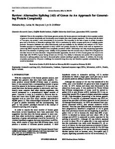
FIGURE 1. Schematic diagram of the human CREM gene showing the two myometrial CREM spliced variants CREM2␣ and CREM␣. A, alternative splicing involves the exons C and G, which encode the Q-rich, glutamine-rich trans-activation domains; P-box, kinase-inducible domain; PI and P2, the alternative promoters; ICERS, inducible cAMP early repressor transcribed from promoter 2. B, Western immunoblot analysis illustrating the switch in the expression of CREM2␣ to CREM␣ that occurs during pregnancy. CREM2␣, a potent transactivator/weak repressor, lacks exon C, whereas CREM␣, a repressor of transcription, lacks both exons C and G, which encode the Q-rich transactivation domains.
regeneration (24); in the heart, where it may play a role in cardiac gene regulation (25); and in the uterus, where there is strong evidence for its role in the regulation of uterine contractility (26, 27). Europe-Finner et al. (19, 20) reported an increased level of cAMP in human myometrial smooth muscle cells during pregnancy, potentiated by altered expression of various components of the cAMP signaling pathway, in particular the G protein G␣s. We have previously reported differential expression of specific CREM protein isoforms within the myometrium tissue throughout pregnancy, namely CREM2␣ and CREM␣ (Fig. 1B), and demonstrated their ability to bind CRE-containing oligonucleotides and activate and/or repress reporter gene transcription (27). Furthermore, through microarray studies, we have recently shown that these potent factors act in myometrial cells to affect the expression of a plethora of genes with defined roles associated with uterine activity during pregnancy (28). Pre-mRNA splicing mechanisms within myometrial cells appear to switch the production of the alternatively spliced CREM2␣ activator that decreases sequentially through the nonpregnant, pregnant nonlaboring, and laboring phases to the synthesis of CREM␣ repressor protein that proceeds from zero expression in the nonpregnant uterus to a high level of expression in the pregnant and laboring myometrium (Fig. 1B). Thus, alternative splicing is responsible for altering the biological consequences, resulting from the transcriptional stimuli of either CREM2␣ or CREM␣ within the human myometrium during fetal maturation. Alternative pre-mRNA splicing is a tightly regulated RNA processing event mediated by a complex interplay of specific cis-elements and trans-acting factors (29 –31). The cis-elements include the 5⬘ and 3⬘ splice sites at exon/intron boundaries and the branch site and polypyrimidine tract preceding the 3⬘ splice site (30, 31). Additional regulatory cis-elements include exon splicing enhancers (ESEs) or exon splicing silencers (ESSs) that have been identified in precursor mRNA sequences from various tissue-specific or developmentally regulated genes (32– 34). ESEs serve as splicing enhancers by contributing to the accuracy and efficiency of alternative splicing when recognized and bound by specific trans-acting factors (34, 35). Numerous trans-acting basal or regulatory splicing factors compete and interact with the various ciselements and indeed with each other, including the ubiquitously expressed U small nuclear ribonucleoprotein particles, the serine-argi-
34522 JOURNAL OF BIOLOGICAL CHEMISTRY
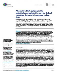
FIGURE 2. Temporal and spatial expression of myometrial SF2/ASF and hnRNPA1 during human gestation and controls for transient transfection and production of SRp40 protein. A, Western immunodetection and subsequent densitometric analysis of SF2/ASF and hnRNPA1 in the upper (US) and lower (LS) myometrium as described by Pollard et al. (42). B, Western immunoblotting demonstrating overexpression of SF2/ASF, hnRNPA1, SC35, and SRp40 after transient transfection. Lanes 1, transfection with pCG control vector; lanes 2, transfection with pCG-SF2, pCG-A1, pCG-SC35, or pCG-SRp40. C, production of SRp40 protein by coupled transcription/translation TnT reaction. Strong staining was observed at ⬃40 kDa after 2 l of TnT product was separated by SDS-PAGE and stained with Coomassie Blue (lane 1). Immunoblotting using anti-SR monoclonal antibody to verify the expression of SRp40 protein by TnT as reflected by the intensity of a single band of 40 kDa (lane 2).
nine (SR) family of nuclear phosphoproteins, and the heterogeneous ribonucleoproteins (hnRNPs) among many others (29, 36, 37). Variations in concentrations and ratios of splicing factors in different cell types and tissues can influence pre-mRNA processing (36 – 40), and individual splicing factors can exhibit unique specificity for particular pre-mRNAs or the tissue type in which they are expressed (29, 30, 37, 38, 41). We have previously reported that the splicing factors SF2/ASF and hnRNPA1 are both spatially and temporally regulated within the human myometrium during pregnancy (Fig. 2A) and that these factors are involved in regulating alternative splicing of the GTP-binding protein G␣s (42, 43). This finding further supports the premise that concentration ratios of trans-splicing factors in vivo may therefore be critical in defining the expression of specific protein isoforms in different tissues. Consequently, in this study, we employed CREM minigene splicing constructs, which incorporate the regulatory cis-elements associated with the alternative splicing of CREM in vivo, and employed transient transfections and in vitro splicing together with RNA electrophoretic mobility shift assays (EMSAs) to investigate the trans- and cis-acting factors and elements involved in regulating the switch in the expression of myometrial CREM2␣ to CREM␣ that occurs throughout pregnancy.
EXPERIMENTAL PROCEDURES Construction of CREM Minigenes—A CREM minigene, pcDNA3.1 CREM-1, was generated from human genomic DNA using CREM-specific PCR primers, containing specific restriction sites, for the individual exon/intron fragments of CREM to facilitate the sequential insertion of each fragment into the pcDNA3.1(⫺) expression vector (Invitrogen). These primers, detailed in TABLE ONE, amplify CREM exons E and F (which encodes the phosphorylation domains), exon G (encoding the second glutamine-rich trans-activation domain), and the downstream exon H, together with truncated versions of their adjacent intronic
VOLUME 280 • NUMBER 41 • OCTOBER 14, 2005
Regulation of CREM by SRp40 TABLE ONE
TABLE TWO
PCR primers for the construction of CREM minigenes Restriction sites are shown in italics.
RT-PCR primers for the amplification of CREM spliced variants Primer name
Exon
Restriction site
DNA sequence (5ⴕ–3ⴕ)
Sense C Antisense C Sense E/F Antisense E/F Sense G Antisense G Sense H Antisense H
Nhe I Xho I Xho I EcoR V EcoR V BamH I BamH I Pme I
GCTAGCTATTAGTGAGTGGTATTACTTA CTCGAGGCGTATAAGCACTTCATA CTCGAGCTATTGCTCAGTTGCTTC GATATCGATACAGTTTAGGTATTAATGA GATATCTTCTCAATTCAGCATAGAA GGATCCTGTGATATAGTAATCCATAG GGATCCTACAAGATCACCTCTTATGA GTTTAAACTCATTAGCCTCAGCTCTC
sequences. PCR products were initially cloned into the TOPOR-TA cloning vector (Invitrogen) and sequenced to confirm exon/intron DNA sequences. Each CREM fragment was then subcloned sequentially into pcDNA3.1 by repeated restriction digestion and ligation using T4 DNA ligase (Promega) to generate the complete pcDNA3.1CREM-1 splicing construct that is under transcriptional control of the cytomegalovirus promoter and contains the bovine growth hormone polyadenylation signal. A second CREM minigene construct, pcDNA3.1CREM-2, which consists of exons C (encoding the first glutamine-rich trans-activation domain) and exons E/F/G/H (and their flanking intronic regions) was also generated in order to reproduce the regulated splicing of the CREM gene in myometrium and ensure that the regulatory factors that splice CREM precursor mRNA into CREM2␣ are present in this model system. Plasmids encoding splicing factors SF2/ASF (pCG-SF2), SRp20 (pCG-20), SRp40 (pCG-40), SRp55 (pCG-55), 9G8 (pCG-9G8), and hnRNPA1 (pCG-A1) have been described (37). Tissues and Cell Culture—Human primary myometrial smooth muscle cell cultures and HeLa cells (LGC-ATTC) were used in this study. Samples of upper and lower segment myometrial tissue were obtained from nonpregnant, pregnant nonlaboring, and spontaneous laboring women undergoing surgery. Written consent was obtained from all women, and ethical approval was granted by the Newcastle and North Tyneside Health Authority Ethics Committee. Primary myocyte isolation was undertaken essentially as described in Ref. 44. Primary myocytes were cultured with complete minimal essential medium-D-valine medium (Cell Culture Technologies), which inhibits fibroblast growth, supplemented with 10% fetal calf serum, penicillin (1 unit/ml), and streptomyocin (1 ng/ml) and cultured under standard conditions at 37 °C with 5% CO2. Transient Transfection of Cultured Myometrial Cells—Transfection experiments on primary myometrial cells (obtained from pregnant tissue samples) were undertaken on subculture passages 2–3. Myometrial and HeLa cells (used as a control) were co-transfected, at 60 –70% confluence (in the absence of antibiotics) using Mirus LT-1 (Cambridge Bioscience) cationic lipid transfection reagent with 0.5 g of pcDNA3.1CREM-1 or pcDNA3.1CREM-2 and 1.5 g of pCG-SF2, pCG-A1, pCG-SC35, pCG-SRp20, pCG-SRp40, pCG-SRp55, pCG9G8, or a pCG control vector. Transfection efficiencies were in the range of 25–30% for all experiments, as determined by transfection with a -galactosidase encoding plasmid, pcDNA3.1 LacZ (Invitrogen) (data not shown). Cells were harvested either 48 h (HeLa cells) or 72 h (myometrial cells) after transfection. Confirmation that the CREM minigene plasmids contain the necessary cis-acting regulatory signals for efficient pre-mRNA splicing was obtained by run-off transcription using a mMESSAGE-mMACHINE capping/transcription kit (described
OCTOBER 14, 2005 • VOLUME 280 • NUMBER 41
pcDNA31 sense Sense E1 Sense E2/V Sense F Antisense F Antisense H/V
Target site Exon E Exon E Exon F Exon F Exon H
DNA sequence (5ⴕ–3ⴕ) CTCACTATAGGGAGACCCAAGC TGCAGAGACAGATGAATCTGCAG CTAGACTCGAGGTAGCAGCAATTGC GAATGAACTGTCCTCTGATGTGC GGTACTGCCATGGTAGCAATAC GGTTTAAACCTGTTTTTCATTAGCCTC
below) and RT-PCR of the CREM spliced products from total RNA extracted from myometrial cells post-transfection. Western Blot Immunodetection—Myometrial tissue homogenates were prepared as previously described (42). Protein concentration was assayed using the DC protein assay kit (Bio-Rad), and SDS-PAGE was performed using 200 g of total protein from each homogenate resolved on 12% polyacrylamide gels. Immunoreactive bands were detected by enhanced chemiluminescence, ECL (Amersham Biosciences), and data obtained where a linear relationship existed between the amount of protein loaded and the intensity of the ECL signal from the immunoblots. Transfection efficiencies were also confirmed in individual experiments by Western immunoblotting using specific monoclonal antibodies to SF2/ASF (anti-SF2; Zymed Laboratories Inc.), hnRNPA1 (4B10; Abcam), and a polyclonal antibody to SRp40 (anti-SRs Zymed Laboratories Inc.). Note the increased protein expression of SF2/ASF, hnRNPA1, SC35, and SRp40 on transfection with their respective plasmids (Fig. 2B), and endogenous levels of these proteins in myometrial cells were also calculated by this procedure (data not shown). Expression of SRp40 Protein—To generate human SRp40 protein, the full-length cDNA coding sequence of the pCG-SRp40 plasmid (described above) was amplified by PCR using Pfx DNA polymerase (Invitrogen). SRp40-specific sense and antisense primers were used that also contain XbaI and BclI restriction sites, respectively, to facilitate the subcloning into a pcDNA3.1 expression vector that has an upstream T7 RNA promoter. SRp40 protein was produced using a transcription/ transcription (TnT) procedure using T7 RNA polymerase (Promega). 2 l of the TnT reaction was resolved on 12% polyacrylamide gels, and production of SRp40 protein was verified by observation of an intense 40-kDa band after Coomassie Blue staining (Fig. 2C, lane 1). Production of SRp40 protein was also confirmed after Western immunoblotting using the polyclonal antibody to SRp40 (anti-SRs Zymed Laboratories Inc.) as shown in Fig. 2C, lane 2. CREM mRNA Splicing Analysis by RT-PCR—CREM mRNA spliced variants generated from the minigenes in transfected cells were analyzed by RT-PCR. Total RNA was isolated from individual transfection experiments using SV total RNA isolation kit (Promega) or Tri-Reagent (Sigma), and first strand cDNA synthesized from 1 g of total RNA using 20 units of Superscript III reverse transcriptase (Invitrogen) with 100 ng of oligo(dT)16 as primer. PCR amplification was carried out using 2 l of cDNA template with CREM-specific primers (detailed in TABLE TWO). PCR analysis was performed with 25 cycles as detailed in the legend to Fig. 3. PCR products were sequenced to confirm their identity. ESE Electrophoretic Mobility Shift Assay (EMSA)—Oligonucleotides containing the individual CREM exon splicing enhancer motifs were designed to be used in RNA EMSAs. Sequences containing the ESE motifs were placed downstream from a T7 RNA promoter to facilitate transcription. Sequences for the oligonucleotides are shown in TABLE THREE. GpppG-capped and [␣-32P]UTP-labeled RNA ESE oligonucleotides were synthesized by run-off transcription for 4 h at 37 °C using
JOURNAL OF BIOLOGICAL CHEMISTRY
34523
Regulation of CREM by SRp40
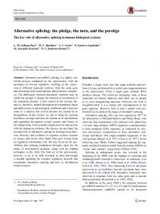
FIGURE 3. Effect of SR proteins and hnRNPA1 on CREM isoform expression. RT-PCR was employed using RNA from transient co-transfection of myometrial cells (A and B) and HeLa cells (C) using pcDNA3.1-CREM-1 and pcDNA3.1-CREM-2 together with plasmids encoding SF2/AF, hnRNPA1, SC35, SRp40, SRp20, SRp55, 9G8, or the pCG control vector. PCRs were performed with an initial hot start cycle at 94 °C (4 min), 55 °C (30 s), and 72 °C (1 min) followed by 20 –25 cycles at 94 °C (1 min), 55 °C (30 s), and 72 °C (1 min). PCRs were carried out using sense primers for the pcDNA 3.1 vector together with antisense primers for exon H plus 9 nucleotides of vector to identify CREM spliced variants expressed from the pcDNA3.1 vector (i). Splicing patterns were consequently confirmed by using the vector sense primers with antisense primers of the exon F of CREM (ii) and sense primers for exon F with antisense primers for exon H (iii). Note that overexpression of SRp40 results in the inclusion of exon G (as indicated by an asterisk). glyceraldehyde-3-phosphate dehydrogenase controls (GAPDH) for each cell type are shown below. TABLE THREE
Sequences for oligonucleotides containing ESE motifs in the exons C and G of CREM Shaded sequence represents the individual ESE motifs. There are overlapping ESE motifs for SRp40 (shaded) and SF2 (boxed) in oligonucleotide 1. Control oligonucleotides with the ESE motifs abolished were also designed. The first 17 bases of each oligonucleotide (in italics) consist of a T7 RNA promoter sequence to facilitate in vitro transcription.
34524 JOURNAL OF BIOLOGICAL CHEMISTRY
VOLUME 280 • NUMBER 41 • OCTOBER 14, 2005
Regulation of CREM by SRp40 the T7 capped transcription kit as described above. 3 l of radiolabeled RNA was incubated for 15 min at 30 °C with either 3 l of SRp-40 protein, 5 units of SF2/ASF, or 5 units of SC35 (or 5 l of nuclear extract) together with 3–5 l of Buffer B (ProteinOne). Both HeLa nuclear extracts (ProteinOne) and nuclear extracts prepared from cultured myometrial cells were used. Myometrial nuclear extracts were prepared using a nuclear extract kit and protocol obtained from ActiveMotif. In supershift experiments, the nuclear extract and transcribed oligonucleotides were supplemented with 1 g of anti-SRs polyclonal antibody (ProteinOne) or 1 g of anti-hnRNPA1 monoclonal antibody (Abcam). RNA band shifts were then analyzed by nondenaturing 10% acrylamide gel electrophoresis followed by autoradiography. In Vitro Splicing Assays—To generate transcripts for in vitro splicing, the pcDNA 3.1CREM minigene constructs, which also harbor a T7 promoter, were first linearized with PmeI. GpppG-capped and [␣-32P]UTP-labeled pre-mRNA substrate was synthesized by run-off transcription using a mMESSAGE-mMACHINE capping/transcription kit with T7 RNA polymerase (Ambion, Inc.). In vitro splicing assays were undertaken essentially as described (45). Briefly, 25–30 fmol of radiolabeled precursor mRNA was incubated for 3 h at 30 °C with 10 l HeLa or myometrial nuclear extract supplemented with 15–25 pmol of SF2/ASF or SC35 (ProteinOne), recombinant hnRNPA1 protein (41, 46), or SRp40 protein, which was produced using a coupled TnT system (Promega) as detailed above (Fig. 2C). Reactions were stopped by the addition of proteinase K, phenol-extracted and then ethanol-precipitated as described (41). RNA products generated from splicing in vitro were then fractionated by denaturing 6% polyacrylamide electrophoresis followed by autoradiography.
RESULTS Effect of Overexpression of SR Proteins on CREM Isoform Expression—Transient co-transfections were undertaken on primary myometrial cell cultures (and HeLa cells) to evaluate the effect of increased levels of several SR proteins and hnRNPA1 on the expression of spliced variants of CREM. CREM mRNA splicedvariantsgeneratedfromthepcDNA3.1-CREMplasmidsco-transfected with SF2/ASF, hnRNPA1, SC35, SRp40, SRp20, SRp55, or 9G8 were analyzed by RT-PCR. Detection of the exogenous CREM mRNA variants CREM2␣ and CREM␣ was undertaken using sense primers for the pcDNA3.1 vector or exon F together with antisense primers for exon F or exon H (which also contained 9 nucleotides of vector). All primers are detailed in TABLE TWO. Data represented here are based on each experiment being performed in triplicate. Representative RT-PCR analyses of transfection experiments are shown in Fig. 3. Co-transfection of pcDNA3.1CREM-1 and pcDNA3.1CREM-2 with the pCG-SRp40 plasmid resulted in increased expression of CREM2␣ mRNA spliced variants, which contain the alternatively spliced exon G, as reflected in the intensity of the 615-bp band compared with the 426-bp band (Fig. 3A (i), lane 4). The retention of exon G as a consequence of overexpression of SRp40 was further confirmed by RT-PCR using specific CREM sense and antisense primers for exons F and H as detailed in Fig. 3, A and B (iii), lanes 4. However, increased levels of SRp40 did not appear to affect the inclusion of exon C into CREM mRNAs, as was observed when the pcDNA3.1CREM-2 plasmid (containing exon C) was transfected. This was reflected by the similar intensities of the 762- and 615-bp PCR bands in Fig. 3B (i), lane 4, and also the 378- and 231-bpbandsinFig.3B(ii),lane4,usingtheCREMsenseandantisenseprimers for exons F and H. Note that in cultured myometrial cells, the basal splicing pattern for pcDNA3.1CREM-1 and -2 in the presence of the control pCG plasmid was similar to the splicing of endogenous CREM in the pregnant myometrium (Fig. 1) in that CREM2␣ and CREM␣ were both expressed, as indicated by the similar intensities of the bands representing these two isoforms (Fig. 3, A and B, lanes 8).
OCTOBER 14, 2005 • VOLUME 280 • NUMBER 41
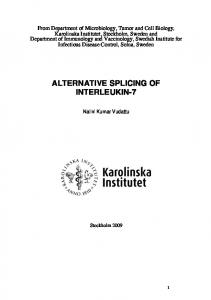
FIGURE 4. Expression profiles for SRp40 and SC35 within the human myometrium during pregnancy. A, Western immunoblotting and densitometric analysis of SRp40 protein expression. Detection of SRp40 was undertaken using an anti-SR polyclonal antibody (Zymed Laboratories Inc.), which recognizes a range of SR proteins but does not react with SR proteins of a similar molecular weight to SRp40. A significant decrease in the levels of SRp40 was observed in the upper and lower uterine regions in all pregnant and laboring samples. Data are shown as the mean ⫾ S.E. (n ⫽ 12 for all nonpregnant (NP), pregnant (P), and laboring (SL) samples. NP versus P/US and versus P/LS ⬍ 0.01; NP versus SL/US and SL/LS ⬍ P 0.001. B, detection of SC35 was undertaken using an anti-SC35 monoclonal antibody (ProteinOne). SC35 protein levels were similar in all nonpregnant, upper uterine pregnant, and laboring samples (n ⫽ 12 for each group). SC35 levels decrease although not significantly in the lower segments in both pregnancy and labor.
In HeLa cells the basal splicing pattern for pcDNA3.1CREM-1 and -2 appeared to be different from that found in myometrial cells. RT-PCR analysis of HeLa cells using pcDNA3.1CREM-1 and -2 and the control plasmid pCG indicated that the predominant CREM mRNA variant expressed in these cells is the small CREM␣ isoform, as indicated by the low intensity of the band representing CREM2␣ and high intensity of the band representing CREM␣. However, a change in the splicing pattern of the CREM minigenes was observed as a consequence of overexpression of SRp40, consistent with the results obtained from myometrial cells, as indicated by an increase in the intensity of a 482-bp band shown in Fig. 3C, lanes 4 and 9. Increased levels of SF2/ASF also appeared to affect the splicing of exon G in HeLa cells, but only when the pcDNA3.1CREM-1 construct was co-transfected (Fig. 3C, lane 6). Expression Profiles of SRp40 and SC35 in the Myometrium during Pregnancy—The expression of SRp40 and SC35 was evaluated by immunoblotting using both nonpregnant and upper and lower myometrium tissues from pregnant and laboring patients. Quantification by densitometric analysis demonstrated that the levels of SRp40 were consistent in all nonpregnant samples (Fig. 4A, lanes 1– 4). However, during pregnancy, myometrial SRp40 expression decreased significantly in both the lower and upper uterine segments (p ⬍ 0.01) and then
JOURNAL OF BIOLOGICAL CHEMISTRY
34525
Regulation of CREM by SRp40
FIGURE 5. Functionality of CREM ESEs by EMSAs. A, EMSAs of SRp40 binding to ESE motifs within the exons C and G of CREM. GpppG-capped and [␣-32P]UTP-labeled RNA oligonucleotides were synthesized by run-off transcription and incubated with either SRp40 TnT reaction or recombinant SC35. RNA-protein complexes were then resolved on nondenaturing 10% acrylamide gels. Band shifts representing the protein䡠ESE complexes were observed for all wild type (wt) oligonucleotides containing ESE motifs for SRp40 and SC35. B, protein/RNA binding was also observed when the oligonucleotides were incubated with myometrial or HeLa nuclear extracts. C, further verification of RNAprotein complexes was accomplished by supershift assays using SRp40 protein with the anti-SR protein antibody. abol, oligonucleotides with the ESE motifs abolished, as detailed in TABLE THREE.
decreased further to undetectable levels at the onset of labor. A different expression profile was observed for SC35. Quantification indicated that SC35 protein levels were comparable in nonpregnant myometrium (Fig. 4B, lanes 1– 4) and the upper uterine region in both pregnancy and labor. However, there was a decrease, although not significant, in the expression of SC35 in the lower uterine segments in both pregnancy and labor. Identification of Multiple ESEs in the Alternatively Spliced Exons C and G of CREM—We analyzed the alternatively spliced CREM exon sequences using ESEfinder (47) (available on the World Wide Web rulai.cshl.edu/tools/ESE/) to identify putative ESE motifs recognized by SF2/ASF, SC35, SRp40, or SRp55. We found multiple high score motifs for all four SR proteins in both the alternatively spliced exons C and G of CREM, as shown in Fig. 5A. One heptameric high score motif (CAGAAGA) for SF2/ASF was identified in exon C, which overlaps with a heptameric SRp40 motif. A second ESE motif for SF2/ASF (CCCAGGA) was identified in exon G. Three octameric motifs for SC35 (GATTTCTA, GACTGCAG, and GGTTGTTG) were found in the exon G sequence, and one hexameric ESE motif for SRp55 (CGCAGC) was also present in exon G. Interestingly, there were seven SRp40 motifs (CCAGAAG, CCACAGC, ACACACC, ATTCAGG, CCACCAG, ACACAGC, and TCCCAGG), the first three in exon C and the last four in exon G, as shown in Fig. 5A. To determine the functionality of these ESEs in SR protein binding, we designed ribooligonucleotides containing the ESE motifs for use in RNA electromo-
34526 JOURNAL OF BIOLOGICAL CHEMISTRY
bility shift assays as detailed in TABLE THREE. To confirm that ESE/SR protein binding was specific, control oligonucleotides with the ESE motifs abolished were also designed. The mutant sequences were designed such that no new SR protein motifs for SF2/ASF, SC35, SRp40, or SRp55 were introduced, according to ESEfinder. SRp40 Functionally Binds Seven Heptameric Exon Splicing Enhancers in CREM—SRp40 was assayed for binding to the individual ESE motif sequences by electrophoretic mobility shift assay. The results demonstrate that SRp40 bound to all seven of the heptamer ESE motifs present in exons C and G of CREM. ESE/SRp40 binding was observed after individual wild type ESE oligonucleotides were incubated with the SRp40 protein, as reflected by the band shifts observed in Fig. 5B. Of note, the intensity of the band shifts for ESE oligonucleotide 6 (which contains two SRp40 binding sites) was particularly strong. SRp40 protein did not bind the control oligonucleotides that had the ESE motifs abolished. SR protein/ESE binding was also observed when the wild type oligonucleotides 4 and 5 were incubated with recombinant SF2/ASF and/or SC35 (Fig. 5B). Similar results were seen when the recombinant proteins were replaced by nuclear extract prepared from cultured myometrial cells. In contrast, when the oligonucleotides were incubated with HeLa nuclear extract, band shifts were observed in some cases when either the wt or mutated ESE oligonucleotides were used (Fig. 5C). It is possible that there are additional unidentified protein binding sites or ESSs present within the sequences of the oligonucleotides. These may bind to other protein factors (such as the SR protein antagonist hnRNPA1) that are present in HeLa nuclear extracts. Alternatively, the mutant sequences may have fortuitously introduced binding sites for other RNA-binding proteins. Sequence-specific binding of SRp40 was also observed when the SRp40䡠ESE complexes were supershifted by using an anti-SR antibody (Fig. 5C). A similar result was observed when the SRp40 protein was replaced with nuclear extracts prepared from cultured myometrial cells (data not shown). hnRNPA1 binding was also observed when both wild type and mutated ESE oligonucleotides 1 (contains overlapping ESE motifs for SRp40 and SF2/ASF) and 4 (contains motifs for both SC35 and SF2/ASF) were used in electromobility shift assays. hnRNPA1 binding was also confirmed when the reactions were incubated with an anti-hnRNPA1 antibody. SRp40 Switches the Splicing Pattern of CREM Spliced Variants—We further evaluated the role of SRp40 in regulating the alternative splicing of CREM by in vitro splicing. Pre-mRNA transcripts from the pcDNA3.1CREM-1 and -2 constructs were spliced in HeLa nuclear extract supplemented with recombinant SF2/ASF, hnRNPA1, SC35, or SRp40 (Fig. 6A). CREM pre-mRNA was spliced under these experimental conditions generating CREM spliced products with and without the inclusion of exons C and G (Fig. 6A). The addition of SRp40 protein to splicing reactions containing precursor RNA from pcDNA3.1CREM-1 generated mRNA products in which the intron between exons F and G was removed and the exon G was retained, corresponding to CREM 2␣ mRNA. Conversely, when the nuclear extract was supplemented with hnRNPA1, the band representing this intron was absent (Fig. 7, lane 2), indicating that exon G was spliced out. This was further evidenced by the presence of the higher molecular weight band representing the lariat intron-exon G complex, which was present only when hnRNPA1 was added but not with SF2/ASF, SC35, or SRp40. Nuclear splicing reactions supplemented with transcribed, unlabeled ESE 6 RNA were also included to further verify the relevance and functionality of the CREM ESEs. ESE 6 was initially chosen because it contains two high score motifs for SRp40. Indeed, ESE 6 competed out the effect of SRp40 and reduced the levels of spliced mRNAs containing exon G (Fig. 7B). Similar results were also observed for ESE oligonucleotides 8 and 9, which
VOLUME 280 • NUMBER 41 • OCTOBER 14, 2005
Regulation of CREM by SRp40
FIGURE 6. SRp40 switches the splicing pattern of CREM spliced variants. A, in vitro splicing of CREM in myometrial nuclear extracts. pcDNA3.1-CREM-1 and pcDNA3.1CREM-2 GpppG-capped and [␣-32P]UTPlabeled precursor-mRNA was supplemented with recombinant SF2/ASF, hnRNPA1, SC35, or SRp40 (TnT reaction). RNA was resolved on 6% PAGE with 8 M urea. Precursors, intermediates, and final spliced products are indicated. B, nuclear splicing reactions supplemented with transcribed, unlabeled ESE 6 RNA were also included to further verify functionality of the CREM ESEs. ESE 6 competed out the effect of SRp40 and reduced the levels of spliced mRNAs containing exon G, suggesting that the SRp40 effect is specific.
also contain high score SRp40 ESE motifs (data not shown). When the ESE 6 control oligonucleotide, which has the ESE motif mutated, was used, no competition was observed, suggesting that the SRp40 effect is specific.
DISCUSSION Alternative splicing promotes a switch in the expression of CREM2␣, an activator, to CREM␣, a repressor, within the human myometrium throughout pregnancy and labor. We have recently shown that this change in the expression of spliced variants of CREM is relevant to the regulation of downstream CRE-responsive target genes involved in uterine activity during gestation and parturition (28). In this present study, we unravel the splicing mechanisms that control the expression of CREM spliced variants in human myometrial cells. We provide evidence to indicate that the switch from synthesis of CREM2␣ to CREM␣ that occurs throughout pregnancy and labor is orchestrated by the down-regulation of an SR protein family member, SRp40. Our data from transfection of CREM minigenes in myometrial and HeLa cells indicate that overexpression of SRp40 favored the proximal alternative 5⬘ splice site, promoting mRNA transcripts in which exon G were included, thus stimulating the synthesis of CREM2␣ within these cells. Overexpression of other members of the SR protein family SF2/ ASF, SC35, SRp20, SRp55, and 9G8 did not appear to affect the splicing patterns of CREM within myometrial cells. In contrast to SRp40, and in keeping with its previously defined role in splice site selection, overex-
OCTOBER 14, 2005 • VOLUME 280 • NUMBER 41
pression of hnRNPA1 favored the selection of distal 5⬘ splice sites such that exon G was skipped out, resulting in the synthesis of CREM␣. We have previously reported that the endogenous alternative splicing pattern of another gene, the adenylyl cyclase stimulatory G-protein G␣s, is different in myometrial cells compared with HeLa cells (43). In myometrial cells, the principal spliced variant was G␣s long, whereas in HeLa cells, the short isoform of G␣s, resulting from the skipping of an internal exon 3, was predominant. A similar pattern of splicing was also observed for CREM, in that in myometrial cells, the principal CREM variant appeared to contain exon G, whereas in HeLa cells, the predominant mRNAs of CREM had exon G skipped out. We propose that the preference for exon skipping in HeLa cells with both G␣s and CREM conforms to the abundance of hnRNPA1, 6 –7 ⫻ 107 copies/cell in these cells (37). Data presented here suggests that SRp40 regulates splice site selection via its interactions with exon splicing enhancers. This is evidenced by the presence and functionality of multiple ESEs present in both of the alternatively spliced exons C and G of CREM. In exon C, there are three SRp40 binding sites, all of which are recognized and bound by SRp40. In exon G, there are four high scoring ESE motifs that specifically bind SRp40, supporting a model in which SRp40 binding is required for the recruitment of the splicing complex to a 5⬘ proximal splice site. The ESEs present in exon G appear to be particularly important in the splice site selection, thus providing a mechanistic explanation as to how SRp40 regulates the switch in CREM2␣ to CREM␣ (Fig. 7).
JOURNAL OF BIOLOGICAL CHEMISTRY
34527
Regulation of CREM by SRp40
FIGURE 7. Model proposing how SRp40 regulates the switch in alternative splicing of CREM2␣ to CREM␣ that occurs in pregnancy and labor. In the nonpregnant state when levels of SRp40 are high, SRp40 promotes the skipping of exon C and inclusion of exon G, resulting in the production of CREM2␣. Conversely, as levels of myometrial SRp40 decrease throughout pregnancy and labor (Fig. 4A), there is a parallel decrease in the expression of CREM2␣ and an increase in CREM␣ (Fig. 1B). Our data indicate that the multiple ESE elements present in CREM affect splicing through initiation by SRp40.
SRp40 has previously been studied for its role in alternative splicing and has been shown to select both proximal and distal splice 5⬘ sites in a substrate-specific manner (29, 39). Our findings indicate that SRp40 appears to concurrently activate the selection of both proximal and distal 5⬘ splice sites within the same precursor mRNA transcript. We suggest that, in vivo, in the nonpregnant state when levels of SRp40 are high, SRp40 promotes the skipping of exon C and inclusion of exon G, resulting in the production of CREM2␣. Conversely, as levels of myometrial SRp40 decrease throughout pregnancy and labor, there is a parallel decrease in the expression of CREM2␣ and an increase in CREM␣. This raises the question of how SRp40 works to regulate the switch in the expression of CREM spliced variants when it is down-regulated. We propose that SRp40 and multiple ESEs present in the exons C and G work in a bidirectional manner to regulate CREM spliced variants. SRp40 via its interactions with ESEs in exon C promotes the proximal splice site, resulting in the inclusion of exon C. Conversely, SRp40 via its interactions with ESEs in exon G promotes the selection of a proximal splice site, resulting in the inclusion of exon G. The down-regulation of SRp40 that occurs throughout pregnancy suggests that when there are only negligible levels of SRp40 present, there is no SRp40䡠ESE complex formation, and consequently, both the alternatively spliced exons C and G are skipped out of the precursor mRNA transcript, thus generating CREM␣. One could speculate that hnRNPA1, a well characterized antagonist to the SR proteins (36), may be a contributory factor and have a silencing affect in regulating CREM. However, hnRNPA1 is clearly spatially regulated in the myometrium during pregnancy, whereas CREM is not. We have previously shown that hnRNPA1 levels are moderate in the nonpregnant myometrium (42), as shown in Fig. 2A, and as such hnRNPA1 may well contribute to the skipping of exon C that
34528 JOURNAL OF BIOLOGICAL CHEMISTRY
occurs in the nonpregnant state. However, throughout pregnancy and at the onset of labor, there is a dramatic switch in the spatial expression of hnRNPA1 within the functionally distinct upper and lower regions of the uterus (Fig. 2A), whereas the expression and switch in the synthesis of CREM2␣ to CREM␣ that occurs is not spatially regulated within the different uterine regions. Moreover, since the expression patterns for SC35 and SF2/ASF within the nonpregnant and pregnant human myometrium are not comparable with the expression of CREM spliced variants, it is unlikely that they regulate the switch in spliced variants of myometrial CREM that occurs in pregnancy. However, SC35 may possibly be a candidate splicing factor in regulating CREM spliced variants in other tissues, since there are three functional SC35-specific ESE motifs present in CREM. Moreover, it could be reasoned that other unidentified protein factors may also be involved. A previous study by Cowper (48) identified SR protein-like repressor factors, named SRrp40 and SRrp35. These factors appear to antagonize authentic SR proteins SF2/ASF and SC35 by selecting the most distal splice sites in the adenovirus pre-mRNA in an activity similar to hnRNPA1. ESEs have been reported to promote either 5⬘ or 3⬘ splice site selection, and there is recent evidence to indicate that in a few examples a single ESE can concurrently promote recognition of both upstream and downstream 5⬘ and 3⬘ splice sites, thus serving as a bidirectional splicing enhancer (49, 50). Caputi et al. (51) reported a novel bidirectional ESE that regulates the expression of the HIV-env, vpu, and nef mRNAs, and interestingly, both SRp40 and SF2/ASF were shown to bind and activate this ESE. What appears to be novel with the CREM gene is the observation that different sets of SRp40 ESEs within different exons within the same gene appear to work bidirectionally to promote distal and proximal 5⬘ splice site selection. To the best of our knowledge, this is the first example of a human gene where multiple ESEs present in two alternatively spliced exons work bidirectionally, resulting in the synthesis of two functionally distinct protein isoforms, namely CREM2␣ and CREM␣. It is widely accepted that alternative splicing is a fundamental mode of gene regulation in the generation of structurally and functionally distinct protein isoforms (52, 53). Furthermore, the accuracy of the splicing machinery is essential for developmental and tissue-specific control of gene expression (24, 53–55). In previous studies, we have demonstrated that alternative splicing regulates expression of different components of the cAMP signaling pathway within the developmentally regulated myometrium throughout pregnancy and labor, namely G␣s and CREM (20, 26). In addition, we have reported that two key splicing factors, SF2/ASF and hnRNPA1, are differentially expressed within the myometrium during gestation (42), and our findings also showed that these pivotal splicing factors regulate the expression of G␣s (43). Data presented here describe the temporal/spatial expression profiles for two more splicing regulators SRp40 and SC35 within the myometrium during pregnancy and labor. In this study, we show that the molecular mechanisms controlling the switch in the expression of a potent transcription activator CREM2␣ to that of a transcription repressor CREM␣ involve the differential expression of SRp40. Hence, our studies emphasize the importance of alternative splicing in controlling the expression of functionally distinct protein isoforms associated with uterine activity during gestation and labor. Evidence from previous studies shows that subtle fluctuations in the concentrations of specific splicing factors can define the formation of different spliced mRNA isoforms derived from a number of precursor mRNA species, and this switching of splice sites also occurs after overexpression of SR proteins in vivo, when tested with a range of reporter genes, promoting aberrant exon skipping and inclusion (29, 38, 39). In this study, we show
VOLUME 280 • NUMBER 41 • OCTOBER 14, 2005
Regulation of CREM by SRp40 that the down-regulation of SRp40 in pregnancy is important for the switch in the splicing pattern of CREM. Since the specificity of SR proteins in regulating the efficiency or pattern of alternative splicing of different genes is attributed in part to the recognition of ESEs (30, 34, 41), it is worthy to note that CREM exons contain multiple copies of SR protein-specific ESE motifs, which raises the question of whether different sets of SR proteins are involved in regulating CREM in vivo. To conclude, our study provides strong evidence that the switch in the alternative splicing of the human CREM gene in the human myometrium during fetal maturation is controlled by SRp40 and involves the use of multiple ESEs. We provide evidence to indicate that the switch in the splicing of CREM2␣ to CREM␣ is controlled by the complex interplay of at least seven SRp40-specific bidirectional splicing enhancers in addition to the consensus splicing cis-elements. In addition, this is the first study to characterize the expression profiles for SRp40 and SC35 in a human organ. We demonstrate that expression of endogenous SRp40 is tightly regulated throughout pregnancy. Moreover, we show that the reduced expression of SRp40 that occurs within the myometrium throughout pregnancy correlates with the levels of CREM2␣ and CREM␣ within these cells. Importantly, differential expression of SRp40 and other members of the SR protein family in various types of tissues, such as the heart, liver, and brain, may under normal physiological and pathophysiological conditions define the expression of functionally distinct isoforms of CREM and their subsequent transcriptional effects on downstream target genes. Acknowledgment—We thank Javier Ca´ceres for kindly supplying the plasmids encoding the splicing factors.
REFERENCES 1. de Groot, R. P., den Hertog, J., Vandenheede, J. R., Goris, J. & Sassone-Corsi, P. (1993) EMBO J. 12, 3903–3911 2. Sassone-Corsi, P. (1998) J. Biochem. Cell Biol. 30, 27–38 3. De Cesare, D., Fimia, G. M. & Sassone-Corsi, P. (1999) Trends Biochem. Sci. 24, 281–285 4. Sun, P., Schoderbek, W. E. & Maurer, R. A. (1992) Mol. Endocrinol. 6, 1858 –1866 5. Habener, J. F., Miller, C. P. & Vallejo, M. (1995) Vitam. Horm. 51, 1–5 6. Landschulz, W. H., Johnson, P. F. & McKnight, S. L. (1988) Science 240, 1759 –1764 7. Ziff, E. B. (1990) Trends Genet. 6, 69 –72 8. Hai, T., Liu, F., Coukos, W. J. & Green, M. R. (1989) Genes Dev. 3, 2083–2090; Correction (1990) Genes Dev. 4, 682 9. Borrelli, E., Montmayeur, J. P., Foulkes, N. S. & Sassone-Corsi, P. (1992) Crit. Rev. Oncog. 3, 321–338 10. Tenbrock, K., Juang, Y-T., Tolnay, M. & Tsokos, G. C. (2003) J. Immunology 170, 2971–2976 11. Isoda T., Paolocci, N., Haghighi, K., Wang, C., Georgakopoulos, D., Servillo, G., Fazia, Agnese, M. A. D., Kranias, E. G., Depaoli-Roach, A. A., Sassone-Corsi, P. & Kass, D. A. (2003) FASEB J. 17, 144 –155 12. Foulkes, N. S., Borreli, E. & Sassone-Corsi, P. (1991) Cell 64, 739 –749 13. Sanborn, B. M., Millan, J. L. & Meistrich, M. L. (1997) J. Androl. 18, 62–70 14. Sanborn, B. M. (2000) Endocrinology 141, 3921–3922 15. Servillo, G., Della Fazia, M. A. & Sassone-Corsi, P. (2002) Exp. Cell Res. 275, 143–154 16. Zuo, J., Lei, Z. M. & Rao, C. V. (1994) J. Clin. Endocrinol. Metab. 79, 907–911 17. Dong, Y. L., Fang, L., Kondapaka, S., Gangula, P. R., Wimalawansa, J. & Yallampalli, C. (1999) J. Clin. Invest. 104, 559 –565
OCTOBER 14, 2005 • VOLUME 280 • NUMBER 41
18. Kofinas, A. D., Rose, J. C., Koritnik, D. R. & Meis, P. J. (1990) J. Reprod. Med. 35, 1045–1050 19. Europe-Finner, G. N., Phaneuf, S., Watson, S. P. & Lopez Bernal, A. (1993) Endocrinology 132, 2484 –2490 20. Europe-Finner, G. N., Phaneuf, S., Tolkovsky, A. M., Watson, S. P. & Lopez Bernal, A. (1994) J. Clin. Endocrinol. Metab. 79, 1835–1839 21. Lopez Bernal, A., Europe-Finner, G. N., Phaneuf, S. & Watson, S. P. (1995) Trends Pharmacol. Sci. 16, 129 –133 22. Yang, X., Lee, F. Y., Sr. & Wand, G. S. (1997) Mol. Endocrinol. 11, 1053–1061 23. Don, J. & Stelzer, G. (2002) Mol. Cell. Endocrinol. 187, 115–124 24. Servillo, G., Fazia, M. A. D. & Sassone-Corsi, P. (1998) Genes Dev. 12, 3639 –3643 25. Isoda, T., Paolocci, N., Haghighi, K., Wang, C., Wang, Y., Georgakoupos, D., Servillo, G., Della Fazia, M. A., Kranias, E. G., Depaoli-Roach, A. A., Sassone-Corsi, P. & Kass, D. A. (2003) FASEB J. 17, 144 –151 26. Bailey, J., Sparey, C., Phillips, R. J., Gilmore, K., Robson, S. C., Dunlop, W. & EuropeFinner, G. N. (2000) Mol. Hum. Reprod. 6, 648 – 660 27. Bailey, J., Phillips, R. J., Pollard, A. J., Gilmore, K., Robson, S. C. & Europe-Finner, G. N. (2002) J. Clin. Endocrinol. Metab. 87, 1717–1728 28. Bailey, J., Tyson-Capper, A. J., Gilmore, K., Robson, S. C. & Europe-Finner, G. N. (2005) J. Mol. Endocrinol. 34, 1–17 29. Ca´ceres, J. F. & Krainer, A. R. (1997) in Eukaryotic mRNA Processing (Krainer, A.R., ed) pp. 174 –212, IRL Press, Oxford, UK 30. Graveley, B. R. (2000) RNA 6, 1197–1211 31. Smith, C. W. & Valcarel, J. (2000) Trends Biochem. Sci. 25, 381–388 32. Tanaka, K., Watakabe, A. & Shimura, Y. (1994) Mol. Cell. Biol. 14, 1347–1354 33. Elrick, L. L., Humphrey, M. B., Cooper, T. A. & Berget, S. M. (1998) Mol. Cell. Biol. 18, 343–352 34. Schaal, T. D. & Maniatis, T. (1999) Mol. Cell. Biol. 19, 1705–1719 35. Sun, Q., Mayeda, A., Hampton, R. K., Kriner, A. R. & Rottman, F. M. (1993) Genes Dev. 7, 2598 –2608 36. Ca´ceres, J. F., Stamm, S., Helfman, D. M. & Krainer, A. R. (1994) Science 265, 1706 –1709 37. Hanamura, A., Ca´ceres, J. F., Mayeda, A., Franza, B. R., Jr. & Krainer, A. R. (1998) RNA 4, 430 – 444 38. Mayeda, A. & Krainer, A. R. (1992) Cell 68, 365–375 39. Zahler, A. M., Neugebauer, K. M., Lane, W. S. & Roth, M. B. (1993) Science 260, 219 –222 40. Wang J. & Manley, J. L. (1995) RNA 1, 335–346 41. Mayeda, A., Screaton, G. R., Chandler, S. D., Fu, X-D. & Krainer, A. R. (1999) Mol. Cell. Biol. 19, 1853–1863 42. Pollard, A. J., Sparey, C., Robson, S. C., Krainer, A. R. & Europe-Finner, G. N. (2000) J. Clin. Endocrinol. Metab. 85, 1928 –1936 43. Pollard, A. J., Krainer, A. R., Robson & Europe-Finner, G. N. (2002) J. Biol. Chem. 277, 15241–15251 44. Phaneuf, S., Europe-Finner, G. N., Varney, N., MacKenzie, I. Z., Watson, S. P. & Lo´pez Bernal, A. (1993) J. Endocrinol. 136, 497–509 45. Mayeda, A. & Krainer, A. R. (1999) Methods Mol. Biol. 118, 315–321 46. Liu, H-X., Zhang, M. Q. & Krainer, A. R. (1998) Genes Dev. 12, 1998 –2012 47. Cartegni, L., Wang, J., Zhu, Z., Zhang, M. Q. & Krainer, A. R. (2003) Nucleic Acids Res. 31, 3568 –3571 48. Cowper, A. E., Ca´ceres, J. F., Mayeda, A. & Screaton, G. R. (2001) J. Biol. Chem. 276, 48908 – 48914 49. Lam, B. J. & Hertel, K. J. RNA 8, 1233–1242 50. Bourgeois, C. F., Popielarz, M., Hildwein, G. & Stevenin, J. (1999) Mol. Cell. Biol. 19, 7347–7356 51. Caputi, M., Freund, M., Kammler, S., Asang, C. & Schaal, H. (2004) J. Virol. 78, 6517– 6526 52. Stamm, S., Ben-Ari, S., Rafalska, I., Tang, Y., Zhang, Z., Toiber, D., Thanararaj, T. A. & Soreq, H. (2005) Gene (Amst.) 344, 1–20 53. Hastings, M. & Krainer, A. R. (2001) Curr. Opin. Cell Biol. 13, 302–309 54. Modrek, B. & Lee, C. (2002) Nat. Genet. 30, 13–19 55. Cooper, T. A. (2004) Cell 120, 1–5
JOURNAL OF BIOLOGICAL CHEMISTRY
34529