Journal of Medical Microbiology (2008), 57, 316–323
DOI 10.1099/jmm.0.47365-0
The TUBEX test detects not only typhoid-specific antibodies but also soluble antigens and whole bacteria Frankie C. H. Tam,1 Thomas K. W. Ling,2 Kam Tak Wong,2 Danny T. M. Leung,1 Raphael C. Y. Chan2 and Pak Leong Lim1 Correspondence
1
Pak-Leong Lim
[email protected]
2
Received 27 April 2007 Accepted 2 December 2007
Clinical Immunology Unit, The Chinese University of Hong Kong, Prince of Wales Hospital, Shatin, New Territories, Hong Kong Department of Microbiology, The Chinese University of Hong Kong, Prince of Wales Hospital, Shatin, New Territories, Hong Kong
TUBEX (IDL Biotech) is a 5 min semiquantitative colorimetric test for typhoid fever, a widely endemic disease. TUBEX detects anti-Salmonella O9 antibodies from a patient’s serum by the ability of these antibodies to inhibit the binding between an indicator antibody-bound particle and a magnetic antigen-bound particle. Herein, we report that TUBEX could also be used to specifically detect soluble O9 lipopolysaccharide in antigen-spiked buffer by the ability of the antigen to inhibit the same binding between the particles. Sensitivity of antigen detection was improved (8–31 mg ml”1) by using a modified protocol in which the test sample was mixed with the indicator particles first, rather than with the magnetic particles as for antibody detection. The antigen was also detectable in spiked serum and urine samples, albeit less well (2–4-fold) than in buffer generally. However, no antigen was detected from six typhoid sera examined, all of which had anti-O9 antibodies. In addition, whole organisms of Salmonella Typhi (15 strains) and Salmonella Enteritidis (6 strains) (both O9+ Salmonella), grown in simulated blood broths or on MacConkey agar, were also detectable by TUBEX when suspended at .9¾108 organisms ml”1. Expectedly, Salmonella Paratyphi A (7 strains), Salmonella Typhimurium (1 strain) and Escherichia coli (2 strains) were negative in the test. Thus, the same TUBEX kit may be used in several ways both serologically and microbiologically for the rapid diagnosis of typhoid fever. However, validation of the newer applications will require the systematic examination of real patient and laboratory materials.
INTRODUCTION Typhoid fever remains a global threat as it affects an estimated 17 million people annually (WHO, 2003). Laboratory diagnosis is crucial to the management and control of the disease since typhoid can be clinically mistaken for other febrile syndromes, such as dengue fever, that often prevail in the affected countries. The laboratory methods commonly used include (1) culture and identification of the causative organism, Salmonella enterica serotype Typhi (S. Typhi), and (2) detection of infectionspecific antibodies from the patient’s circulation (WHO, 2003). The problem with culture is that the method is laborious and long. In this approach, blood, bone marrow or stool is normally inoculated into an enrichment broth, and when growth appears, subcultures are made on solid agar. Colonies thus obtained are identified by biochemical testing and confirmed by slide agglutination with appropriate antisera. The whole process can thus take several days. 316
Serology based on antibody detection has been widely used to diagnose typhoid fever since the introduction of the Widal test (Widal, 1896). This test has remained the mainstay of serological tests in many of the endemic countries despite numerous accounts of its shortcomings (Quiroga et al., 1992; Parry et al., 1999; Nsutebu et al., 2002). The main reason for its continued use is the fact that it is simple – requiring just a single step and no instrumentation – and is affordable by the affected countries. In addition, when performed on slides (instead of tubes), this simple bacterial agglutination test takes only a few minutes to perform. In contrast, more accurate and more sophisticated tests developed over the years, such as those based on the multi-step ELISA method (Nardiello et al., 1984; Sippel et al., 1989; House et al., 2005), have not found widespread use in these countries. An antibody-detection test kit recently introduced to the typhoid market in several countries, including Pakistan, Vietnam and the Philippines, which is user-friendly like the 47365 G 2008 SGM Printed in Great Britain
Rapid immunodiagnosis of typhoid
Widal test and which can be used for point-of-care applications, is TUBEX (produced by IDL Biotech, Sollentuna, Sweden). TUBEX is similar to the slide latex agglutination test in being simple and rapid, both employing efficient mixing of the analyte with reagent latex particles, and both utilizing an easy, visual readout of the results (Lim et al., 1998; Tam & Lim, 2003). However, improvements have been made to enhance the sensitivity and specificity in the TUBEX system in various ways, including the use of specially designed V-shaped tubes instead of slides, and of two types of reagent particles instead of one – a coloured indicator particle and a magnetic particle. Importantly, too, an inhibition assay
format is used to achieve good specificity. Thus, a patient’s S. Typhi-specific antibodies are detected by their ability to inhibit the binding between coloured indicator particles that are coated with a monoclonal antibody (mAb) specific for the S. Typhi O9 lipopolysaccharide (LPS) antigen, and magnetic particles that are coated with S. Typhi LPS (see Fig. 1a). The results are read after 5 min following sedimentation of the magnetic particles (and any bound indicator particles) with a magnet (Fig. 1b). Good sensitivities (75–85 %) and specificities (75–90 %) were observed with the test when used in several clinical trials recently (House et al., 2001; Olsen et al., 2004; Kawano et al., 2007).
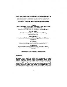
Fig. 1. The TUBEX test. (a) Cartoon illustrating how TUBEX works for detecting anti-O9 antibodies or for detecting O9 antigen. Shown are the antibody-bound indicator particles and the antigen-bound magnetic particles that are inhibited from binding to each other by the patient’s antibodies or antigen. (b) Photograph of four sets of reaction wells (six wells each) containing various reaction mixtures, showing the possible result outcomes, which can be read against the colour chart provided on the magnet stand (score 0, most red and most negative; score 10, most blue and most positive). Results are read similarly for both antibody and antigen detection. Shown here are the results for antigen detection in buffer, serum or urine samples (corresponding to cases listed in Table 2) that were spiked with various amounts of S. Typhi (batch 1) or S. Typhimurium LPS. (c) Detection of S. Typhi LPS by TUBEX using the original protocol for antibody detection, or the new protocol for antigen detection. Two different batches of LPS were examined, each prepared in varying amounts in buffer. Results were scored against the colour chart and are representative of duplicate experiments. The shaded bar denotes the cutoff (score 2) between positive and negative results. http://jmm.sgmjournals.org
317
F. C. H. Tam and others
By virtue of the mechanism employed in TUBEX, we reasoned that the same test could also be used to detect antigen instead of antibody. Antigen could presumably bind to the indicator particles and block the reaction between the two reagent particles (Fig. 1a). Moreover, the antigen to be detected might not necessarily be small and soluble, but also in the form of whole bacteria. We investigated these possibilities in the present study, including their potential clinical applications.
METHODS Antigens and antibodies. Purified LPS was obtained commercially: S. Typhi, ‘Salmonella typhosa’ (lot 0901) and Salmonella Enteritidis LPS (Difco); Salmonella Typhimurium LPS (Calbiochem); and Escherichia coli LPS (Sigma). Thus two batches of S. Typhi LPS made at different times (including ‘S. typhosa’) were used in the study. Crude Salmonella Paratyphi A LPS was extracted from freshly prepared cells using trichloroacetic acid (Staub, 1965), and purified by precipitation with 6 vols ethanol. Crude antigenic extract was obtained from laboratory isolates (strain #13, S. Typhi and S. Paratyphi A) as the culture supernatant of cells grown overnight in Luria–Bertani (LB) broth (Invitrogen) and heated at 100 uC for 5 min in a thermocycler (MJ Research). The protein content was determined using the BCA Protein Assay kit (Pierce).
Mouse mAbs to the O9 antigen (Lim & Ho, 1983) and the O12 antigen (produced in our laboratory, unpublished), both IgM, were purified from ascites material by cryoprecipitation. Serum and urine samples. Blood (serum) and urine samples were
obtained freshly from healthy laboratory workers with due consent. These were kept at 4 uC and used within 3 days.
The TUBEX test. For antibody detection, the instructions of the
manufacturer (IDL Biotech) were followed. Briefly, the test serum sample (25 ml) was mixed in a chamber of the reaction container provided (which contains a set of six identical V-shaped chambers: Fig. 1) with the Brown reagent (antigen-coated magnetic particles; 25 ml) for 2 min, before mixing with the Blue reagent (antigen-coated indicator particles; 50 ml) for another 2 min (Tam & Lim, 2003). The reaction mixture was then placed on the magnet stand, and the resultant colour was read immediately and scored against the colour chart (score 0–10). Scores 0–2 were considered negative, and scores 3–10 positive. For antigen detection, the test sample (25 ml) was mixed with the Blue reagent (50 ml) in the reaction well for 2 min, and then with the Brown reagent (25 ml) for another 2 min. The results were then read after sedimentation of the magnetic particles. ELISA. The method described previously (Wun et al., 2001) was followed. Briefly, Immulon-2 microplates (Dynex) coated with fivefold serial dilutions of the crude antigen extract (starting concentration 40 mg ml21) were incubated with the anti-O9 or anti-O12 mAb for 1 h at room temperature. After washing, peroxidase-labelled goat anti-mouse Ig (all classes) (BD Sciences) was added and incubated for 30 min. Following substrate (3,39,5,59tetramethylbenzidine) development, the results were read in a Dynex MRII reader. Western blot analysis. The crude antigen extract (40 ml, 4 mg ml21;
heated at 100 uC for 5 min in loading buffer containing 20 % 2mercaptoethanol, 40 % glycerol and 8 % SDS) was electrophoresed (100 V) on 12 % polyacrylamide gel under reducing conditions, and electroblotted onto 0.22 mm PVDF membrane (Bio-Rad). After blocking with 5 % skim milk, the membrane was incubated with the anti-O9 or anti-O12 mAb overnight at 4 uC. The assay was developed using peroxidase-labelled anti-mouse Ig (BD Biosciences) and ECL chemiluminescence (Amersham Biotech).
Bacterial strains. All strains were isolated and identified by the
routine laboratory of the Department of Microbiology at the Prince of Wales Hospital, Hong Kong. Standard procedures were followed, including the use of API 20E (bioMe´rieux) for biochemical identification, and slide bacterial agglutination tests for serotyping Salmonella isolates using polyclonal rabbit anti-O and anti-H antisera (Remel Europe). Stock cultures of these were kept on nutrient agar slopes. Bacterial culture. Bacteria were grown in LB broth with shaking
overnight at 37 uC. The cells were washed in saline by centrifugation (500 g, 10 min) and resuspended in saline. The bacterial cell concentration was estimated by comparing the turbidity of the suspension with those of the McFarland standards. The suspension was (i) concentrated using Microcon YM-10 (Millipore), or (ii) acetone-fixed by adding 3 vols acetone to the 10 % (w/v) suspension, or (iii) heated at 100 uC for 5 min in a thermocycler. In simulated blood culture, LB broth supplemented with 20 % blood (obtained from a healthy laboratory worker) was used. Briefly, a single colony of the test organism obtained from a purity agar plate was inoculated into 5 ml of the blood broth and incubated overnight at 37 uC with shaking (250 r.p.m.). The culture was centrifuged at 300 g for 2 min to pellet the red blood cells. The supernatant thus obtained was heated at 100 uC for 5 min in a water bath and used in the studies. In other studies, three colonies were randomly selected from the test organism grown by purity plating on MacConkey agar, and resuspended in 4 ml saline. The turbidity of the suspension was scored against the McFarland standards (5 ml solutions). The suspension was heated at 100 uC for 5 min in a water bath and used. 318
RESULTS Detection of soluble antigen in buffer by TUBEX Buffer preparations containing various amounts of S. Typhi LPS were examined for their ability to react (inhibit) in the TUBEX test. The original protocol used for antibody detection was initially followed, in which the LPS preparation was mixed with the LPS-coated magnetic beads prior to incubation with the antibody-coated indicator particles. Concentrations ¢63 mg ml21 (LPS batch 1) or 31 mg ml21 (LPS batch 2) were found to be reactive (Fig. 1c). Reactivity was arbitrarily defined as TUBEX score ¢3 (Fig. 1), based on the general cutoff used for antibody detection as advocated by the manufacturer, although to detect antigen in buffer, the background reactivity for antigen detection in buffer in the present study was virtually totally absent (score 0). However, when the protocol was modified to specifically detect antigen instead of antibody (mixing test sample with indicator particles before adding the magnetic particles), the sensitivity of detection was increased 2–4-fold (Fig. 1c). Specificity of the antigen detection was examined using LPS (crude or purified) obtained from various bacteria, including several serotypes of Salmonella. Only the group Journal of Medical Microbiology 57
Rapid immunodiagnosis of typhoid
Table 1. TUBEX detection of various LPS and various bacteria prepared in various ways P. mirabilis, Proteus mirabilis; o/n, overnight; Bacterium
S. Typhi S. Enteritidis S. Paratyphi A S. Typhimurium E. coli P. mirabilis
ND,
not done; –, not relevant.
O antigens H antigens
9, 9, 2, 4, – –
12, Vi 12 12 (5), 12
d g a i – –
TUBEX results* LPS
o/n culture
o/n culture conc. 10¾
o/n culture acetone-fixed
o/n culture heated 100 6C/5 min
MacConkey colonies
10 10 0 0 0
0 2 2 2 0
5 4 0 2 2
8 6 0
8 (6)D
2
8 6 2 2 2
ND
ND
ND
ND
ND
ND
ND
0 ND
0 0
*Score 0–2, negative; 3–10, positive. DAnother S. Typhi isolate; single strain used in other cases.
D salmonellae (S. Typhi and S. Enteritidis) were found to be reactive (TUBEX score, 10; Table 1). Presumably, LPS from other group D organisms would also react, but this was not tested. Antigen detection was repeated using five urine and four serum samples obtained from healthy individuals, each spiked with various amounts of S. Typhi LPS (batch 1) or S. Typhimurium LPS. With both urine and serum, the sensitivity of detection was slightly affected, being 2–4-fold lower than in buffer (Table 2; Fig. 1b). The reactivity was more pronouncedly affected at the lower antigen concentrations (,125 mg ml21) than at higher concentrations in all cases. The effect depended on the individual urine or serum sample, but not the pH of the sample. No reactivity was observed when the urine or serum samples were spiked with S. Typhimurium LPS. Real specimens from typhoid patients were next examined to see whether S. Typhi LPS could be found in these. Sera from nine typhoid patients were randomly selected from a
collection of sera kept frozen in our laboratory that were known to be antibody-positive by TUBEX, and heated in the presence of 1 % SDS at 90 uC in a water bath for 5 min. (This heat treatment was found in preliminary studies to destroy all inherent antibody activity in the serum but only had minimal – about 25 % – effect on the antigenic activity of the S. Typhi LPS when used to spike the serum; both activities determined by TUBEX.) Of the nine sera, three became heat-coagulated and were discarded. The remaining six sera were examined for both antibody and antigen activity by TUBEX. All were found to be negative for both activities. This suggests that, if any, no more than 80 mg ml21 of S. Typhi LPS (approx., see Table 2) was present in these sera. Detection of whole bacteria in buffer or broth by TUBEX The ability of whole bacterial cells to react (inhibit) in TUBEX was next investigated, using the protocol
Table 2. Detection of S. Typhi or S. Typhimurium LPS by TUBEX in buffer and in spiked urine or serum samples The values shown are TUBEX scores: 0–2, negative; 3–10, positive. S. Typhi LPS batch 1 (mg ml”1)
Medium
Buffer (pH 8.2) Urine 1 (pH 6.1) Urine 2 (pH 6.4) Urine 3 (pH 7.7) Urine 4 (pH 8.0) Urine 5 (pH 6.8) Serum 1 (pH 8.4) Serum 2 (pH 8.4) Serum 3 (pH 8.4) Serum 4 (pH 8.4)
http://jmm.sgmjournals.org
S. Typhimurium LPS (mg ml”1)
500
250
125
62.5
31.25
500
9 9 9 9 9 9 9 9 9 9
9 8 9 8 8 8 9 8 9 9
9 6 8 6 6 6 8 6 6 8
7 4 6 4 2 4 6 4 4 7
4 2 4 2 0 2 4 2 2 5
0 0 0 0 0 0 0 0 0 0
319
F. C. H. Tam and others
established for antigen detection. Broth cultures of various Salmonella serotypes, including E. coli, obtained by overnight growth in shaking culture, were used undiluted in the test. None of the cultures were reactive (Table 1). However, when the cultures were concentrated 10-fold, both S. Typhi and S. Enteritidis became weakly reactive (TUBEX score 4–5), while S. Paratyphi A, S. Typhimurium and E. coli remained negative (Table 1). In addition, when the unconcentrated cultures were pre-fixed with acetone or pre-heated over a flame (until boiling) or in a thermal cycler (100 uC for 5 min), both S. Typhi and S. Enteritidis became strongly positive in TUBEX while the other bacteria remained negative (Table 1). Titration of the pre-heated S. Typhi overnight culture showed that bacterial concentrations greater than 96108 organsims ml21 (equivalent to McFarland standard no. 3) were required for the TUBEX test. We next investigated whether TUBEX could be used to directly identify S. Typhi organisms grown on MacConkey agar. Laboratory strains of S. Typhi (2 isolates), S. Paratyphi A, Proteus mirabilis and E. coli were purity plated on MacConkey agar, and colonies obtained were randomly selected to make a suspension. The bacterial suspensions (all having cell densities .1010 organsims ml21) were pre-heated and used in TUBEX. Only the two strains of S. Typhi were positive in the test (Table 1). The applicability of TUBEX to detecting Salmonella group D whole organisms in simulated blood culture broths was next examined. Laboratory strains of S. Typhi (15 isolates), S. Enteritidis (6 isolates), S. Paratyphi A (7 isolates) and E. coli (2 isolates) were grown overnight with gentle shaking in LB broth, supplemented with 20 % human blood. A sample from each culture was then examined for reactivity in TUBEX by two investigators independently. The results revealed 13 out of 15 S. Typhi cultures, and all 6 S. Enteritidis cultures, to be positive in TUBEX (data not shown). All the other cultures were negative. The two S. Typhi isolates that were negative in TUBEX were examined further. Biochemical characterization using API 20E indicated that one of the isolates (#8) was 99.9 % Salmonella species (not S. Typhi), while the other (#13) was 63.5 % S. Typhi. Antigenic testing with relevant anti-O and anti-H antisera identified isolate #8 to be S. Typhimurium (positive for O antigens [4, 5] and H antigens [phase 1, i]), and isolate #13 to be S. Typhi (positive for O antigen [9] and H antigens [phase 1, d]). Isolate #13 was investigated in more detail. A cell-wall extract (crude LPS) was prepared and examined by ELISA using anti-O9 and anti-O12 mAbs. Compared to known S. Typhi LPS, the extract from isolate #13 revealed very little antigenic activity with the anti-O9 mAb, and no activity with anti-O12 (Fig. 2a). Confirmation of these results was obtained by Western blot analysis using both anti-O9 and anti-O12 mAbs (Fig. 2b). Thus, isolate #13 is not a typical S. Typhi strain. 320
Fig. 2. Antigenic analysis of unknown isolate #13. (a) ELISA results (means±SD, n53) of antigenic extracts obtained from isolate #13 (X), isolate #1 (known S. Typhi, #) and isolate #22 (known S. Paratyphi A, g), developed with anti-O9 or anti-O12 mAb. (b) Western blot analysis of the same antigenic extracts probed with the same mAbs.
Effect of antigen presence on antibody detection by TUBEX To simulate possible clinical situations in patients where both disease-specific antigen and antibody could be simultaneously present, we investigated the effect on the TUBEX reaction of adding varying amounts of S. Typhi LPS (batch 1, Fig. 1c) to various amounts of anti-O9 mAb in buffer, using the antibody-detection protocol. At a low antibody concentration (40 mg ml21), where the TUBEX score was 4 in the absence of any antigen, adding antigen had no effect until 31 mg ml21, when the reactivity dropped to score 0 (Fig. 3). However, when more antigen (125– 500 mg ml21) was added, increasing reactivities (score 2–7) were seen (Fig. 3). At a higher antibody concentration (100 mg ml21), where the TUBEX score was 7 in the absence of any antigen, no Journal of Medical Microbiology 57
Rapid immunodiagnosis of typhoid
Salmonella O9 antigen (group D LPS) in solution, and the ability to identify Salmonella group D organisms directly from agar colonies or from blood culture broths. This makes TUBEX a unique test. The ability to detect both antibody and antigen is theoretically important for the serological diagnosis of acute infectious diseases, because antigen – but not antibody – is expected to be present at the start of an infection, while the converse is true for later stages of the illness (see below). This could be one of several reasons why TUBEX has been found to be quite sensitive for serum antibody detection (House et al., 2001; Olsen et al., 2004; Kawano et al., 2007), since it is possible that antigen rather than antibody might account for the reactivity seen in some typhoid patients. This is notwithstanding the fact that of the few typhoid sera examined by TUBEX in the present study, none had detectable antigen activity. The sera used may not be appropriate, as they were presumably obtained late in the infection when antibodies were generated abundantly, and antigen produced would have been cleared by the body in the form of immunecomplexes. It may be more successful to examine early sera. Previous investigators using counter-immunoelectrophoresis (Gupta & Rao, 1979; Tsang & Chau, 1981; Sundararaj et al., 1983; Sharma et al., 1997) or co-agglutination (Sivadasan et al., 1984) have found variable (25–100 %) success in detecting serum antigen from typhoid patients.
Fig. 3. Effect of antigen presence on antibody detection by TUBEX. The antibody-detection protocol was used to detect different amounts (0, 40, 100 or 250 mg ml”1) of the anti-O9 mouse antibody (mAb) in buffer. At each concentration, varying amounts (0–500 mg ml”1) of S. Typhi LPS (O9 antigen) were added. Results were read against the colour chart and are representative of duplicate experiments. The shaded bar denotes the cutoff between positive and negative results.
effect was seen when 7.8 mg antigen ml21 was added (Fig. 3). Increasing the antigen concentration caused an increasing loss of TUBEX reactivity, resulting in a zero score at 63–250 mg antigen ml21. However, a slight improvement in reactivity (score 2) was observed when the antigen concentration was increased further to 500 mg ml21 (Fig. 3). A similar trend was observed when the antibody concentration was increased further (250 mg ml21; Fig. 3). However, the antigen effect was generally less marked than when 100 mg ml21 antibody was used.
DISCUSSION We have described two new applications of TUBEX besides its use in detecting anti-O9 antibodies: the ability to detect http://jmm.sgmjournals.org
Although we have not investigated this in the present study, urine may be a more promising specimen than serum in which to look for antigen. Whereas antigen is often rapidly cleared from the circulation, it is, on the other hand, continually or intermittently excreted as free antigen in the urine during the course of the infection. Another advantage of using urine is that it can be concentrated many-fold by simple means from large volumes to improve the detection. Indeed, Rockhill et al. (1980) were able to detect the O9 LPS, Vi and flagellar Hd antigens from the urine in 97 % (59/61) of typhoid patients. The method used was co-agglutination, which like slide latex agglutination is less easy to read and more vulnerable to influence by non-specific effects than TUBEX. More recently, using a mAb-based sandwich ELISA, Chaicumpa et al. (1992) detected O9 antigen from 65 % of typhoid patients who were blood culture-positive when a single urine sample was examined, but the sensitivity increased to 95 % (21/22) when using multiple specimens collected serially over weeks. The latter success was ascribed to the pathogenesis of typhoid fever, where antigens could be intermittently produced and excreted. Using similar mAb-based ELISAs, Fadeel et al. (2004) confirmed that the O9 and Hd antigens, as well as Vi, could be found in the urine of typhoid patients, and success in the detection improved when serial samples were used. Vi detection was found to give the best results (32/44 or 73 %, based on first specimen), but the specificity of detection (3/12 or 25 %) was rather poor based on control subjects who had brucellosis. The corresponding sensitivity and specificity for O9 antigen detection were 44 % and 83 %, respectively. 321
F. C. H. Tam and others
We entertained the possible scenario in typhoid patients that the O9 antigen and the O9-specific antibodies could co-exist in the circulation at certain stages of the infection in a person. Although this is presumptuous as there is no formal proof of it at present, it is mechanistically interesting, nevertheless, whether the presence of antigen would affect or even enhance the antibody detection by TUBEX. We demonstrated (Fig. 3) that the effect depends on the relative amounts of antigen and antibody present. In situations where there is either antibody or antigen excess, TUBEX remains positive. However, when there are equivalent amounts of antibody and antigen present, regardless of whether the absolute concentration of each is high (e.g. 250 mg antibody ml21) or low (e.g. 40 mg antibody ml21), TUBEX reactivity can be reduced markedly, in some situations to nil. This implies the formation of immune-complexes at the equivalent concentrations, in which, presumably, all reactive antigenic groups and antibody-combining sites are effectively occupied or shielded from the outside. These immunecomplexes would also not, in fact, be detectable by other immunoassay systems designed to detect antibody or antigen. This may be the reason why serological tests are rarely 100 % sensitive. However, although this may conceivably be an important factor when diagnosing chronic infections such as malaria and hepatitis, the problem is probably less severe in the case of typhoid fever and other acute infections. In another aspect of the study, we demonstrated the potential application of TUBEX in diagnostic microbiology. We showed, firstly, that if S. Typhi colonies are suspected on a primary MacConkey agar plate, these can be quickly checked by TUBEX. If positive, the organisms can be serotyped further for the presence of Hd (and Vi) antigens (Table 1) so that a presumptive identification can be made and reported. The identity can be subsequently confirmed by biochemical analysis. Valuable information can thus be relayed early to the clinician. Similarly, if S. Typhi is suspected in a blood culture broth, this can be quickly ruled in or out by TUBEX. If necessary, the bacterial cells can be concentrated. Haemolysis, when present, does not pose a problem because pre-heating the sample removes the redness. Again, the clinical utility of these approaches needs to be proven in a routine laboratory. Nonetheless, agglutination tests employing antibody-coated latex particles have been successfully used previously to identify S. Typhi bacteria from blood broths (Lim & Fok, 1987; Jesudason et al., 1994), or to identify other Salmonella organisms from stool enrichment broths (Metzler & Nachamkin, 1988; McGowan & Rubenstein, 1989), from food enrichment broths (Cheesbrough & Donnelly, 1996), or from solid agar (Geers & Backes, 1989; Kocka et al., 1992). TUBEX is theoretically more attractive and reliable than the latex agglutination tests used in these studies because it is (a) less prone to nonspecific reactions due to interfering substances or pH changes in the medium since it utilizes an inhibition two-particle assay system, 322
(b) easier to read since it is colour-based, and (c) biologically safer to use since it is a closed system. Although not investigated herein, stool and food enrichment broth cultures would seem just as suitable for testing by TUBEX as the blood cultures. The preliminary data obtained in this study suggest that TUBEX is both sensitive and specific for identifying Salmonella group D bacteria (Table 1). Thirteen out of 15 S. Typhi isolates were TUBEX positive. The two isolates that were negative turned out to be S. Typhimurium and an aberrant strain of S. Typhi that had very little O9 antigen and no O12 antigen. In the TUBEX test, it is necessary that the bacterial cell suspension be heated or acetone-fixed to expose the O9 antigen from the Vi capsular envelope. If a thermocycler is not available, a simple procedure is to hold the bacterial suspension over an open flame intermittently, or by constantly rotating the tube, for a few minutes, until the suspension starts to boil. In conclusion, the TUBEX kit can potentially be used in several ways to diagnose typhoid fever, including antibody and antigen detection from serum, antigen detection from urine to complement the serum detection, and the detection or identification of whole organisms from primary plate cultures or from blood (or stool) broth cultures. However, proof that there are sufficient amounts of soluble antigen in the serum or urine of typhoid patients for detection will require detailed investigation with real patient specimens.
ACKNOWLEDGEMENTS We thank Ms Peggy Fung amply for her secretarial assistance. This work was presented in part at the Sixth International Conference on Typhoid Fever and Other Salmonelloses, Guilin, China, 12–14 November, 2005 (abstract in proceedings).
REFERENCES Chaicumpa, W., Ruangkunaporn, Y., Burr, D., Chongsa-Nguan, M. & Echeverria, P. (1992). Diagnosis of typhoid fever by detection of
Salmonella typhi antigen in urine. J Clin Microbiol 30, 2513–2515. Cheesbrough, S. & Donnelly, C. (1996). The use of a rapid
Salmonella latex serogrouping test (Spectate) to assist in the confirmation of ELISA-based rapid Salmonella screening tests. Lett Appl Microbiol 22, 378–380. Fadeel, M. A., Crump, J. A., Mahoney, F. J., Nakhla, I. A., Mansour, A. M., Reyad, B., El Melegi, D., Sultan, Y., Mintz, E. D. & Bibb, W. F. (2004). Rapid diagnosis of typhoid fever by enzyme-linked immu-
nosorbent assay detection of Salmonella serotype Typhi antigens in urine. Am J Trop Med Hyg 70, 323–328. Geers, T. A. & Backes, B. A. (1989). Evaluation of two rapid methods
to screen pathogens from stool specimens. Am J Clin Pathol 91, 327–330. Gupta, A. K. & Rao, K. M. (1979). Simultaneous detection of Salmonella typhi antigen and antibody in serum by counterimmunoelectrophoresis for an early and rapid diagnosis of typhoid fever. J Immunol Methods 30, 349–353.
Journal of Medical Microbiology 57
Rapid immunodiagnosis of typhoid
House, D., Wain, J., Ho, V. A., Diep, T. S., Chinh, N. T., Bay, P. V., Vinh, H., Duc, M., Parry, C. M. & other authors (2001). Serology of typhoid fever
Parry, C. M., Hoa, N. T., Diep, T. S., Wain, J., Chinh, N. T., Vinh, H., Hien, T. T., White, N. J. & Farrar, J. J. (1999). Value of a single-tube
in an area of endemicity and its relevance to diagnosis. J Clin Microbiol 39, 1002–1007.
Widal test in diagnosis of typhoid fever in Vietnam. J Clin Microbiol 37, 2882–2886.
House, D., Chinh, N. T., Diep, T. S., Parry, C. M., Wain, J., Dougan, G., White, N. J., Hien, T. T. & Farrar, J. J. (2005). Use of paired
Quiroga, T., Goycoolea, M., Tagle, R., Gonzalez, F., Rodriguez, L. & Villarroel, L. (1992). Diagnosis of typhoid fever by two serologic
serum samples for serodiagnosis of typhoid fever. J Clin Microbiol 43, 4889–4890.
methods. Enzyme-linked immunosorbent assay of antilipopolysaccharide of Salmonella typhi antibodies and Widal test. Diagn Microbiol Infect Dis 15, 651–656.
Jesudason, M. V., Sridharan, G., Mukundan, S. & John, T. J. (1994).
Vi-specific latex agglutination for early and rapid detection of Salmonella serotype typhi in blood cultures. Diagn Microbiol Infect Dis 18, 75–78. Kawano, R. L., Leano, S. A. & Agdamag, D. M. (2007). Comparison of
serological test kits for diagnosis of typhoid fever in the Philippines. J Clin Microbiol 45, 246–247. Kocka, F. E., Dorigan, F. D., Abbasi, Q. T., Swiatlo, A. L. & HubbardShepard, M. (1992). Clinical evaluations of the Wellcolex Colour
Shigella and Salmonella tests. Diagn Microbiol Infect Dis 15, 1–4.
Rockhill, R. C., Rumans, L. W., Lesmana, M. & Dennis, D. T. (1980).
Detection of Salmonella typhi D, Vi, and d antigens, by slide coagglutination, in urine from patients with typhoid fever. J Clin Microbiol 11, 213–216. Sharma, M., Datta, U., Roy, P., Verma, S. & Sehgal, S. (1997). Low
sensitivity of counter-current immuno-electrophoresis for serodiagnosis of typhoid fever. J Med Microbiol 46, 1039–1042. Sippel, J., Bukhtiari, N., Awan, M. B., Krieg, R., Duncan, J. F., Karamat, K. A., Malik, I. A., Igbal, L. M. & Legters, L. (1989). Indirect
blood culture broth and of soluble antigen by tube agglutination using an O-9 monoclonal antibody latex conjugate. J Clin Microbiol 25, 1165–1168.
immunoglobulin G (IgG) and IgM enzyme-linked immunosorbent assays (ELISAs) and IgM capture ELISA for detection of antibodies to lipopolysaccharide in adult typhoid fever patients in Pakistan. J Clin Microbiol 27, 1298–1302.
Lim, P. L. & Ho, M. Y. (1983). Diagnosis of enteric fever by inhibition
Sivadasan, K., Kurien, B. & John, T. J. (1984). Rapid diagnosis of
assay using peroxidase-labelled monoclonal antibody and Salmonella typhi lipopolysaccharide. Aust J Exp Biol Med Sci 61, 687–704.
Staub, A. M. (1965). Bacterial lipido-proteino-polysaccharides (‘O’
Lim, P. L. & Fok, Y. P. (1987). Detection of group D salmonellae in
Lim, P. L., Tam, F. C., Cheong, Y. M. & Jegathesan, M. (1998). One-
step 2-minute test to detect typhoid-specific antibodies based on particle separation in tubes. J Clin Microbiol 36, 2271–2278. McGowan, K. L. & Rubenstein, M. T. (1989). Use of a rapid latex
agglutination test to detect Salmonella and Shigella antigens from gram-negative enrichment broth. Am J Clin Pathol 92, 679–682.
typhoid fever by antigen detection. Lancet 1, 134–135. somatic antigens). Extraction with trichloroacetic acid. Methods Carbohydr Chem 5, 92–93. Sundararaj, T., Ilango, B. & Subramanian, S. (1983). A study on the
usefulness of counter immuno-electrophoresis for the detection of Salmonella typhi antigen in the sera of suspected cases of enteric fever. Trans R Soc Trop Med Hyg 77, 194–197.
Metzler, J. & Nachamkin, I. (1988). Evaluation of a latex agglutination test for the detection of Salmonella and Shigella spp. by using broth enrichment. J Clin Microbiol 26, 2501–2504.
Tam, F. C. H. & Lim, P. L. (2003). The TUBEX typhoid test based on
Nardiello, S., Pizzella, T., Russo, M. & Galanti, B. (1984).
Tsang, R. S. & Chau, P. Y. (1981). Serological diagnosis of typhoid
Serodiagnosis of typhoid fever by enzyme-linked immunosorbent assay determination of anti-Salmonella typhi lipopolysaccharide antibodies. J Clin Microbiol 20, 718–721.
WHO (2003). Background Document: the Diagnosis, Treatment and
particle-inhibition immunoassay detects IgM but not IgG anti-O9 antibodies. J Immunol Methods 282, 83–91. fever by counterimmunoelectrophoresis. Br Med J (Clin Res Ed) 282, 1505–1507.
Nsutebu, E. F., Ndumbe, P. M. & Koulla, S. (2002). The increase in
Prevention of Typhoid Fever. Geneva: World Health Organization.
occurrence of typhoid fever in Cameroon: overdiagnosis due to misuse of the Widal test? Trans R Soc Trop Med Hyg 96, 64–67.
Widal, F. (1896). Serodiagnostique de la fie`vre typhoı¨de. Bull Soc Med
Olsen, S. J., Pruckler, J., Bibb, W., Nguyen, T. M., Tran, M. T., Nguyen, T. M., Sivapalasingam, S., Gupta, A., Phan, T. P. & other authors (2004). Evaluation of rapid diagnostic tests for typhoid fever. J Clin
Wun, H. L., Leung, D. T. M., Wong, K. C., Chui, Y. L. & Lim, P. L. (2001).
Microbiol 42, 1885–1889.
http://jmm.sgmjournals.org
16, 259. Molecular mimicry: anti-DNA antibodies may arise inadvertently as a response to antibodies generated to microorganisms. Int Immunol 13, 1099–1107.
323