graphy: a new technique for biliary tract imaging. 1A GILLAMS ... at the liver hilum in eight, the common bile duct in five and the common hepatic duct in one.
1994, The British Journal of Radiology, 67, 445-448
Three-dimensional computed tomography cholangiography: a new technique for biliary tract imaging 1 1
A GILLAMS, FRCR, 2 J GARDENER, PhD, 2R RICHARDS, PhD, 2 A C TAN, PhD, 2 A LINNEY, PhD and W R LEES, FRCR
department of Radiology, The Middlesex and University College Hospital, Mortimer Street, London W1, and department of Medical Physics and Bioengineering, University College, 11-20 Capper Street, London WC1A6AJ, UK Abstract
Knowledge of the segmental anatomy and intersegmental biliary connections is an essential prerequisite to the effective management of patients with complex biliary strictures. Three dimensional (3D) imaging has the ability to demonstrate complex anatomical relationships that are difficult to appreciate on simple noninvasive two dimensional (2D) imaging. Our aim was to develop a technique for accurate, non-invasive 3D computed tomography (CT) cholangiography. Contiguous 4 mm CT sections were obtained through the liver during a dynamic bolus of 200 ml IV contrast. 3D surface reconstructions were then performed, the biliary system was isolated from surrounding hepatic parenchyma using segmentation and contrast threshold algorithms. 14 patients (six females, eight males, median age 68 years (range 48-82)) were studied. 13/14 had malignant biliary obstruction and one had obstruction secondary to a pancreatic pseudocyst. Obstruction was at the liver hilum in eight, the common bile duct in five and the common hepatic duct in one. Four patients had biliary endoprostheses but were symptomatic from inadequate drainage. There was good demonstration of the biliary anatomy, obstructed segments and intersegmental biliary connections in 13/14; irregular biliary dilatation secondary to primary sclerosing cholangitis rendered interpretation difficult in one. 3D cholangiography provided a useful adjunct to other imaging techniques. In particular, in patients with complex hilar strictures it aided implementation of appropriate interventional drainage procedures.
Clinical applications for three-dimensional (3D) imaging are both varied and diverse, examples would include radiation planning, reconstructive facial surgery, endoprosthesis manufacture, the display of surface brain anatomy and superimposition multimodality imaging [1-6]. The value of 3D reconstructions in musculoskeletal imaging of the pelvis and the shoulder girdle is already well established [7-9]. Here the bony skeleton curves, traversing multiple planes so that on simple planar imaging there is inevitable partial representation on any given slice and partial volume effects. 3D imaging effectively circumvents this problem. The biliary tract has a similar spatial disposition, not optimally displayed on non-invasive planar imaging, therefore a technique for producing 3D computed tomography (CT) reconstructions of biliary anatomy was developed. Received 14 May 1993 and in revised form 26 November 1993, accepted 6 December 1993.
Address correspondence to A Gillams, FRCR, 55 Sewall Avenue, Brookline, MA 02146, USA. Vol. 67, No. 797
Patients and methods
3D CT cholangiograms were performed in 14 patients who had been diagnosed as having biliary obstruction. CT scans were obtained on a conventional Somatom DRH scanner (Siemens Ehrlangen, Germany). Dynamic scanning was performed through the liver during a bolus of 200 ml of intravenous contrast (Omnipaque 240). Scanning was commenced after the first 100 ml had been injected. Contiguous 4 mm sections were obtained and the patient was asked to reproduce each breath-hold as closely as possible. The data was transferred to the Imaging workstation (Medical Graphics and Imaging Workstation, The Department of Medical Physics and Bioengineering, University College, London) for 3D post-processing. The workstation consists of a multitransputer based system which uses ray tracing surface reconstruction techniques. The ray tracing algorithm involves generating an intensity profile by passing a ray through the data set. This process is repeated for each pixel of the final image, objects of interest can be selected and projected as a 3D image with a shading algorithm. The workstation was designed to maximize user options and 445
A Gillams, J Gardener, R Richards et al
includes a number of editing tools, variable magnification, colouring, threshold values, depth shading and viewing options. In addition to being able to postprocess CT data this workstation can also handle raw data from ultrasound and magnetic resonance imaging (MRI). The data was initially inverted so dark areas appeared bright and vice versa. The hepatic area was isolated using the editing tools. A broad shading brush was used to demarcate the area of interest and the remainder of the data was then discarded. Isolation of the liver from the other abdominal structures could be performed by two different methods. The liver could be "shaded in" on one slice and this area extrapolated to the remainder of the data volume or the liver could be defined on each slice. While the latter technique produced a more pleasing end result it was time-consuming taking on average 20 min as opposed to 5 min for the first technique. The overall post-processing time including segmentation and thresholding was, on average, 40 min. Segmentation of the pancreatic duct required an additional 10 min, and while aesthetically pleasing was probably not necessary. An alternative to dedicated hepatic isolation was to edit out only the anterior abdominal wall, providing in effect a window looking in onto the biliary system. Rotation of these reconstructions was less informative as overlying structures quickly intervened between the biliary system and the viewer but as a shortcut, this technique provided quick answers. Once the hepatic area had been isolated, a threshold attenuation value was then selected and all tissues with an attenuation value equal to or higher were isolated, in effect separating the bile ducts from hepatic parenchyma. The final 3D image can be rotated in any direction and viewed from any angle. Routinely the reconstruction was rotated around a cranio-caudal axis and then a transverse axis in small increments of 5-10°. The whole imaging sequence could then be displayed as a cine loop to improve appreciation of the anatomical relationships. Additional views were necessary in three patients for optimal display of the biliary anatomy. One patient had an external biliary drain in situ so a positive CT cholangiogram was obtained by injecting very dilute contrast (1% Omnipaque 240) via the drain, the 3D reconstruction was then performed in the usual manner but without data inversion. The final images were analysed with respect to ultrasound, the two dimensional (2D) CT data that formed the raw data for the 3D reconstruction, endoscopic retrograde cholangiopancreatography (ERCP) or percutaneous transhepatic cholangiography (PTC). The images were assessed for their ability to demonstrate the level of obstruction, the contiguity of the ducts between different hepatic segments (intersegmental communications) and the presence/absence of isolated hepatic segments. Two radiologists analysed the 3D reconstruction and then compared the accuracy of this data with the information available from the other imaging modalities. 446
Results
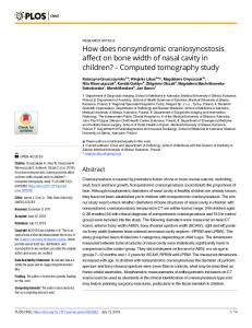
14 patients (eight male; six female) with a median age of 68 years (range 48-82) were studied. The cause of obstruction was pancreatic or ampullary carcinoma in four, cholangiocarcinoma in eight and nodal metastases in one. The fourteenth patient had biliary obstruction secondary to a large pancreatic pseudocyst. The biliary tract was obstructed at the level of the liver hilum in eight, in the mid-common hepatic duct in one and in the distal common bile duct in five. All patients underwent ultrasound, eight had ERCP and six PTC as ERCP was not technically possible. The 2D CT data obtained as the basis for the 3D reconstructions were also included in the evaluation. Two patients subsequently underwent surgery and the operative findings were also included in the analysis. Four patients had had a biliary endoprosthesis inserted previously and one patient had an external biliary drain. In two of the patients with endoprostheses and complex hilar strictures there was only partial decompression of the biliary tree, further intervention was planned for relief of persistent obstructive symptomatology. In the other two the obstructing process had extended beyond the limits of the endoprosthesis or the endoprosthesis itself was blocked. As with 2D CT or MR cholangiography, narrowed or normal sized ducts were beyond the resolution of the technique. In 13 of the 14 patients the 3D cholangiogram demonstrated the dilated ducts, the level of obstruction and the intersegmental anatomy (Figures 1, 2). There was complete agreement between the 3D cholangiogram and the "interventional cholangiogram" obtained either at ERCP or PTC in 13 patients. In the other patient the 3D cholangiogram appeared patchy and incomplete. This patient had a cholangiocarcinoma secondary to primary sclerosing cholangitis and had diffusely irregular, beaded ducts. The 3D image demonstrated the dilated ducts but the narrowed segments were not depicted, interpretation of the final cholangiogram was therefore difficult. In three patients the pancreatic duct was also obstructed and dilated and this was included in the 3D reconstruction. Although the threshold value was selected for each individual patient it was noted that there was very little interpatient variability in the attenuation value of obstructed bile approximately ±3 HU. Structures or lesions with similar attenuation values to the bile ducts were also isolated using the threshold algorithm, in our group of patients this included intrahepatic metastatic deposits and abscesses and a large pancreatic pseudocyst. The relationship between these lesions and the biliary tree was therefore delineated, for example the pancreatic pseudocyst was the cause of biliary obstruction, the 3D image showed how the cyst displaced, distorted and obstructed the bile duct (Figure 2). Discussion
3D CT cholangiography provides an innovative, noninvasive comprehensive demonstration of biliary anatomy. In 13 of 14 patients studied the obstructed biliary system and intersegmental biliary anatomy were The British Journal of Radiology, May 1994
3D CT cholangiography
Figure 2. 3D CT cholangiogram of a large pancreatic pseudocyst (arrow) which is causing obstruction of the distal common bile duct (open arrow) and gross biliary dilatation.
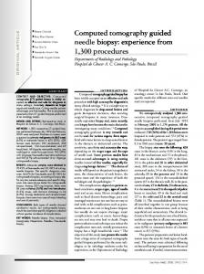
Figure 1. (a) Positive contrast 3D CT cholangiogram showing an irregular occlusion of the mid common hepatic duct (arrow). The open arrow indicates the external biliary drain through which contrast was injected, (b) Conventional cholangiogram confirming the mid hepatic occlusion (arrow).
clearly displayed. In one patient with a cholangiocarcinoma secondary to primary sclerosing cholangitis, the 3D CT cholangiogram accurately depicted the dilated ducts but as neither narrowed nor normal sized ducts were shown, the cholangiogram appeared patchy and interpretation was difficult. To date there has been little reported work on 3D CT imaging of the liver. One study compared the accuracy of segmental localization of metastases using 2D CT during arterial portography (CTAP) and 3D CTAP [10]. All patients subsequently underwent surgery, 3D CTAP was shown to be significantly more accurate. Attempts have been made to perform 3D ultrasound cholangiography but the authors concluded that the images were Vol. 67, No. 797
not diagnostic [11]. Improvements in image registration via a direct digital input could make this a more viable technique. Maximum intensity projection MR cholangiography has recently been described and appears to provide similar information [12]. The drawbacks of this technique include availability, cost and the need for good patient cooperation. 3D reconstruction techniques can be broadly divided into surface or volumetric rendering. Surface rendering techniques operate on a binary system whereby a voxel is assumed to contain all or none of one type of tissue. However, some voxels inevitably contain a mixture of duct and hepatic parenchyma. At segmentation a threshold attenuation value is selected which includes some, all or none of the mixed tissue voxels. Inclusion will result in slightly larger ducts and exclusion in underestimation of duct size, or in smaller ducts in the periphery of the liver exclusion could result in false gaps. The smaller the voxel size, the fewer the voxels containing a mixture of tissues, hence the advantage of using a 4 mm CT slice thickness rather than standard 8 mm slices. In order to aid segmentation further a large difference in attenuation value between duct and parenchyma was generated by injecting 200 ml of intravenous contrast and commencing scanning after the first 100 ml when the parenchyma was already enhanced. False gaps did not occur in the liver hilum in any patient and this technical limitation did not interfere with the demonstration of intersegmental communications. Theoretically a 3D system utilizing volumetric rendering which makes some allowance for voxels containing mixed tissue would provide more accurate images. In reality the surface rendering technique proved satisfactory, costs less and is more widely available. The most optimal images were obtained in those patients who were able to breath-hold in a reproducible 447
A Gillams, J Gardener, R Richards et al
At the present time 3D CT cholangiography is a useful adjunct to other imaging modalities in certain clinical situations. If technical advances in spiral CT result in improved resolution/coverage it is feasible that 3D CT cholangiography will supplant other more invasive procedures such as diagnostic ERCP or PTC. If this is the case then even with the added expense (as compared with standard CT) the benefits of a noninvasive procedure would outweigh the disadvantages. At the present time we anticipate it will be of particular value in patients with complex hilar strictures where knowledge of intersegmental communications is needed. Acknowledgments
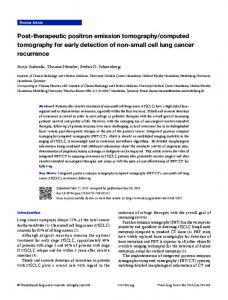
Support for this project was gratefully received from the Information Technology Division, Department of Health, UK and The Wolfson Foundation. Figure 3. A hilar cholangiocarcinoma separates the biliary tract into three groups of ducts. The smallest segment (arrow) was drained as the most readily accessible at ERCP, but obstructive symptoms persisted requiring further intervention. Scrutiny of the 3D CT cholangiogram prior to intervention would have predicted this outcome.
fashion. Spiral CT with its ability to rapidly acquire volumetric data would particularly lend itself to this technique. To date the necessity of sacrificing either spatial resolution or the volume covered has hampered implementation, but improved spiral CT that will overcome these difficulties has been promised in the near future [13]. The major application for our technique lies in the demonstration of intersegmental anatomy. In patients with obstructive jaundice it has been estimated that a minimum of 30% of liver volume needs to be decompressed in order to relieve symptoms of jaundice and pruritus. Patients with type III Klatskin tumours may have several isolated hepatic segments, drainage of the larger segments will provide satisfactory symptomatic relief where drainage of the smaller segments would not. In this situation knowledge of the intersegmental biliary connections can prove invaluable in planning treatment (Figure 3). Similarly in patients with biliary obstruction, multiple isolated segments and sepsis it is often not known which segment or segments are the origin of the septic focus. There are two approaches to this problem: to decompress as much of the liver as possible even if this involves multiple procedures, and insertion of multiple endoprostheses. Alternatively, the septic focus can be identified by ultrasound guided percutaneous fine needle aspiration of bile from different groups of ducts and microbiological analysis can be undertaken. Once again knowledge of intersegmental communication is helpful. 3D CT cholangiography would not necessarily be indicated in every patient with biliary obstruction but in those specific situations where there is a particular need to identify the intersegmental anatomy. In patients with a simple biliary obstruction 2D imaging is usually adequate and the added expense of 3D CT cholangiography is probably not warranted. 448
References 1. SHALEY, S, HAHN, P, BARTEL, L ET AL, The quantitative evaluation of alternative treatment plans. In Proceedings of the Ninth International Conference on the Use of Computers in Radiation Therapy, ed. by I A D Bruinvis, P H van der Giessen, H J van Kleffens and F W Wittkamper. Elsevier Science, Amsterdam, pp. 115-118 (1987). 2. ZONNEVELD, F W, LOBREGT, S, VAN DER MEULEN, J C and VAANDRAGER, J M, Three-dimensional imaging in craniofacial surgery, World J. Surg., 13, 328-342 (1989). 3. FISHMAN, E, MAGID, D, NEY, D ET AL, Three-dimensional imaging, Radiology, 181, 321-327 (1991). 4. LEVIN, D N, HU, X, TAN, K K and GALHOTRA, S, Surface of the brain: three-dimensional MR images created with volume rendering, Radiology, 171, 277-280 (1989). 5. PELIZZARI, C A, CHEN, G T Y, SPELBRING, D R ET AL, Accurate three-dimensional registration of CT, PET and/or MR images of the brain, JCAT, 13, 20-26 (1989). 6. LEVIN, D, HU, X, TAN, K K ET AL, The brain: integrated three-dimensional display of MR and PET images, Radiology, 172 783-789 (1989). 7. MAGID, D, FISHMAN, E K, Imaging of musculoskeletal trauma in three dimensions. An integrated 2-dimensional/ 3-dimensional approach with CT. Radiol. Clin. North Am., 27, 945-956 (1989). 8. SCOTT, JR W W, FISHMAN, E K and MAGID, D, Acetabular fractures: optimal imaging, Radiology, 165, 537-539 (1987). 9. KUHLMAN, J E, FISHMAN, E K, NEY, D R and MAGID, D, Complex shoulder trauma: three-dimensional CT imaging, Orthopedics, 11, 1561-1563 (1988). 10. SOYER, P, ROCHE, A, GAD, M ET AL, Preoperative segmental localization of hepatic metastases: utility of 3 dimensional CT during arterial portography, Radiology, 180, 653-658 (1991). 11. FINE, D, PERRING, S, HERBETKO, J ET AL, Three-dimensional (3D) ultrasound imaging of the gallbladder and dilated biliary tree: reconstruction from real-time B-scans, Br. J. Radiol. 64, 1056-1057 (1991). 12. MORIMOTO, K, SHIMOI, M, SHIRAKAWA, T ET AL, Biliary obstruction: evaluation with three-dimensional MR cholangiography, Radiology 183, 578-580 (1992). 13. FISHMAN, E K, SYATT, S H, NEY, D R ET AL, Spiral CT of the pancreas with multiplanar display, AJR 159, 1209-1215 (1992). The British Journal of Radiology, May 1994


![usa.siemens.com/computed-tomography [PDF]](https://m.moam.info/img/260x300/usasiemenscom-computed-tomography-pdf_6479f808098a9ec7448b4651.jpg)