e-Herz: Case study Herz 2013 DOI 10.1007/s00059-013-3861-8 Received: 28 March 2013 Revised: 19 May 2013 Accepted: 23 May 2013 © Urban & Vogel 2013
e-Herz
Additional material online his article includes four additional Videos. You will find this supplemental at dx.doi. org/10.1007/s00059-013-3861-8.
The term cor triatriatum, also known as “triatrial heart,” usually refers to cor triatriatum sinister or “divided left atrium.” It is a rare congenital anomaly in which the left or right atrium is divided into two parts by a fold of tissue, a membrane, or a fibromuscular band. Classically, the proximal portion of the corresponding atrium receives venous blood, whereas the distal portion is in contact with the atrioventricular valve and contains the atrial appendage [1, 2]. These patients are usually diagnosed early in childhood; however, they are occasional-
M.A. Astarcioglu · M.O. Gürsoy · H. Kaya · S. Karakoyun · M. Kalcik · M. Ozkan Department of Cardiology, Koşuyolu Kartal Heart Training and Research Hospital, Istanbul
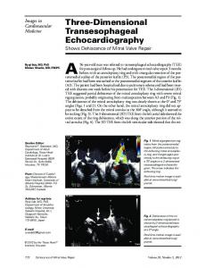
Three-dimensional transesophageal echocardiographic evaluation of cor triatriatum in adults
ly discovered later in adulthood [2]. The membrane that separates the atrium into two parts varies significantly in size and shape. The course and prognosis of cor triatriatum depend on the degree of obstruction and magnitude of the gradient across the membrane fenestrations. We present the cases of two adult patients diagnosed with cor triatriatum. The diagnosis was made with transesophageal echocardiography, and the anatomic characteristics of the embryonic remnant were determined precisely by realtime three-dimensional transesophageal echocardiography, and finally confirmed at surgery.
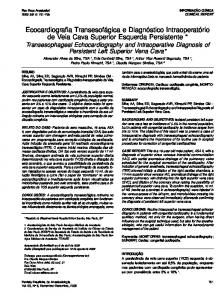
Case presentations Patient 1 A 69-year-old woman presented with the complaint of exertional shortness of breath lasting several months. Her medical history included systemic hypertension and recent peripheral pitting edema of the lower extremities. A soft systolic heart murmur with a split S2 was heard on physical examination. Two-dimensional transthoracic echocardiography (TTE) revealed normal left ventricular systolic function and severe tricuspid regurgitation with an estimated pulmonary artery systolic pressure of 80 mmHg. The right atrium (RA) was dilated and divided into two distinct chambers by a membranous septum. Transesophageal echocardiography (TEE) was performed to confirm the diagnosis and exclude
Fig. 1 8 a Transthoracic two-dimensional echocardiography (see also Video 1 online) and b real-time three-dimensional transesophageal echocardiography demonstrating the right atrium divided into two distinct chambers by a membranous septum and secundum ASD (see also Video 2 online). ASD atrial septal defect, LA left atrium, RA right atrium, LV left ventricle, IAS interatrial septum, arrowheads a communication orifice Herz 2013
| 1
e-Herz: Case study
Abstract · Zusammenfassung Herz 2013 · [jvn]:[afp]–[alp] DOI 10.1007/s00059-013-3861-8 © Urban & Vogel 2013 M.A. Astarcioglu · M.O. Gürsoy · H. Kaya · S. Karakoyun · M. Kalcik · M. Ozkan
Three-dimensional transesophageal echocardiographic evaluation of cor triatriatum in adults Abstract We present the cases of two adult patients with cor triatriatum due to left atrial membrane with atrioventricular septal defect and right atrial membrane. Two-dimensional and real-time three-dimensional transthoracic echocardiography were performed. These noninvasive modalities provided a comprehensive anatomic and hemodynamic evaluation of the anomaly. Fig. 2 8 Transesophageal two-dimensional echocardiography demonstrates ostium primum type ASD and two left-sided distinct chambers. ASD atrial septal defect
other associated congenital abnormalities. A membrane extended from the inferior vena cava to the interatrial septum, which was defined as cor triatriatum dexter. A secundum type atrial septal defect (ASD) with a 2-cm drop-out was also detected. Real-time three-dimensional transesophageal echocardiography (RT-3D TEE) demonstrated a membrane that separated the RA and the secundum ASD in“en face” views (. Fig. 1, Videos 1 and 2). A cross-sectional plane of the membrane allowed measurement of the maximal fenestration orifice area of 1.6 cm2. The patient was referred for surgery. The excised membrane was identical to the RT-3DTEE image. The secundum ASD was closed with a pericardial patch and the postoperative course was uneventful. Postoperative TEE revealed an intact interatrial septum with no residual membrane (Video 3).
Patient 2 A 43-year-old man presented with pedal edema and dyspnea at rest. His jugular venous pressure was 10 cm. A grade 3/6 holosystolic murmur was heard at the 4th left intercostal space and apex. Auscultation of the chest revealed bilateral fine crackles on both lower lung fields. His electrocardiogram (ECG) showed sinus rhythm. A chest X-ray revealed an in-
2 |
Herz 2013
creased cardiothoracic ratio and bilateral pleural effusion. TTE and TEE showed dilated right-sided heart chambers, mild tricuspid and moderate-to-severe mitral regurgitation with mitral cleft, and an ostium primum type ASD. There was no ventricular septal defect (. Fig. 2, Video 4). A membrane separating the left atrium (LA) into an anterior and a posterior chamber was seen (. Fig. 3). RT3D TEE revealed that the membrane was not intact, and that there was a communication between the two LA chambers (. Fig. 4). There was no significant pressure gradient across the membranous septum and no color flow acceleration was seen across the membrane. The patient was referred to surgery. The membrane was excised, and the primum ASD and mitral valve were repaired. The postoperative course was uneventful. Postoperative TEE revealed an intact interatrial septum with no residual membrane.
Discussion Cor triatriatum is an uncommon anomaly with an estimated incidence of 0.1– 0.4% of all congenital cardiac malformations [3]. In cor triatriatum sinister, the left atrium is divided by a membrane into a posterior–superior chamber that receives the four pulmonary veins and an anterior–inferior chamber that connects
Keywords Two-dimensional transesophageal echocardiography · Real-time threedimensional transesophageal echocardiography · Cor triatriatum · Congenital heart disease · Atrioventricular septal defect
Dreidimensionale transösophageale echokardiographische Untersuchung eines Cor triatriatum bei Erwachsenen Zusammenfassung Vorgestellt werden die Fälle zweier erwach sener Patienten mit Cor triatriatum auf grund einer linksatrialen Membran mit atrioventrikulärem Septumdefekt und ei ner rechtsatrialen Membran. Es wurden eine zweidimensionale Echokardiographie und eine dreidimensionale transthorakale Echt zeitkardiographie durchgeführt. Mit diesen nichtinvasiven Verfahren ließen sich die Anatomie und die Hämodynamik bei dieser Anomalie umfassend untersuchen. Schlüsselwörter Zweidimensionale transösophageale Echokardiographie · Dreidimensionale transösophageale Echtzeitechokardiographie · Cor triatriatum · Kongenitale Herzerkrankung · Atrioventrikulärer Septumdefekt
to the left ventricle through the mitral valve. In cor triatriatum dexter, a membrane divides the right atrium into two chambers. It is believed that cor triatri-
Fig. 3 9 Transthoracic two-dimensional echocardiography demonstrates a membrane dividing the left atrium (LA) into two distinct chambers (see also Video 4)
Fig. 4 8 Three-dimensional en face view of the membrane from the posterosuperior left atrium chamber showed the whole of the abnormal membrane and its fenestration orifice (arrowheads). RA right atrium, Ao aortic valve
atum dexter results from persistence of the right valve of the sinus venosus [4]. In 1949, Loeffler classified cor triatriatum into three groups based on the number and the size of the fibromuscular septum: group 1 has no opening; group 2 has one or more small openings in the septum, leading to a high grade of obstruction; and group 3 has a wide opening in the membrane resulting in little or no obstruction [5]. Cor triatriatum has various clinical manifestations depending on the degree of communication between the two chambers. Adult patients typically present with dyspnea, hemoptysis, and orthopnea. Cor triatriatum can be frequently misdiagnosed as mitral valve disease or primary pulmo-
nary hypertension. Transthoracic echocardiography can accurately help diagnose the majority of cases of cor triatriatum. TEE has the advantage of enhanced visualization of the LA, LA appendage, membrane, and the pulmonary veins, thus providing higher sensitivity. Threedimensional echocardiography provides unique and better spatial orientation, and allows visualization of the size and number of fenestrations on the partitioning membrane as in our patients. There are few reports in the literature describing the role of 3D-TEE in the assessment of cor triatriatum [6, 7]. We emphasize that RT-3D TEE is useful for diagnosing cor triatriatum dexter, a membrane with a large opening separating the vena ca-
va superior from the right atrium inferiorly, such as in patient 1. The secundum type ASD was also clearly demonstrated on RT-3D TEE. The second patient represents an uncommon case of cor triatriatum with partial atrioventricular septal defect (AVSD). Association of cor triatriatum with AVSD is rare; only three cases with complete AVSD and ten with partial AVSD have been reported to the best of our knowledge [8]. Whether there is a common embryologic link between the two is not clear. In general, patients with AVSD and cor triatriatum have high right ventricular, pulmonary artery, and pulmonary wedge pressures. Pre- and intraoperative echocardiography is essential to achieve successful surgical results for cor triatriatum with AVSD. Management of cor triatriatum depends on the grade of obstruction between the chambers. Surgery is generally reserved for those patients with significant obstruction. Excision of the dividing membrane often provides symptomatic relief, as in our patients.
Conclusion There are multiple imaging techniques used in the diagnosis of cor triatriatum, such as 2D echocardiography, TEE, computed tomography, and magnetic resonance imaging. Cor triatriatum can be accurately diagnosed with 3D echocardiography, which allows direct visualization and planimetric measurement of the fenestrations. 3D echocardiography is an excellent noninvasive method that provides a rapid bedside diagnosis for Herz 2013
| 3
e-Herz: Case study this uncommon congenital heart disease compared to magnetic resonance imaging. Finally, cor triatriatum in association with AVSD is an infrequent but life-threatening anomaly that should be considered as a cause of heart failure.
Corresponding address M.A. Astarcioglu Department of Cardiology, Koşuyolu Kartal Heart Training and Research Hospital Istanbul Turkey
[email protected] Conflict of interest. On behalf of all authors, the corresponding author states that there are no conflicts of interest.
References 1. Godoy I, Tantibhedhyangkul W, Karp R et al (1998) Images in cardiovascular medicine: cor triatriatum. Circulation 98:2781 2. Sen T, Guray Y, Demirkan B et al (2010) Cor triatriatum sinister in a 67-year-old man with atrial fibrillation. Tex Heart Inst J 37(2):246–247 3. Arrants JE, Riopel DA, Catalano PW (1973) Cor triatriatum: preoperative diagnos is and successful surgical correction in a ten-week-old infant. Chest 63:1027–1028 4. Schutte DA, Rowland DG, Allen HD et al (1997) Prominent venous valves in hypoplastic right hearts. Am Heart J 134:527–531 5. Loeffler E (1949) Unusual malformation of the left atrium: pulmonary sinus. Arch Pathol 48:371–376 6. Melzer C, Bartel T, Muller S et al (1997) Dynamic three dimensional echocardiography in the assessment of cor triatriatum. Clin Cardiol 20:82–83 7. D’Aloia A, Vizzardi E, Caretta G et al (2011) Diagnosis of cor triatriatum sinister in patient with pulmonary edema and severe pulmonary arterial hypertension: assessment by three-dimensional transesophageal echocardiography. Echocardiography 28:198–201 8. Goel AK, Saxena A, Kothari SS (1998) Atrioventricular septal defect with cor triatriatum: case report and review of the literature. Pediatr Cardiol 19:243–245
4 |
Herz 2013