The British Journal of Radiology, 83 (2010), 882–887
SHORT COMMUNICATION
Time-resolved imaging of contrast kinetics three-dimensional (3D) magnetic resonance venography in patients with pelvic congestion syndrome E A DICK, MRCP, FRCR, MD, C BURNETT, A ANSTEE, and W M W GEDROYC, MRCP, FRCR
MRCP, FRCR,
M HAMADY,
FRCS, FRCR,
D BLACK,
FRCS
Department of Radiology and Vascular Surgery, Imperial College NHS Trust, St Mary’s Hospital, Praed St, London W2 1NY, UK
ABSTRACT. The purpose of this study was to assess the role of magnetic resonance venography (MRV) with time-resolved imaging of contrast kinetics (TRICKS) in dynamically evaluating ovarian vein dilation, reflux and direction of flow in patients with suspected pelvic congestion syndrome (PCS). The hypotheses tested were: (i) That conspicuity scores of the ovarian veins across three raters was greater using TRICKS MRV compared with T2W or T2* imaging; (ii) That three key MR variables (ovarian vein diameter, timing and grade of reflux) correlated across all raters. We carried out a retrospective study of 13 patients undergoing T2W and TRICKS MRI and pelvic sonography (n54) or catheter venography (n55). Three observers rated conspicuity, vessel diameter, timing and grade of ovarian vein reflux for T2/T2*W and TRICKS MRI. The mean left ovarian diameter for all patients with reflux was 7.9 mm (range 2.2– 12 mm). There was high inter-observer agreement for ovarian vein diameter for both sequences. TRICKS showed significantly greater conspicuity than T2/T2*W imaging (TRICKS: T2/T2* mean (SD)57.80 (3.20):5.50 (1.97), F (1,12)55.80, p , 0.05). TRICKS MRV demonstrated high inter-observer correlation for timing and grade of reflux (r (36) 50.77,0.71,0.79, p , 0.01). TRICKS MRA/V was significantly degraded by breathing artefact in two patients. We conclude that TRICKS MRV accurately and dynamically demonstrates ovarian vein reflux in patients with PCS but requires quiet respiration. TRICKS MRV has better image conspicuity than T2/T2*W imaging and sufficient temporal resolution to distinguish between Grade I, II and III reflux.
The purpose of this study was to assess the role of time-resolved imaging of contrast kinetics magnetic resonance venography (TRICKS MRV) in dynamically evaluating ovarian vein dilation, reflux and direction of flow in patients with suspected pelvic congestion syndrome (PCS). PCS is the presence of long-standing pelvic pain due to a variety of causes, including ovarian vein incompetence [1]. The most important aetiological factor is parity, which is associated with ovarian vein dilation and valvular incompetence in 50–73% of women; although it is important to note that not all women with ovarian vein incompetence are symptomatic [1–3]. In women whose PCS is caused by ovarian vein incompetence, surgical ligation or transcatheter ovarian vein embolisation can be successful [4, 5]. To select patients suitable for surgical or interventional therapy, the ideal diagnostic technique has high spatial and temporal resolution, is non-invasive and does not involve ionising radiation. One such technique may be Address correspondence to: Dr Elizabeth A Dick, Department of Radiology, Imperial NHS Trust, St Mary’s Hospital, Praed St, London W2 1NY, UK. E-mail:
[email protected] Financial Disclosures/Funding: None
882
Received 24 December 2009 Revised 7 March 2010 Accepted 1 April 2010 DOI: 10.1259/bjr/82417499 ’ 2010 The British Institute of Radiology
TRICKS MRV, a modified 3D fast gradient echo (GRE) pulse sequence [6]. Here we evaluate the role of TRICKS MRV in assessing ovarian veins, including image conspicuity, spatial and temporal resolution.
Methods and materials Patients All procedures were carried out in accordance with the ethical standards of the World Medical Association (Declaration of Helsinki). Our institutional Ethics Committee considered that an ethical opinion was not needed for this retrospective study. The study population consisted of 13 women with a clinical diagnosis of PCS (mean age 42.6 years, range 27–72 years old), referred over a 12-month period by the Department of Vascular Surgery for Magnetic Resonance Imaging of the pelvis. All patients underwent T2 or T2*W axial and coronal imaging and TRICKS MRV. Five patients underwent ovarian vein embolisation, four underwent pelvic ultrasonography. A retrospective review of imaging and case notes was conducted in all cases. The British Journal of Radiology, October 2010
Short communication: TRICKS MRA/V in pelvic congestion syndrome
Imaging techniques Axial MR imaging was performed with a 1.5T unit (GE Medical Systems, Milwaukee, WI) using a four-element phased-array torso coil. All protocols included the following sequences: a three-plane single-shot fast spinecho (SSFSE) localiser followed by axial and coronal T2W chemically selective fat saturated imaging – either T2 fast spin-echo (FSE) or fast imaging employing steady-state acquisition (FIESTA). Imaging parameters for the axial T2 FSE fat saturated sequence (repetition time (TR)/echo time (TE) echo train54060 ms/70.6 ms/10, field of view (FOV)530 6 30 cm, matrix5384 6 224, CLASSIC fat saturation, GE Medical Systems, Milwaukee, WI) resulted in a total of 29 slices with slice thickness of 10 mm with a 2 mm gap in 4.39 minutes. Coronal T2 FSE fat saturated sequences were acquired (TR/TE/echo train5 3,400 ms/75 ms/10, FOV530 6 30 cm, matrix5384 6 224) with a total of 14 slices with slice thickness of 8 mm with a 2 mm gap in 4.16 minutes. Imaging parameters for the FIESTA sequence were: TR calculated/TE/ flip angle/band width (BW)53.6/1.5/70 ˚/83.33 kHz, FOV538 6 38 cm, matrix5288 6 192, frequency5192, phase5288, number of excitations (NEX)51, phase FOV 1, resulting in 26 slices with a slice thickness of 4 mm, with a 0.5 mm gap and acquisition time of 90 seconds. 3D volumetric coronal TRICKS MR angiography and venography (TRICKS MRA and MRV) were performed in quiet respiration (TR/TE/flip/BW55.4/ 2.0/45 ˚/ 62.5 kHz, FOV543 6 30 cm, equivalent slice thickness of 1.8 mm, 3/4 phase FOV, matrix5512 6 192, 25 6 6.9 second time frames). Contrast medium (15 ml gadopenate dimeglumine) was administered by hand injection (Bracco UK, High Wycombe, Bucks, UK) followed by a normal saline flush of 20 ml. One coronal volume prescription from the level of T12 to the pubic symphysis achieved full venous coverage. A mask was acquired prior to intravenous contrast injection [7], followed by 25 post-contrast phases, each taking 6.9 seconds to acquire the entire abdominopelvic volume. Hence, the complete post-contrast acquisition took 2.87 minutes (6.9 6 25 second time windows). The study was post-processed into 25 collapsed maximumintensity projection (MIP) images, but review emphasised the source images. Pelvic ultrasonography was performed transvaginally or transabdominally in quiet respiration and with the Valsalva manoeuvre. The presence and diameter of ovarian and uterine veins and the presence of reversed flow on the Valsalva manoeuvre was recorded. Conventional digital subtraction venography (DSV) studies were performed in five patients (six studies in total) using the Seldinger approach via a right internal jugular vein or right common femoral vein puncture, 0.035 inch guidewire (Boston Scientific, Natick, MA), 4 Fr or 5 Fr modified varicocele embolisation kit catheter (William Cook Europe, Bjaeverskov, Denmark) and selective left (or right) ovarian vein catheterisation, opacification and subsequent embolisation with multiple coils (platinum-free spirales 6 6 30 mm to 10 6 120 mm, BALT Extrusion, Montmerency, France). DSV images were acquired with a 38 cm FOV (Philips INTEGRIS, Philips Medical Systems, Reigate, UK). Injections with a non-ionic contrast agent (Omnipaque, The British Journal of Radiology, October 2010
Nycomed Amersham, Buckinghamshire, UK) were performed by hand injection and sequential DSV images were obtained.
Image analysis Source, MIP and rotational MIP images were retrospectively independently reviewed in random order on a picture archive computer station (PACS) workstation by three radiologists (‘raters’) with 20, 10 and 5 years’ experience of MRI and/or vascular radiology. The raters were unaware of the results of other examinations. T2W, FSE or FIESTA axial and coronal images were compared with TRICKS MRV for image conspicuity, vessel diameter and grade and timing of reflux. Conspicuity of ovarian vein visualisation was assessed on a 10-point scale (05not visible, 105maximum conspicuity). The maximum ovarian vein and parauterine vein diameter (of the largest vessel if there were multiple vessels) was recorded for the axial and coronal T2/T2*W images and the coronal TRICKS sequences. The presence of an ovarian vein of a diameter 6 mm or greater was considered diagnostic of dilation and likely incompetence, while parauterine veins were considered abnormal if tortuous and of a diameter greater than 4 mm [8]. The grade of reflux was assessed as follows, based on Hiromura et al’s classification: grade I (Figure 1a): retrograde flow remains in the left ovarian vein; grade II (Figure 1b): retrograde flow advances into the ipsilateral parauterine vessels; grade III (Figure 1c): the retrograde flow crosses the midline via the uterine plexus [9]. The grade of reflux was indirectly assessed on static T2/ T2*W images from the degree of ovarian and parauterine venous dilation [9]. For dynamic TRICKS MRA/V the retrograde direction of flow was directly visualised and confirmed or refuted in each case. The first frame in which the upper left ovarian vein opacified (grade I), the first frame in which the left parauterine veins opacified (grade II) and the first frame in which the right parauterine or right ovarian vein opacified where this occurred (grade III) were recorded [9]. In patients undergoing DSV, grade of reflux was recorded for comparison to T2/T2*W or TRICKS MRV.
Statistical analysis All statistical calculations were performed using SPSS v12 (SPSS Inc, Chicago, IL). In order to test the conspicuity of T2/T2*W vs TRICKS MRV, ANOVA was performed with conspicuity scores entered as the dependent variable. Three categorical explanatory variables were entered: i) T2/T2*W vs TRICKS; ii) rater (n53); iii) patients (n513). The first two variables were entered as fixed factors, while the patients were entered as a random factor. Three separate analyses designed to explore correlations between the three raters were also performed: i) ovarian vein diameter values, as assessed on T2/T2*W images; ii) ovarian vein diameter values, as assessed on TRICKS images; iii) grading of reflux (grades I–III) as assessed on TRICKS MRV images. Inter-observer agreement across these variables was tested using a nonparametric test, Pearson correlation (two-tailed). 883
E A Dick, C Burnett, A Anstee et al
(a)
(b)
(c)
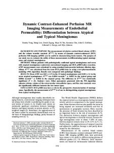
Figure 1. Line diagram of female abdominopelvic venous anatomy. The left ovarian vein drains to the left renal vein, while the right ovarian vein usually drains into the anterolateral IVC (but occasionally drains to the right renal vein). The ovarian veins form part of a complex of venous interconnections, which includes the uterine plexus. Three grades of reflux have been described [9]. (a) Grade I ovarian vein reflux: retrograde flow remains in the left ovarian vein. IVC, inferior vena cava; LK, left kidney; U, uterus. (b) Grade II ovarian vein reflux: flow advances into the ipsilateral parauterine veins. (c) Grade III ovarian vein reflux: retrograde flow crosses the midline across the uterine plexus ¡ into the right ovarian vein.
A paired t-test was performed for each rater to compare the left ovarian diameter measurements from T2/T2*W images with those from TRICKS MRV. Correlation between DSV or sonography and T2/T2*W or TRICKS for grade of reflux was compared for all patients who underwent both. The effects of embolisation on those who underwent interventional therapy were also recorded.
Results MRI was performed on 13 consecutive women. No patients were excluded from analysis. Of these, 11 patients had ovarian vein incompetence with grade II or grade III reflux and a left ovarian vein diameter of 6 mm or greater, or a parauterine vein diameter of 4 mm or greater in the presence of multiple vessels. The mean left ovarian vein diameter for patients with reflux for all observers was 7.9 mm (range 2.2–12 mm). Two patients did not have ovarian vein incompetence: one had a gluteal arteriovenous malformation causing vulval varices, the second had no evidence of reflux and a single left ovarian vein of diameter 5 mm. In two patients, TRICKS MRV images were degraded by movement artefact due to breathing, in both cases the T2/T2*W imaging was adequate. The ANOVA performed on the conspicuity scores (rated 0–10) demonstrated a significant difference in the values obtained for the left ovarian vein for TRICKS MRV vs standard T2 imaging across all three raters (TRICKS: T2 mean (SD)57.80 (3.20):5.50 (1.97), F (1,12)5 5.80, p , 0.05). 884
There were significant correlations between all three observers on the parameters examined. For left ovarian vein diameter there was a significant correlation between all three observers on TRICKS imaging, r(10)50.99, 0.89, 0.84, p , 0.01. For left ovarian vein diameter measurements assessed on T2/T2*W imaging there was a similarly significant effect, r(10)50.98, 0.82, 0.76, p , 0.01. Mean left ovarian vein diameters for each observer are recorded in Table 1. Note that the mean diameter is over a narrower range for TRICKS than T2/T2*W imaging (range of 5.36–5.59 mm for TRICKS, range of 5.85– 7.05 mm for T2/T2*W imaging). There was a trend towards the T2/T2*W imaging yielding greater left ovarian vein diameter measurements than the TRICKS MRA/V imaging but this was non-significant (p . 0.05 for each radiologist). Inter-observer agreement on the first frames that each grade (I–III) of reflux was not assessed on T2W or T2* imaging, since they lack a temporal component, but was assessed on TRICKS MRV since this has multiple sequential acquisitions. For TRICKS MRV there was a significant correlation between raters across all three Table 1. Mean left ovarian vein diameter ¡ standard deviation (mm) for each observer for all patients enrolled in study (n513) measured on T2W/FIESTA and TRICKS MRV Observer No.
T2W/FIESTA*
TRICKS MRV*
1 2 3
6.35 mm (3.5) 5.85 mm (3.6) 7.05 mm (3.8)
5.52 mm (3.3) 5.36 mm (3.4) 5.59 mm (3.9)
*For both T2/T2*W and TRICKS MRCV there is a statistically significant correlation between observers.
The British Journal of Radiology, October 2010
Short communication: TRICKS MRA/V in pelvic congestion syndrome
(a)
(b)
(c)
(d)
(e)
(f)
The British Journal of Radiology, October 2010
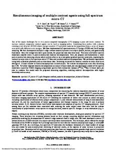
Figure 2. 34-year-old woman with pelvic congestion symptoms and grade III ovarian vein reflux treated successfully with bilateral ovarian vein embolisation. (a)(b) Coronal T2W fast imaging employing steady-state acquisition (FIESTA) demonstrates dilated left ovarian vein (arrow, 2a) and dilated parauterine plexus (arrow, 2b). (c)(d)(e) Coronal sequential TRICKS MRV demonstrates retrograde filling of the left ovarian vein (arrow, 2c), followed by the uterine plexus (arrow, 2d) and, finally, by filling of the right ovarian vein via the uterine plexus (arrow, 2e), equating to grade III reflux. (f) Transfemoral percutaneous ovarian venography demonstrated grade III reflux, subsequently coil embolisation of the left and right ovarian veins was performed. Left ovarian vein coils in situ (arrow). Right ovarian venogram demonstrates dilated venous structures (arrowheads).
885
E A Dick, C Burnett, A Anstee et al
grades, r(36)50.77,0.71,0.79, p , 0.01, implying that there was good inter-observer agreement on the first frame in which each grade of reflux was seen. Both T2/T2*W imaging and TRICKS MRV correlated with pelvic sonography (n54) or venography (n55) in assessing the presence of ovarian vein dilation and/or the grade of reflux. All five patients who underwent direct digital subtraction venography and ovarian vein embolisation (six procedures, one left and one right ovarian vein coiling in one patient), reported good to excellent relief of their pelvic congestion symptoms (4–10 months after embolisation) (see Figure 2).
Discussion To our knowledge, this study is the first to use a multiphase MRA/V technique to dynamically image the ovarian veins and confirm or refute the presence of reflux in women with clinically diagnosed PCS. Our study clearly demonstrates that TRICKS MRA/V can delineate ovarian vein and parauterine venous dilation with greater conspicuity than that achieved with conventional T2/ T2*W imaging. Both T2/T2*W and TRICKS MRV imaging yielded high inter-observer correlation for left ovarian vein diameter, implying good spatial resolution for both techniques. The close correlation between all three observers, as to the timing of the arrival of the contrast bolus within the ovarian vein and parauterine veins on TRICKS MRV, implies sufficient temporal resolution to assess dynamic filling and grade of reflux in a way that single-phase CT or even three-phase MRA could not do. While for conventional contrast-enhanced MRA it is critical to co-ordinate the acquisition of central k-space with contrast arrival, TRICKS is less dependent on bolus arrival and, therefore, there is no need to co-ordinate bolus arrival with image acquisition [7, 10]. Like ultrafast/ subsecond contrast-enhanced MRA, TRICKS MRA/V can achieve temporal sampling rapid enough to depict shortlived vascular processes such as shunts. However, unlike ultrafast contrast-enhanced MRA, in TRICKS MRA/V there is no trade-off of low spatial for good temporal resolution [11]. In TRICKS, spatial resolution remains comparable with conventional MRA [6]. TRICKS combines elliptical centric (EC) view order with undersampled Cartesian-based acquisition techniques to generate high spatial and high temporal resolution MRV [10]. EC view ordering ensures high temporal resolution with an effective frame rate of one volume every 2–6 seconds, ensuring good arterial–venous discrimination [11–13]. The undersampled Cartesian-based technique of k-space segmentation samples the entire k-space for each time point. K-space is divided along the phase encoding direction with each region (A, B and C), sampling the entire set of section encoding values. The central region A, responsible for contrast resolution, is sampled more frequently than regions B and C (peripheral k-space, responsible for higher spatial frequencies). This ensures the central k-space segment is acquired during peak arterial or venous enhancement [7]. Since TRICKS is a 3D imaging technique, image acquisition is near isotropic and, therefore, rotational MIP images can be produced in addition to volume rendered images [6]. Spatial resolution is around 1 mm2 in-plane and 1–2 mm2 through-plane. 886
TRICKS has a role in several anatomical areas: for detecting significant arterial stenosis, it has a sensitivity of 87–94% and specificity of 90–92% when compared to conventional transcatheter peripheral angiography [6, 14]. Initial reports on time-resolved dynamic contrastenhanced 3D MR urography, peripheral venography and early studies of time-resolved large FOV 3D MRA at 3T also show promise [15, 16]. TRICKS has obvious advantages over more traditional methods of diagnosing ovarian vein incompetence, such as transfemoral venography, ultrasonography, CT and MRI, including lack of ionising radiation or intervention and the production of 3D volume images dynamically over multiple phases [9]. However, there are some drawbacks to TRICKS MRV. Firstly, the mask is acquired without producing an image for the operator, therefore, the operator cannot confirm that the area required has been covered, although T2W imaging can overcome this. A second disadvantage is that patients cannot breath-hold for the entire image acquisition of 25 phases (2.87 minutes). So there may potentially be some mis-registration of mask and data imaging due to breathing – a problem that we encountered in 2 out of 13 patients. TRICKS MRA/V has the further disadvantage of generating large data sets requiring intensive image processing [10]. Our study has some inherent weaknesses including: a relatively small patient group; that interventional DSV and sonography was not performed in all patients; and that TRICKS MRA/V was non-diagnostic in two patients due to respiratory motion. In addition, all imaging was performed with the patient supine during quiet respiration, without a Valsalva manoeuvre.
Conclusions In summary, we have found that TRICKS MRV is a useful addition to T2W or T2* axial and coronal imaging since it provides significantly greater conspicuity than T2/T2*W images with sufficient temporal resolution to distinguish between grade I, II and III reflux. The radiological findings of ovarian vein dilation and reflux must be taken within the context of underlying symptoms, but TRICKS MRA/V may be of value in the diagnosis of pelvic vein dilation and reflux as a cause of PCS. It is non-invasive, does not require ionizing radiation and has good spatial and temporal resolution. Furthermore, TRICKS MRA/V may enable suitable patients to go onto definitive one-stage venographic embolisation or surgical ligation.
Acknowledgments We would like to thank Dr A Leff and Dr F Gordon for their helpful guidance.
References 1. Pui M. Imaging of vascular disorders of the female pelvis. Australas Radiol 2006;50:405–11. 2. Tarazov PG, Prozorovskij KV, Ryzhkov VK. Pelvic pain syndrome caused by ovarian varices. Treatment by transcatheter embolization. Acta Radiol 1997;38:1023–5.
The British Journal of Radiology, October 2010
Short communication: TRICKS MRA/V in pelvic congestion syndrome 3. Kim C, Miller MJ Jr, Merkle EM. Time-resolved MR angiography as a useful sequence for the assessment of ovarian vein reflux. American Journal of Roentgenology 2009;193:W458–63. 4. Capasso P, Simons C, Trotteur G, Dondelinger RF, Henroteaux D, Gaspard U. Treatment of symptomatic pelvic varices by ovarian vein embolization. Cardiovasc Intervent Radiol 1997;20:107–11. 5. Gandini R, Chiocchi M, Konda D, Pampana E, Fabiano S, Simonetti G. Transcatheter foam sclerotherapy of symptomatic female varicocele with sodium-tetradecyl-sulfate foam. Cardiovasc Intervent Radiol 2008;31:778–84. 6. Mell M, Tefera G, Thornton F, Siepman D, Turnipseed W. Clinical utility of time-resolved imaging of contrast kinetics (TRICKS) magnetic resonance angiography for infrageniculate arterial occlusive disease. J Vasc Surg 2007;45::543–8; discussion 548. 7. Carroll TJ, Korosec FR, Petermann GM, Grist TM, Turski PA. Carotid bifurcation: evaluation of time-resolved threedimensional contrast-enhanced MR angiography. Radiology 2001;220:525–532. 8. Coakley FV, Varghese SL, Hricak H. CT and MRI of pelvic varices in women. J Comput Assist Tomogr 1999;23:429–34. 9. Hiromura T, Nishioka T, Nishioka S, Ikeda H, Tomita K. Reflux in the left ovarian vein: analysis of MDCT findings in asymptomatic women. Am J Roentgenol 2004;183: 1411–15.
The British Journal of Radiology, October 2010
10. Du J, Bydder M. High-resolution time-resolved contrastenhanced MR abdominal and pulmonary angiography using a spiral-TRICKS sequence. Magn Reson Med 2007; 58:631–5. 11. Salanitri J. MR angiography of aberrant left subclavian artery arising from right-sided thoracic aortic arch. Br J Radiol 2005;78:961–6. 12. Madhuranthakam AJ, Hu HH, Barger AV, Haider CR, Kruger DG, Glockner JF, et al. Undersampled elliptical centric view-order for improved spatial resolution in contrast-enhanced MR angiography. Magn Reson Med 2006;55:50–8. 13. Korosec FR, Frayne R, Grist TM, Mistretta CA. Timeresolved contrast-enhanced 3D MR angiography. Magn Reson Med 1996;36:345–51. 14. Swan JS, Carroll TJ, Kennell TW, Heisey DM, Korosec FR, Frayne R, et al. Time-resolved three-dimensional contrastenhanced MR angiography of the peripheral vessels. Radiology 2002;225:43–52. 15. Frydrychowicz A, Bley T, Zadeh ZA, Harloff A, Winterer J, Hennig J, et al. Image analysis in time-resolved large field of view 3D MR-angiography at 3T. Journal of Magnetic Resonance Imaging 2008;28:1116–24. 16. Kim S, Jacob J, Kim D, Rivera R, Lim R, Lee V. Timeresolved dynamic contrast-enhanced MR urography for the evaluation of ureteral peristalsis: initial experience. Journal of Magnetic Resonance Imaging 2008;28:1293–8.
887