FIO/ LS Technical Digest @ 2011 OSA
Movies of nanoscale dynamics using soft x-ray laser illumination S. Carbajo1,2†, I. D. Howlett1,2, A. Sakdinawat1,4, Y. Liu1,4, W. Chao1,3, E.H. Anderson1,3, A. V. Vinogradov5, I. A. Artioukov5, D.T. Attwood1,4, M. C. Marconi1,2, J.J. Rocca1,2, and C.S. Menoni1,2 1NSF ERC for Extreme Ultraviolet Science and Technology 2Electrical and Computer Engineering, Colorado State University, Fort Collins, USA 3Center for X-ray Optics, Lawrence Berkeley National Laboratory, Berkeley, USA 4ECE Department, University of California, Berkeley, USA 5P. N. Lebedev Physical Institute, Moscow, Russia †Corresponding author:
[email protected]
Abstract: Movies of magnetic force microscope tips oscillating at 65.5 kHz were acquired using flash soft x-ray laser illumination. Changes in the oscillation amplitude of 11 ± 4 nanometers were detected. OCIS codes: (180.0180); (120.0120)
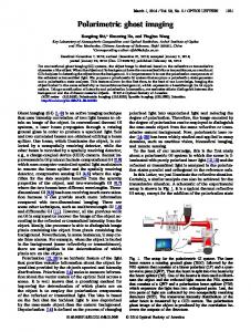
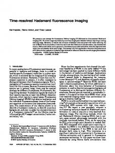
With pulses of tens of microjoule energy and picosecond to nanosecond pulse duration, table-top soft xray lasers offer the capabilities to record time-resolved images with high spatial resolution [1-2]. The implementation of full-field microscopes exploiting this technology have proven to be capable of capturing images of nanostructures with 50 nm spatial resolution using a single laser shot [3]. Implicit to such demonstration, these microscopes have the ability to freeze the motion of nanoscale processes and render animations of their dynamics through a sequence of single-shot acquisitions. To demonstrate his capability, we have mapped the motion of a magnetic force microscope tip interacting with a magnetic sample using a microscope that employs a desk-top size soft x-ray laser source for illumination. Single-shot real-space sequential full-field images of a resonating magnetic cantilever were acquired employing a zone plate objective to image objects with 1200x magnification onto a CCD detector (Figure 1). The illumination is provided by a desktop-size λ=46.9 nm soft x-ray laser with 10 μJ pulse energy (~1012 photons/pulse) of ~ 1.5 ns pulse duration [2-3]. Amplitude shifts as small as ~10 nm in the natural oscillation of the magnetic tip, originated from an external magnetic force, were measured by capturing synchronized single-shot images of the tip motion (Figure2). To obtain these data the magnetic cantilever was driven at its resonance frequency of 65.5 kHz while the magnetic interaction conditions were reproduced to resemble those in standard magnetic force microscope (MFM) setups. For this experiment, the stray magnetic fields from permalloy stripes were used as the perturbing agent and tip-to-magnetic surface separations of 100-200 nanometer were selected. This technique opens new imaging possibilities to freeze the dynamics of nanoscale processes. Furthermore, this proof-of-principle demonstration of dynamic imaging is wavelength scalable. The recent demonstration of pulse energies of ~ 10 μJ at λ = 13.9 nm from table-top lasers has potential to extend time-resolved imaging to picosecond time resolution and better than 38 nm spatial resolution [4]. This work was supported by the Engineering Research Centers Program of the National Science Foundation under NSF Award Number EEC-0310717.
1 FTuL3.pdf 1
9/30/2011 10:13:02 AM
FIO/ LS Technical Digest @ 2011 OSA
Fig. 1. Experimental setup of synchronized single-shot flash imaging
Natural Oscillation Perturbed Oscillation
Fig. 2. Time-resolved displacement a magnetic cantilever showing its natural oscillation and the perturbed oscillation by magnetic stray fields at 65.5 kHz drive frequency.
[1] G. Vaschenko et al, “Sub-38 nm resolution tabletop microscopy with 13 nm wavelength laser light,” Opt. Lett. 9, 1214-1216 (2006). [2] S. Heinbuch et al., “Demonstration of a desk-top size high repetition rate soft x-ray laser,” Opt. Exp. 13, 4050-4055 (2005). [3] C. A. Brewer et al., “Single-shot extreme ultraviolet laser imaging of nanostructures with wavelength resolution,” Optics Letters 33, 518-520 (2008). [4] D. H. Martz et al., “High-energy 13.9 nm table-top soft-x-ray laser at 2.5 Hz repetition rate excited by a slap-pumped Ti:sapphire laser,” Opt. Lett. 10, 1632-1634 (2010).
2 FTuL3.pdf 2
9/30/2011 10:13:02 AM