CUNY Brooklyn College, 2900 Bedford Avenue, Brooklyn, NY. 11210. Phone: 718-951-5000 ... subway platforms, playground equipment, and other com- mon urban .... In the second and third set of columns the students have been grouped by ...
MCQ-based assessments are common in large-class modules, given their cost-effectiveness (12). Consequently, students in large cohorts are often exposed to ...
POGIL is one type of small group activity in which students are ... the activity's exploration, concept development, and new ... who keeps written answers to the assigned questions, and a ... two strains of S. pneumoniae are different, one virulent,
Richard Feynman. As any student in an undergraduate genetics course can attest, it is a challenging subject. Genetics has a vocabulary built upon, but distinct ...
Financial System Tips & Tools. TIPS. TOOLS. How to view/print an invoice* in
Financials system: • Go to Procurement Inquiry Home Page. • Select Voucher ...
Dec 15, 2017 - and were given one worksheet per group (Appendix 2). The worksheet had a ... The answer key is available from the ... based on a data table about pikas. Students ... in which we grouped related responses into subcategories.
plants-fungi/lichen-id-guide/index.dsml. Accessed May 2012. 4. Randler, C. 2008. Teaching species identification â a prerequisite for learning biodiversity and ...
One Anatomy and Physiology (A&P) and three Micro- biology professors participated in the project over three semesters. The faculty selected the STEC theme, ...
more and more important for forensic science students to ... The historical nature of forensic science programs being ... on entomology, serology, and hair. Finally ...
interactive worksheet-based activity to improve student ... a âdehydration synthesis reactionâ to form peptide bonds. (see Fig. 1). Once they ... (e.g. ionic bonds).
IP: 107.172.251.188. On: Thu, 12 May 2016 16:38:43 ... Volume 17, Number 2. 286. *Corresponding .... cast, Everyday Einstein), or that give ideas to fuse STEM.
JOURNAL OF MICROBIOLOGY & BIOLOGY EDUCATION, December 2013, p. 260-262 ... The instructor constructed a variety of PowerPoint slides, each ...
JOURNAL OF MICROBIOLOGY & BIOLOGY EDUCATION, May 2014, p. 45-46 ... 2Society for General Microbiology, Reading, UK. FIGURE 1. Diagram of ...
on students' ability to: (a) develop testable hypotheses; (b) determine suitable ... data to draw evidence-based claims; and (d) pose questions for future research. ... example, one of these game decks might contain a picture of an overweight ...
INTRODUCTION. Microbial activities are widely exploited in the manufac- ture of valuable products. Indeed, industrial microbiology employs microbial ...
The study of cellular respiration in organisms is one of the most important but often difficult subjects to teach at the high school, undergraduate, or continuing ...
observing mosses in their natural habitats, 3) a PowerPoint presenting basic needs ... moss-associated invertebrates play in local and global ecosystems, and ...
â Supplemental materials available at http://jmbe.asm.org. INTRODUCTION ... the table of elements is differentiated by size, starting at .... incorporate the kits to teach chemistry concepts in biology classes. ... Appendix 2: Periodic table key.
the respiration metabolic pathways (Glycolysis, Krebs Cycle, and Electron Transport .... because they could memorize the enzymes and intermedi- ates often ...
â Supplemental materials available at http://jmbe.asm.org. INTRODUCTION ... the table of elements is differentiated by size, starting at .... incorporate the kits to teach chemistry concepts in biology classes. ... Appendix 2: Periodic table key.
Published by the American Society for Microbiology. This is an Open Access article ... the form of scripted PowerPoint lectures that emphasize instructor-centered learning ... of their MBA program and with first-year medical students to illustrate ..
Online social networking services such as Facebook. (www.facebook.com) have continued to increase in popu- larity, likely due to the accessibility of mobile ...
supplemental tutorial website to engage students and help ... in Table 1 (Appendix 2). Briefly, more ... saw a high correlation between students who never visited.
Previous work on CUREs in traditionally field-based disci- plines ... complex research questions (7). ... station, Itasca Biological Station and Laboratories (IBSL),.
Keys are provided in Appendices. 2 and 4. Possible modifications to this lab ... and gel electrophoresis â answer key. Appendix 3: Student handout â Real-time ...
Tips & Tools
JOURNAL OF MICROBIOLOGY & BIOLOGY EDUCATION, May 2014, p. 51-52 DOI: http://dx.doi.org/10.1128/jmbe.v15i1.665
Two Laboratory Activities Using Conventional or Real-Time PCR to Simulate Pathogenic E. coli Detection † Joanna R. Klein University of Northwestern – St. Paul, St. Paul, MN 55113 INTRODUCTION This lab activity is designed as an extension to a case study activity in which students design a PCR-based diagnostic test for a pathogenic strain of E. coli (3). In the case study, students use bioinformatics to discover that the Shiga toxin genes (stx1 and stx2) are unique to the Enterohemorrhagic E. coli (EHEC) strain O157:H7 and come to the conclusion that doing PCR with primers designed for Shiga toxin will differentiate O157:H7 from other strains of E. coli. In this activity, students actually perform the PCR assay. Two lab activities are provided, one using conventional PCR and gel electrophoresis and the other using real-time PCR. The activities are designed to complement one another, but either could be used alone. Each activity takes two lab or class periods, although there is enough overlap that when both are done the time needed can be as little as two lab periods plus one lecture (see Appendix 5). The intended audience is Microbiology and Biology majors taking an introductory level course in general microbiology or biology, as well as upper level students. While conventional PCR and gel electrophoresis are easily within the scope of many teaching laboratories, real-time PCR is used less frequently. However, real-time PCR has widespread applications in disease diagnosis and management, gene expression studies, food testing, and forensics (1, 4) and students are likely to encounter it in the workplace or in subsequent schooling. Therefore, it is a valuable addition to any curriculum. The real-time PCR lab uses SYBR green to detect the amount of PCR product produced. SYBR green is a dye that fluoresces when bound to double-stranded DNA. Fluorescence is detected by a camera attached to a thermocycler after each round of amplification, providing a measure of the relative number of amplicons. While real-time PCR using SYBR green lacks the sensitivity of other detection methods, such as TAQMAN probes, its straightforward methodology
Corresponding author. Mailing address: University of Northwestern – St. Paul, 3003 Snelling Ave. N., St. Paul, MN 55113. Phone: 651-286-7468. Fax: 651-286-7532. E-mail: [email protected]. †Supplemental materials available at http://jmbe.asm.org
makes for a good introduction to the technique, especially for undergraduates. This lab continues a case study scenario in which students are asked to determine if any E. coli isolates from a local beach are the strain O157:H7. Students perform colony PCR on various strains of E. coli using primers specific to the stx1 gene. Students can test isolates they obtain from the environment, or the E. coli strains can be provided for them, depending on the situation (see Appendix 5).
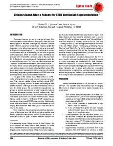
PROCEDURE In this lab, colony-PCR using non-pathogenic strains of E. coli is employed. E. coli O157:H7 has two Shiga toxin genes, stx1 and stx (5); testing could be done for either one. Testing for stx1 is described here, since a nonpathogenic strain of E. coli with stx1 cloned on a plasmid, TOP10 pSTX1 (2), is available. It was used as a positive control strain and as a mock positive unknown, eliminating the risk associated with using O157:H7. E. coli TOP10 was used as the negative control. Detailed instructions for PCR, gel electrophoresis, and real-time PCR are provided on the student and instructor handouts (Appendices 1 and 5). Typical student results for conventional and real-time PCR are provided in the instructor handout (Appendix 5). Safety issues involve proper handling of microorganisms and general precautions surrounding gel electrophoresis and are discussed in the instructor handout (Appendix 5).
CONCLUSION The conventional PCR and gel electrophoresis lab has been used twice in an undergraduate microbiology course for biology majors. When used in conjunction with the virtual PCR case study (3), students found this lab reinforced the concepts of PCR and gel electrophoresis. Comments included “This step solidified what we had been learning in the previous parts. Virtually doing PCR is good, but the hands-on experience is even better. The more times I do PCR, the better I understand the process,” and “I enjoyed this very much, it was interesting and exciting.” When compared with students who had only done the virtual PCR, students who did the PCR laboratory
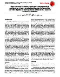
performed better on assessment questions testing their understanding of PCR and gel electrophoresis. The average class score on a question asking what happens at 95oC during PCR was 75% for students who had only done the virtual PCR, while it was 95% for students who had done both the virtual and hands-on lab. Similarly, when asked to identify the correct size of a band compared to a molecular mass ruler run on an agarose gel, the average scores were 75% and 100%, respectively. On one occasion, students performed the real-time PCR lab following the conventional PCR lab. Between the two labs, instruction was provided on the theory of realtime PCR. These students, half of whom were freshmen, found real-time PCR to be challenging to understand, but comparing the real-time PCR results with the gel electrophoresis results increased comprehension. Therefore, if time allows, it is recommended that students perform both lab activities and that sufficient in-class time be allowed for explanation and interpretation of the results. The lab handouts have several questions pertaining to PCR and gel electrophoresis that can be used to assess student learning. Keys are provided in Appendices 2 and 4. Possible modifications to this lab could include having students design primers to amplify the Shiga toxin gene rather than providing pre-designed primers for the students. In addition, the lab could easily be modified by the instructor to incorporate alternate scenarios, such as testing patient specimens or food products. Students could also find, read, and summarize primary literature in which real-time PCR is utilized.
ACKNOWLEDGMENTS Thanks to Matt Takata for his technical assistance in preliminary real-time PCR trials. The author declares that there are no conflicts of interest.
REFERENCES 1. BioRad. 2006. Real-time PCR applications guide. Bulletin 5279. 2. Grys, T. E., L. M. Sloan, J. M. Rosenblatt, and R. Patel. 2009. Rapid and sensitive detection of Shiga toxin-producing Escherichia coli from nonenriched stool specimens by real-time PCR in comparison to enzyme immunoassay and culture. J. Clin. Microbiol. 47:2008–2012. 3. Klein, J. R., and T. Gulsvig. 2012. Using bioinformatics to develop and test hypotheses: E. coli specific virulence determinants. J. Microbiol. Biol. Educ. 13:161–169. 4. Madigan, M. T., J. M. Martinko, P. V. Dunlap, and D. P. Clark. 2009. Brock biology of microorganisms, 12th ed. Pearson Benjamin Cummings, San Francisco, CA. 5. Mellies, J. L., A. M. S. Barron, and A. M. Carmona. 2007. Enteropathogenic and enterohemorrhagic Escherichia coli virulence gene regulation. Infect. Immun. 75:4199–4210.