Myoclonic epilepsy and ragged-red fibers (MERRF) is a maternally inherited syndrome associated with an A-to-G transition at the nucleotide position (np).
SHORT REPORT
Key words: tissue distribution MERRF mtDNA neurilemoma mitotic segregation MUSCLE 81 NERVE 19:519-521 1996
TISSUE DISTRIBUTION OF MUTANT MITOCHONDRIAL DNA IN A PATIENT WITH MERRF SYNDROME ROU-SHAYN CHEN, MD, CHIN-CHANG HUANG, MD, NAI-SHIN CHU, PhD, CHUN-CHE CHU, MD, KWANG-DAR SHIH, CHENG-YOONG PANG, and YAU-HUE1 WEI, PhD
Myoclonic epilepsy and ragged-red fibers (MERRF) is a maternally inherited syndrome associated with an A-to-G transition at the nucleotide position (np) 8344 in the tRNALYSgeneof the mitochondria1 DNA (mtDNA).5,6,13,19 In view of the heteroplasmy of mtDNA, different proportions of mutant mtDNA among different tissues or individuals have been proposed to be responsible for high phenotypic variability.16J9,21,22 However, two questions deserve further elucidation: (1) Is tissue distribution of the mutant mtDNA uniform or uneven in patients with the MERRF syndrome? (2) Does mitotic segregation play a major role in determination of tissue distribution of the mutant mtDNA? Several studies have reported conflicting results on heteroplasm of the mutant mtDNA in different tissues.'2,16s20 Four patients reported by Seibel et a1.16 showed variable proportions in different tissues, whereas 3 patients in the series of LombCs et al.l2 and Tanno et a1.20showed little variation. However, the proportion of mutant mtDNA was much lower in the report of LombCs. We hereby report a female patient with the M E W syndrome associated with a pelvic retroperitoneal neurilemoma. We performed a quantitative
From the Department of Neurology, Chang Gung Medical College and Memorial Hospital (Drs. Chen, Huang, N.-S. Chu, and C,-C. Chu); Depart ment of Biochemistry, School of Life Science, National Yang-Ming University (Ms. Shih, Pang, and Dr. Wei), Taipei, Taiwan. Acknowledgments: The work was supported by research grants (NSC 830412-010-083 and NSC 83-0412-8-182-015) from the National Science Council, Republic of China. Address reprint requests to Chin-Chang Huang, MD, Department of Neurology, Chang Gung Memorial Hospital, 199, Tung Hwa North Road, Taipei, Taiwan, R.O.C.
analysis of the mutant mtDNA from 10 different tissues of ectodermal and mesodermal orgins and from the neurilemoma. CASE REPORT
The 67-year-old woman experienced occasionally generalized muscle jerks since age 30. At age 40, she noted fatigue, muscle cramps, and sometimes myoclonicjerks which made her unable to hold chopsticks and fall down easily. She was the eldest proband (1-1) of the M E W family previously reported by US.^ Physical examinations on admission revealed a well-communicating, thin, and short woman with a height of 145 cm and a weight of 35 kg, and a palpable left low abdominal mass. Cafe-au-lait spots, axillary freckles, and cutaneous neurofibromas were not found. Neurological evaluations showed multifocal jerky movements, gait dyssynergia, proximal muscle weakness, and bilateral sensorineural hearing impairment. High cortical functions, ocular movement, and funduscopic examinations were normal. Laboratory studies revealed normal concentration of serum lactate and pyruvate. Electroencephalogram disclosed diffuse intermittent theta waves. Magnetic resonance imaging of the brian showed cortical atrophy of both cerebrum and cerebellum. Although there were no ragged-red fibers in the muscle biopsies, analysis of mtDNA from blood cells showed the point mutation at the 8344th nucleotide position of the tRNALYsgene. Gynecological ultrasonography disclosed a left adnexal mass with dominant solid component. At laparotomy, the mass was a 12 X 8 cm well-encapsulated retroperitoneal neurilemoma. At the request of the patient, incidental total hysterectomy and bilateral oophorectomy were also performed. The histopathology of all these tissues showed no pathological changes and mtDNA analysis was sent for.
Accepted for publication October 2, 1995 CCC 0148-639W96/040519-03 0 1996 John Wiley & Sons, Inc
Short Reports
Total DNA was extracted from 11 tissues and the mutant mtDNA was quantitatively ana-
DNA Analysis.
MUSCLE & NERVE
April 1996
519
lyzed according to the standard method described p r e v i o u ~ l y . These ~ ~ ~ ~ 'tissues ~ included three ectoderma1 tissues of skin, hair follicles, and the tumor cells of neurilemoma, and eight mesodermal tissues of muscle, blood cells, cervix, ovary, oviduct, and adventitia, myometrium, and endometrium of the uterus. To avoid sampling error, we examined two samples of the same tissue from two separate regions (approximately 1 cm apart). An A-to-G transition at the nucleotide position 8344 in the tRNALy5 gene of the mtDNA was confirmed in all tissues studied, and the proportions of mutant mtDNA in various tissues showed little variation (Fig. 1). The mean proportion of mutant mtDNA was 91.5 t 5.3% for all tissues combined, 92.6 5 5.4% (range, 87-99%) for tissues from the ectoderm, and 91.1 -C 5.4% (range, 81-99%) for tissues from the mesoderm. DISCUSSION
Our study demonstrated that high percentages of mutant mtDNA were widely and evenly distributed in various tissues with concentrations ranging from 83% to 99%. These results were similar to those of previous reports. Tanno et a1." documented a uniform distribution of the mutant mtDNA in 14 tissues (93-99'70). 1,ombi.s et al.I2 reported an even, but lower, distribution in four tissues (54-56%). Although Seibel et a1.16reported a wide range of the mutant mtDNA from 31% to 8796, the tissues were usually blood cells and fibroblasts. It is unclear why the percentage of mutant mtDNA is generally lower and highly variable in blood cells; a selective advan-
tage for rapid replicating cells has been suggested." The uniform tissue distribution of mutant mtDNA in M E W was also found in MELAS (mitochondria1 myopathy, encephalopathy, lactic acidosis, and strokelike episodes) syndrome, although some exceptions have been reported.'^^ l4 l5 However, this phenomenon is in contrast to different distributions of the deleted mtDNA in Kearns-Sayre syndrome." The relatively even distribution of mutant mtDNA among different tissues supports the notion that preferential clinical involvement of certain tissues in the M E W syndrome cannot be attributed to different tissue distributions of mutant mtDNA only. Other factors such as different thresholds of mutant mtDNA in various tissues may also be involved. It is intriguing to find that the proportions of mutant mtDNA in M E W was not different in tissues deriving from ectodermal and mesodermal germ layers. Although sample size was small, our finding was conformable to the results of patients with MELAS ~yndrome.'~ Besides, the proportion of mutant mtDNA (88%) in the neurilemoma was similar to the other tissues. These findings indicate that the mutant mtDNA at np 8344 did not undergo rapid mitotic segregation after the three germ layers were established in the early embryogenesis. However, further longitudinal studies with large series and more tissue samples are needed to clarify the tissue. To our knowledge, retroperitoneal neurilemoma had not been reported in MERRF syndrome."'(' Berkovic et al.' have described axial lipomas occurring in patients with the M E W syndrome and in asymp-
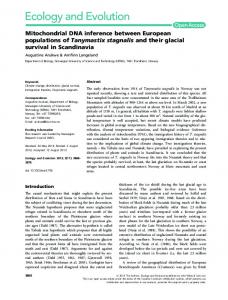
Mu Sk ON OT OV Ce UA UM UE ---__________-M 1 2 1 2 1 2 1 2 1 2 1 2 1 2 1 2 1 2 ~
310 c 271,281 * 234 c 194 F
118
0
223 197
*
Yo of mutant mtDNA
81 84 88 94 83 89 89 87 91 94 90 92 95 95 92 89
96 92
FIGURE 1. Restriction analysis for quantitative study of the 8344 point mutation of mtDNA in various tissues of the MERRF patient. A 223-bp fragment of mtDNA harboringthe putative 8344 A-to-G point mutationwas amplified from each tissue sample DNA. The polymerase chain reaction products were digested with Nae I and then the digested DNA mixtures were subjected to electrophoresis. The DNA bands were stained with ethidium bromide and photographed under short ultraviolet transillumination. The relative proportion of the mutant mtDNA was determined with densitometer in all tissues. However, the proportions of mutant mtDNA of blood cells and hair follicles are not shown in the figure (blood cells, 99%; hairfollicles, 99Y0).Abbreviations: Mu = muscle, Sk = skin, ON = ovary, OT = neurilemoma (tumor), OV = oviduct, Ce = cervix, UA = uterine adventitia, UM = uterine myometrium, UE = uterine endometrium, M = marker.
520
Short Reports
MUSCLE & NERVE
April 1996
tomatic carriers with the 8344th tRNALysmutation. The occurrence of neurilemoma in our patient could be coincidental. However, a high percentage of mutant mtDNA was found in neurilemoma (88%) as well as in lipoma (go%).'
REFERENCES 1. Berkovic SF, Andermann F, Shoubridge EA, Carpenter S, Robitaille Y, Andermann E, Melmed C, Karpati G Mitochondrial dysfunction in multiple symmetrical lipomatosis. Ann Neurol 1991;29:566-569. 2. Ciafaloni E, Ricci E, Servidei S, Shanske S, SilvestriG, Manfredi G, Schon EA, DiMauro S: Widespread tissue distribution of a tRNA1.eU(c'UR) mutation in the mitochondrial DNA of a patient with MELAS syndrome. Neurology 1991;41:1663-1665. 3. Ciafaloni E, Ricci E, Shanske S, Moraes CT, Silvestri G, Hirano M, Simonetti S, Angelini C, Donati MA, Garcia C, Martinuzzi A, Mosewich R, Servidei S, Zammarchi E, Bonilla E, DeVivo DC, Rowland LP, Schon EA, DiMauro S: MELAS: clinical features, biochemistry, and molecular genetics. Ann Neurol 1992;31:391-398. 4. Fang W, Huang CC, Chu NS, Lee CC, Chen, RS, Pang CY, Shih KD, Wei YH: Myoclonic epilepsy with ragged-red fibers (MERRF) syndrome: report of a Chinese family with mitochondrial DNA point mutation in tRNALj' gene. Muscle N m e 1994;17:52-57. 5. Fukuhara N, Tokiguchi S, Shirakawa K, Tsubaki T: Myoclonus epilepsy associated with ragged-red fibers (mitochondrial abnormalities): disease entity or a syndrome? Light- and electronmicroscopic studies of two cases and review of literature. J Neurol Sci 1980;47:117-133. 6. Hammans SR, Sweeney MG, Brockington M, Morgan-Hughes JA, Harding AE: Mitochondrial encephalopathies: molecular genetic diagnosis from blood samples. Lancet 1991;337:13111313. 7. Holme E, Larsson NG, Oldfors A, Tulinius M, Sahlin P, Stenman G: Multiple symmetric lipomas with high levels of mtDNAwith the tRNALvsA + G("'"4'mutation as the only manifestation of disease in a carrier of myoclonus epilepsy and ragged-red fibers ( M E W ) syndrome. Am J Hum Genet 1993;52:551-556. 8. Huang CC, Chen RS, Chen CM, Wang HS, Lee CC, Pang CY, Hsu HS, Lee HC, Wei TH: MELAS syndrome with mitochondrial tRNALeU'"UR' gene mutation in a Chinese family. J Neurol Neurosurg Psychiatly 1994;57:586-589. 9. Hunter W, Burke TW,Crooks LA. Retroperitoneal nerve sheath tumors: an unusual cause of pelvic mass. Obstet Gynecol 1988;71:1050-1052. 10. Khatib RA, Khalil AM, Saba MI, Aswad NK, Mroueh AM: A pelvic retroperitoneal schwannoma presenting as an adnexal mass. Gynecol Oncol 1994;53:242-244.
Short Reports
11. Larsson NG, Tulinius MH, Holme E, Oldfors A, Andersen 0, Wahlstrom J, Aasly J: Segregation and manifestations of the mtDNA tRNALysA G'83444' mutation of myoclonus epilepsy and ragged-red fibers (MERRF) syndrome. Am J Hum Genet 1992;51:1201-1212. 12. LombCs A, Diaz C, Romero NB, Ziegler F, Fardeau M: Analysis of the tissue distribution and inheritance of heteroplasmic mitochondrial DNA point mutation by denaturing gradient gel electrophoresis in MERRF syndrome. Neuromusc Disurd 1992;2:323-330. 13. LombCs A, Mendell JR, Nakase H, Barohn JR, Bonilla E, Zeviani M, Yates AJ, OmerzaJ, Gales TL, Nakahara K, Rizzuto R, Engel WK, DiMauro S: Myoclonic epilepsy and raggedred fibers with cytochrome oxidase deficiency: neuropathology, biochemistry, and molecular genetics. Ann Neurol 1989;26:20-33. 14. Macmillan C, Lach B, Shoubridge EA: Variable distribution of mutant mitochondrial DNAs (tRNALeU[s2431) in tissues of symptomatic relatives with MELAS: the role of mitotic segregation. Neurology 1993;43:1586- 1590. 15. Obermaier-Kusser B, Paetzke-Bmnner I, Enter C, MullerHocker J, Zierz S, Ruitenbeek W, Gerbitz KD: Respiratory chain activity in tissues from patients (MELAS) with a point mutation of the mitochondrial genome [tRNALcU(UUR)]. FEBS Lett 1991;286:67-70. 16. Seibel P, Degoul F, Bonne G , Romero N, Francois D, Jouas MP, Ziegler F, Eymard B, Fardeau M, Marsac C, Kadenbach B: Genetic, biochemical and pathophysiological characterization of a familial mitochondrial encephalomyopathy (MERRF). J Neurol Sci 1991;105:217-224. 17. Shanske S, Moraes CT, Lombts A, Miranda AF, Bonilla E, Lewis P, Whelan MA, Ellsworth CA, DiMauro S: Widespread tissue distribution of mitochondrial DNA deletions in Kearns Sayre syndrome. Neurology 1990;40:24-28. 18. Shih KD, Yen TC, Pang CY, Wei YH: Mitochondrial DNA mutation in a Chinese family with myoclonic epilepsy and ragged-red fiber disease. Biochem Biophys Res Commun 1991; 1741109-1116. 19. Shoffer JM, Lott MT, Lezza AMS, Seibel P, Ballinger SW, Wallace DC: Myoclonic epilepsy and ragged-red fiber disease (MERRF) is associated with a mitochondrial DNA tRNALp mutation. Cell 1990;61:931-937. 20. Tanno Y, Yoneda M, Tanaka K, Kondo R, Hozumi I, Wakabayashi K, Yamada M, Fukubara N, Ikuta F, Tsuji S Uniform tissue distribution of tRNAL"mutation in mitochondrial NDA in MERRF patients. Neurology 1993;43:1198-2000. 21. Wallace DC, Zheng X, Lott MT, ShoffnerJM, Hodge JA, Kelley FI, Epstein CM, Hopkins LC: Familial mitochondrial encephalomyopathy (MERRF): genetic, pathophysiological, and b i e chemical characterization of a mitochondrial DNA disease. Cell 1988;55:601-610. 22. Zeviani M, Amati P, Bresolin N, Antozzi C, Piccolo G, Toscano A, DiDonato S: Rapid detection of the A -+ G(8"4)mutation of mtDNA in Italian families with myoclonus epilepsy and ragged-red fibers (MERRF). AmJHum Genet1991;48:203-211.
MUSCLE & NERVE
April 1996
521