Dec 2, 1992 - search (to P. A. B.) and the Damon Runyon-Walter Winchell Cancer. Research Fund (to S. 0. M.). The costs of publication of this article.
THEJOURNALOF BIOLOGICAL CHEMISTRY Q 1993 by The American Society for Biochemistry and Molecular Biology, Inc.
Vol. 268, No.20, Issue of July 15, pp. 15150-15157, 1993 Printed in U.S.A.
Tissue-specific Alternative Splicing Generates Two Isoforms of the trkA Receptor* (Received for publication, December 2, 1992, and in revised form, March 19, 1993)
Philip A. Barker*, Catherine Lomen-HoerthS, Erin M. GenschS, Susan 0. MeakinS, David J. Glass& and Eric M. Shooter*ll From the $Department of Neurobiology, Stanford university, Stanford, California94305-5401 and SRegeneron Pharmaceuticals Znc., Tarrytown, New York 10591
The trkA receptor functions as a signal transducing receptor for nerve growth factor. In this report, we show that alternative splicing results the in production of two distinct trkA isoforms in both rats and humans. These isoforms differ by virtue of a 6-amino acid insertion in their extracellular domain, the placement of which corresponds exactly with the breakpoint found in several human trkA oncogenes. When tested in fibroblasts, the presence (trkAII) or absence (trlzAI) of the 6-amino acid insert does not affect the receptor’s ligand binding specificity or its ability to transduce functional signals in response to nerve growth factor. In rats and humans, trkAII is the only isoform expressed within neuronal tissues at appreciable levels whereas trkAI, the form of trkA originally cloned, appears to be expressed mainly in non-neuronal tissues.
respond to each of these factors in vitro have been identified, suggesting that each neurotrophin may play important roles in the development andmaintenance of specific neuronal population in vivo. Neurotrophins bind two types of cell surface receptors, the p75 NGF receptor ( ~ 7 5and,the ~ ~ ~trk~receptors. ) p75NGFR is a transmembrane glycoprotein that binds all neurotrophins with affinities in the nanomolar range (Johnson et al., 1986; Radeke et al., 1987; Rodriguez-Tebar et al., 1990; RodriguezTebar et al., 1992). This receptor contains no obvious signaling motif and its role in transduction of the neurotrophinmediated signal cascade remains uncertain. In contrast, the three trk receptors, termed trkA (Martin-Zanca et al., 1989), trkB (Middlemas et al., 1991; Klein et al., 1989, 1990), and trkC (Lamballe et al., 1991) are all transmembrane tyrosine kinases which are activated upon binding of neurotrophins (Glass et al., 1991; Kaplan et al., 1991a; Klein et al., 1991a; Lamballe et al., 1991; Squinto et al., 1991). In contrast to p75NGFR, each of the trk receptors shows a highdegree of The neurotrophins are a family of closely related proteins discrimination in binding members of the neurotrophin family. When ectopically expressed in fibroblasts, the preferred that play important roles in the development and maintenance of the nervous system. Nerve growth factor (NGF)’ was ligand(s) for the p140trkAreceptor is NGF (Cordon-Cardo et ~ ’ ~ ~BDNF (Klein et al., 1991b; the first neurotrophin to be discovered and remains the best al., 1991), for the ~ 1 4 5receptor characterized at the biochemical, molecular, and biological Soppet et al., 1991; Squinto et al., 1991) and NT-4/5 (Berkelevels. It plays a role in the proliferation of neuroblasts meier et al., 1991; Ip et al., 1992; Klein et al., 1992), and for (Cattaneo andMcKay, 1990),in neuronal maturation (Wright the ~ 1 4 5 receptor ~ ’ ~ ~NT-3 (Lamballe et al., 1991). In PC12 et al., 1992), in affecting neuronal phenotype (Miller et al., cells or in fibroblasts, activation of the signal transduction 1991; Diamond et al., 1992) and in maintainingneuronal cascade initiated by each of the trkreceptors results in similar survival (Barde, 1989). Three other neurotrophins have also biological consequences (Kaplan et al., 1991a; Glass et al., been described brain-derived neurotrophic factor(BDNF) 1991; Ip et al., 1992). Thus, it would appear likely that the (Barde et al., 1982; Leibrock et al., 1989), neurotrophin-3 neurotrophin requirements of distinct neuronal populations (NT-3)(Hohn 1990; Maisonpierre et al., 1990; Rosenthal et may be dictated by highly specific regulation of trk receptor al., 1990), andneurotrophin-4/5(NT-4/5)(Ip et al., 1992; expression. Berkemeier et al., 1991). Distinct neuronal populationswhich The first trkreceptor identified was t r M . It was originally discovered asatransforming allele in which most of its * This work was supported by National Institutes of Neurological extracellular domain was replaced with a 221-amino acid and Disorders and Stroke Grant NS04270 (to E. M. S.), postdoctoral segment of a tropomyosin pseudogene (Martin-Zanca et al., fellowships from the Alberta Heritage Foundation for Medical Research (to P. A. B.) and the Damon Runyon-Walter Winchell Cancer 1986). Subsequently, the trkA proto-oncogene was cloned Research Fund (to S. 0. M.). The costs of publication of this article from K562 cells, a human erythroleukemic cell line (Martinwere defrayed in part by the payment of page charges. This article Zanca et al., 1989). In situ hybridization studies revealed that must therefore be hereby marked“advertisement” in accordance with in vivo trkA expression was limited to neurons presentwithin 18 U.S.C. Section 1734 solely to indicate this fact. dorsal root and cranial sensory ganglia (Martin-Zanca et al., The nucleotide sequence(s) reportedin thispaper has been submitted 1990). These results provided a crucial clue leading to the accessionnumber(s) to the GenBankTM/EMBLDataBankwith identification of t r k A as a receptor for NGF which displays L12225. ll To whom correspondence should be addressed Dept. of Neuro- some of the properties of the high affinity, or Type I NGF biology, Stanford University School of Medicine, Stanford University, receptor (Klein et al., 1991a; Kaplan et al., 1991a;Meakin and Stanford, CA 94305-5401. Tel.: 415-723-5811; Fax: 415-725-0388. Shooter, 1991).Activation of the trkA tyrosine kinase by NGF The abbreviations used are: NGF, nerve growth factor; DMEM, does not require coexpression of the p75NGFR, at least in Dulbecco’s modified Eagle’s medium; PCR, polymerase chain reaction; BDNF, brain-derived neurotrophic factor; NT-3 or -415, neu- Xenopus oocytes (Nebreda et al., 1991) or mouse fibroblasts rotrophin-3 or -4/5; MTT, 3-[4,5-dimethylthiazol-2-yl]-2,5-diphen-(Klein et al., 1991a). Fibroblasts expressing the t r M receptor undergo ligand-dependent activation of c-fos, DNA synthesis, yltetrazolium bromide.
15150
Alternative Splicing of trkA
15151
run for 30 cycles of 94 "C X 40 5, 60 "C X 60 9, 72 "C X 120 8. To and cell division (Cordon-Cardo et al., 19911, in a manner analyze intron/exon boundaries, genomic fragments were generated analogous to that observed with other tyrosine kinase recep- by PCR using rat genomic DNA as template for 30 cycles of 94 "cX tors. 40 S, 55 "C x 60 s, 72 "C x 180 s with primer sets T63/T69 or T38/ The functional diversity of products derived from a single T70. Prior to subsequent analysis, all fragments were purified using gene may be increased by alternative splicing of primary Geneclean (Bio 101) and subcloned into PCRlOOO (Invitrogen). transcripts. The timing and location of the expression of Multiple replicate clones were sequenced using Sequenase (u. s. alternatively spliced mRNAs are often highly regulated by Biochemical Corp.) according tothe manufacturer'sinstructions. PCR primers used are shown as follows. BA9, GACTCG AGT CGA mechanisms that remain poorly understood. Several types of CAT CGA TTT TTT TTT TTT TTT T T BA45, AT ACC TGG transmembranous cell surface proteins have been shown to CGA CAT CTG; BA46, AACAA CGG CAA CTA CAC; T36, AGC undergo differential mRNA splicing events which result in GTA CGA TGT GTTGG; T38, GCT CCC ACTTGA GAA TG, T62, different receptor isoforms. The functional consequences of CAC TGG GAC ACA ACA AC;T63: ATG AGA CCA GCT TCA TC; T70, CTC CTT CTC GCC theseeventsare diverse and include alteration of ligand T69, CCA CTG GCGAGAAGGAG; binding affinity or specificity (Mosthaf et al., 1990; Miki et AGT GG. Expression Constructs and Creation of Stable Sublines-A clone al., 1992; Werner et al., 1992; Attisano et al., 19921, shifts in (pTrk6) containing the entire rattrkAI open reading frame has been intracellular trafficking patterns (Paietta et al., 1992) and previously characterized in our laboratory (Meakin et al., 1992). To alterations in adhesive properties (Arch et al., 1992). Alter- generate an expression construct containing trkAI, a portion of the native splicing has also been observed within the trk family open reading frame containing the alternatively spliced domain was of proteins where trkB mRNA is alternatively spliced to obtained using nested PCR on rat kidney cDNA, using primers T32 produce receptor isoforms lacking a functional tyrosine kinaseand T57 for the first PCR and using primers T36 and T62 for the second PCR. The PCR product was digested with BamHI/ScaI and (Klein et al., 1990; Middlemas et al., 1991). However, the the resulting fragment was exchanged for the same fragment in physiological role of these truncated receptors remains unpTrk6. The fidelity of sequence obtained by PCR was confirmed by known. DNA sequencing. For expression, each of the open reading frames In thisreport, we show that alternative splicing of the trkA were subcloned into the XbaI site of CMX (Davis et al., 1991) to primarytranscriptresults in the production of two trkA generate pPB139 (trkAIl) and pPB146 (trkAI). Each of these plasreceptor mRNA isoforms. Two forms of trkA cDNA cloned mids was transfected into variant NIH 3T3 fibroblasts designated MG87 (Zhan and Goldfarb, 1986; Glass et al., 1991) by calcium previously from human (Martin-Zanca et aL, 1989) and rat phosphate precipitationto create pools of sublines expressing each of (Meakin et al., 1992)are now shown to represent alternatively the trkA splice variants, MG87 fibroblasts were transfected with a spliced mRNA species which differ by virtue of a 6-amino 5:l ratio of trkSV2neo plasmids by calcium phosphate precipitation and selected in 500 pg/ml G418 to produce cell lines MG139-2 acid stretch within the extracellular domain. (expressing pl4Ot"") and MG146-2 (expressing pl4OtrkA1). Immunoprecipitation and Western Blotting"1O6 fibroblasts from pools expressing each of the trkA splice variants were placed on 100Hybridization Probe~-[~~P]CTP-labeled cRNA probes were gen- mm plates 24 h prior to addition of neurotrophins. The next day, erated from plasmid pSPtrk 5-7 and plasmid pSPtrk 5-2, which cells were washed twice in Dulbecco's modified Eagle's medium contain nucleotides 934-1531 of the rat trkAZZcDNA (Meakin et al., (DMEM) containing 0.1% bovine serum albumin (DMEB) and left 1992) subcloned in opposite orientations into theXhoI site of pSP72 for 1 h at 37 'C in 5 ml of DMEB. A prewarmed 2 X concentrated (Promega). For probe a, plasmid pSPtrk 5-7 was linearized with stock of neurotrophin in DMEBwas then added to each dish and left AuaII (cuts at nucleotide 1234) and transcribed with T7 RNA polym- for various times. Plates were then rinsed twice with ice-cold phoserase. For probe b, plasmid pSPtrk5-2 was linearized with HincII phate-buffered saline and the cells were lysed with RIPA (10 mM (cuts a t nucleotide 1129) and transcribed with SP6 RNA polymerase. Tris, pH 7.4, 150 mM NaC1, 1%Nonidet P-40, 0.5% sodium deoxyProbe a had a total length of397 nucleotides, 294 nucleotides of cholate, 0.1% SDS) supplemented with 1mM sodium orthovanadate. which were complementary to rattrkAZI (i.e. nucleotides 1234-1531). Lysates were incubated first with 2.5 pg of a polyclonal antipeptide Probe b had a total length of409 nucleotides, 405 nucleotides of antibody directed against the COOH terminus of trkA (Santa Cruz which were complementary to rattrkAII (i.e. nucleotides 1129-1534). Biotechnology) followed by 60 pl of goat anti-rabbit IgG coupled to For production of [32P]CTP-labeledcRNA probes for human trk4 agarose (Sigma). Beads were washed twice in RIPA containing 1mM RNA analyses (probe c), a 279-base pair fragment containing nucle- sodium orthovanadate and then boiled for 5 min in 100 gl of sample otides 1183-1443 of human trkAIZ was produced using PCR (see buffer. Immunoprecipitates were separated on 8% Laemmli acrylbelow) and subcloned into the EcoRI/HindIIIsites of pGEM4Z amide gels and transferred to nitrocellulose. Blots were blocked in (Promega) to generate pPB136. pPB136 was linearized with EcoRI Blotto (10 mM Tris, pH7.4,150 mM NaC1,5% dry skim milk powder) and transcribed with T7 RNA polymerase to generate a probe of 350 containing 0.2% Tween 20 and then incubated with an anti-pTyr nucleotides which contained 279 nucleotides complementary to hu- monoclonal antibody (UBI) followed by a horseradish peroxidaseman trkAII (Le. nucleotides 1183-1443 plus the 18-nucleotide insert). conjugated goat anti-mouse antibody (Sigma). Reactive bands were RNA Preparation, RNase Protection, and Northern Blots-RNA detected using enhanced chemiluminescence according to themanuwas isolated from tissues and cells using either guanidinium isothio- facturer's instructions (Amersham). cyanate lysis followed by cesium chloride gradient centrifugation '"I-NGF Cross-linking-2.5 S NGF (Bioproducts for Science) was (Chirgwin et al., 1979), or by the one-step guanidinium isothiocyanate radioiodinated using lactoperoxidase to a specific activity of about 60 procedure (Chomczynski and Sacchi, 1987). RNase protection analy- cpm/pg. COS cells or fibroblasts expressing each form of trkA were sis was performed as described previously (Barker etal., 1991) except harvested from plates using Versene and counted. Cells were washed that hybridizations were performed at 50 "C and only RNase A (40 once in binding buffer and then resuspended at a concentration of pg/ml) was used in the RNA digestion step. 500,000 cells/ml. "'1-NGF was added to 1-ml aliquots of cells to Cloning and Sequence Analysis-For production of cDNA clones, produce a final concentration of 1 nM (assuming 1 M = 26,500 d). total rat kidney RNA or total human DRG RNAwere reversed Where required, unlabeled NGF was used at 250nM. Cells were transcribed with Moloney murine leukemia virus reverse transcrip- incubated for 3 h on ice and then cross-linked using 1 mM disuccintase (Superscript, Bethesda Research Laboratories) in a 50-pl reac- imidyl suberate (Pierce Chemical Co.) for 30 min a t room temperation containing 10 mM Tris-HC1 (pH 8.0), 1.25 mM deoxynucleotides, ture. The reaction was terminated by addition of Tris-HC1 (pH 7.5) 10 mM dithiothreitol, 50 mM KCl, 15 mM MgCl,, 0.02% gelatin, and to afinalconcentration of 1 mM. Cells were then collected by 200 pmol of primer BA9 for 60 min at 42 "C. The reaction mixture centrifugation and either resuspended in 100 p1 of sample buffer (COS was diluted to 500 pl with water and 5 pl was used for subsequent cells) or suspended in 0.5 ml of RIPA and immunoprecipitated using PCR. For PCR of rat kidney cDNA, the initial reactions were run antibodies against trkA as described above. using primers T62 and T36 and nested PCR were performed using Cell Culture"PC12 cells were maintained in DMEM containing primers T63 and T38. For PCR analysis of human DRG cDNA, the 6% horse serum and 6% bovine calf serum in 7% CO, at 37 "C. For initial PCR reactionswere run using primers T63 andT37 and nested NGF treatment, cells were washed twice in DMEB and then incuPCR was performed using primers BA45 and BA46. All PCR were hated for 3 days in DMEB containing 10 ng/ml NGF. MG87 fibroEXPERIMENTALPROCEDURES
Alternative Splicing of trkA
15152
blasts were maintained in DMEM containing10% bovine calf serum in 7% C 0 2 a37 t "C. Stably transfected MG87 fibroblasts were maintained in DMEM containing 10% calf serum and 200 pg/ml G418 in 7% COZ a t 37 "C. Stably transfected fibroblasts containing each of the splice variants were always compared using cells at the same passage number and were not used after passage 20. MTT Survival Assay-Fibroblast cell survival was assayed using an MTT assay (Ip et al. 1993). Fibroblasts were washed twice in DMEM and 2500 cells were plated in wells of 96-well plates in 3:1 DMEM/F-12 media supplemented with insulin,transferrin, selenium, linoleic acid, and bovine serum albumin and the appropriate amount of neurotrophin. After 4 days, 15 pl of MTT dye solution (Promega) was added to each well and 4 h later, the reaction was terminated by the addition of 100 pl of solubilization reagent (Promega). Samples were incubated with shaking for 4 h to solubilize formazan crystals and then read on an enzyme-linked immunosorbent assay reader, using a test wavelength of 570 nm and a reference wavelength of 630 nm. The survival experiments were repeated 3 times and within each experiment, 4 data points were analyzed a t each neurotrophin concentration. RESULTS
I
1100
W
1600
Transmembrane Domain
C
350 114
RobeC
147
279
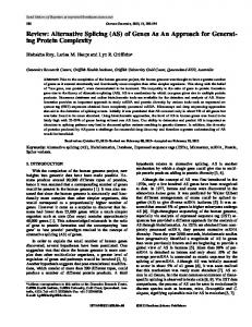
TMl TMll
FIG. 1. cRNA probes used for RNase protection analysis. Top, a portion of the trkA open reading frame is indicated by the heavy line. The location of thetransmembrane domain andthe alternatively spliced insert are indicated by raised bars. The three RNase protection probes (a, b, and c) and thefragments protected by them are shown in corresponding panels A, B, and C. In each panel, the top line represents the protection probe, with the thick portion corresponding to cRNA sequence and the thinportion corresponding to probe sequence contributed by the transcription vector. The next line shows the fragment(s)expected from protection by trkAI mRNA transcript,andthe bottom line shows fragment(s) expected from protection by the trk.411 mRNA. Numbers shown above each line representthe length (in nucleotides) of each of the probes and protected fragments.
Identification of Alternatively SplicedtrkA mRNA SpeciestrkA cloned from rat PC12cells (Meakin etal., 1992) is highly homologous to human trkA cloned from K562 cells (MartinZanca et dl., 1989), particularly in the juxtamembraneregion and in the tyrosine kinase domain.However, we have previtrkA ously noted that a 6-amino acid insert present in the rat extracellular domain, located 15 amino acids from thebeginning of the putative transmembrane domain, is not present in the cloned human trkA cDNA. The position of this insert corresponds precisely with the point atwhich seven of eight exons of a non-muscle tropomyosin gene are fused with the LL (3 transmembrane and cytoplasmic portions of the trkA protoz oncogene to produce the oncogene, OncD (Martin-Zanca et al., 1986). Other trkA oncogenes that occur naturally or that are generated by gene transfer experiments have been characterized. In severalcases, the breakpoint corresponds exactly to the position of the 6-amino acid insert in the rat trkA cDNA (Kozma et al., 1988; Barbacid et al., 1991). This conservation of the breakpoint between the oncogenes together 391 with the otherwise almost perfecthomology between human and rat trkA in this region suggested that the 6 amino acids 294 present within the rat trkA sequence might be due to the 263 insertion of a small exon and that the difference between the rat and human cDNA might actually reflect an alternative splicing event. RNase protection analysis was performed to determine if alternatively spliced trkA mRNAtranscriptsarepresent within PC12 cells. An antisense cRNA (probe a) encompassing nucleotides 1234-1531 of the rat trkA sequence (Fig. 1) was hybridized to RNA isolated from untreated PC12cells or FIG.2. RNase protection analysis of PC12 cell RNA. Probe from PC12 cells exposed to NGF for3 days. Fig. 2 shows that in addition to the fully protected cRNA, a smaller species of a was used in RNase protection analysis on 20 pg of yeast tRNA or on 1 pg of total RNA isolated from native PC12 cells or from PC12 263 base pairs was present in both RNA samples. Interestcells grown for 72 h in serum-free media supplemented with 10 ng/ ingly, the NGF-dependent increase of trk mRNA that we ml NGF. Size determinations in this and subsequent RNase protecpreviously detected by Northern blot (Meakin et al., 1991) tion analyses were made by comparison with lanes containing 3zPreflects mainly an increase in thefully protected species. T o end labeled fragments of HinfI -digested 6x174. determine how the two protected fragmentsdiffer, additional protectionanalysis was performedusingantisensecRNA protected fragment was present a t relatively high levels in containing nucleotides 1130-1533 (probe b). Again, a fully kidney (Fig. 4A). Therefore, kidney cDNA was synthesized protected cRNA anda smaller species were observed and the and used as template in PCR designed to identify the variable smaller protected fragmentproduced by probe b corresponded region. The first PCR generatedcDNA encoding nucleotides exactly insize with that produced by probe a (data notshown, 618-1791. This was thenused as template fora second PCR see Fig. 1). Therefore, the two rat trkA mRNA transcripts to generate cDNA representing nucleotides 1035-1598. Rediffer near the 5'-end of the portion protectedby probe a. actions products from this second reaction were subcloned Characterization of trkAZ and trkAIZ Zsoforms-RNase pro- into PCRlOOO and sequenced. Of nine clones generated from tection analysis of trkA expression within several rat tissues the kidney cDNA, the sequence of one was identical to that of the rat trkA sequence characterized previously (Meakin et revealed that the trkA isoform represented by the smaller
Alternative Splicing of trkA
15153
the insert-lacking and insert-containing forms of trkA, respectively. The Expression of the trkA Splice Variants Is Tissue-specific-To determine whether the expression of trkA isoforms is tissue-specific, rat tissues were examined for trkA expression by RNase protectionanalysis with probe a. Fig. 4A shows that although PC12 cells express both trkAI and trkAII, only trkAZI was detected in RNA isolated from total brain, dorsal root ganglia, and sympathetic ganglia. The trkAI isoform is also expressed in vivo, both in kidney, and toa lesser degree, in lung. Longer exposure of the RNase protection gel shows that although low amounts of trkAIZ are produced by kidney and lung, trkAI was not detected within the neuronal tissues. Thus, it would appear that in rats, the form of trkA lacking the 6-amino acid insert (trkAI) is expressed primarily outside of the nervous system, whereas the form of trkA containing the insert (trkAII) is primarily, althoughnot exclusively, expressed within neuronal tissues. The distribution of trkA isoforms within human tissueswas determined using RNase protection analyses with probe c (Fig. 1). Expression of the trkAI isoform is indicated by protected fragmentsof 114 and 147 nucleotides and thetrkAII isoform is indicated by a protected fragment of 279 nucleotides. Fig. 4B shows that trkAIZ is expressed at high levels in several neuronal tissues. Humansympathetic, dorsal root, and trigeminal ganglia all express abundant trMII. Longer exposures have shown that these same tissues also express trkAI but only at very low levels. As in the rat, trkAI is also expressed in uivo within human kidney. Thus, the expression patterns of the trkA isoforms appear to bewell conserved between species. We also examined human cell lines in which trkQ expression had been previously studied. K562 cells, from which the trkAZ isoform was originally cloned, express trkAI at moderate levels but express only very low levels of trkAII (Fig. 4B). The SHSY-5Y neuroblastoma cell line responds to NGF by extending neurites (Sonnenfeld and Ishii, 1982) and has been shown to contain p140tTkA(Kaplan et al., 1991b), whereas the A875 melanoma cell line contains high levels of p75NGFR(Marano etal., 1987) but undetectable levels of p140trkA(Kaplan et al., 1991b). Surprisingly, trMZ is readily detected within both of these cells and both express low but A detectable levels of trkAII. TrkAl ...GAC CCC ATC CCT GpC ACT AAC AGC ACA... Functional Characterization of the trkA Splice VariantsTo assay the functional role of the alternatively spliced domain, open reading frames of trkAZI and trkAI were subcloned TrkAll ...GAC CCC ATC into theexpression vector CMX (Davis et al.,1991) to produce expression vectors pPB139 and pPB146, respectively. The NGF binding properties of the rat trkA isoforms were then compared by cross-linking in COS cells. Both trkA isoforms B were cross-linked to lZ5I-NGFto produce a complex of about V S F S P V 160 kDa (Fig. 5). The labeled band of approximately 75 kDa ...cctaclacaaITCTCCTTCTCGCCAGTGGIataaattacc .... is not competed by unlabeled NGF and therefore likely represents as nonspecifically labeled background protein. Additional cross-linking studies of the receptor isoforms expressed in COS cells revealed slow dissociation characteristics at 0 "C and resistance to trypsin (data not shown), propertiespreviD P I P D T N S T S ously ascribed to the Type I, or high affinity NGF receptor ...GAC CCC ATC CCT G lgtgtgagagc .... ...,gtccccacag b C ACT AAC AGC ACA TC ... (Meakin and Shooter, 1991). For further functional analysis of the trkA isoforms, we FIG. 3. Schematic of trM isofornu generated by alternative splicing.A, differences in the nucleotide sequence of the trkisoforma employed variantNIH3T3 fibroblasts designated MG87 are shown. The bored region represents the exon which is selectively (Zhan and Goldfarb, 1986).MG87 cells were stably transincluded in rat trkA22 and excluded from rat trkA2. The human and fected with either pPB139 or pPB146 to produce cell pools rat sequences are identical in the sequence shown, except for the tranalationally silent substitution(A to G ) indicated at position 1261. MG139-2 and MG146-2, respectively, and then analyzed for B, intron/exon junctions areshown for the alternatively spliced exon expression of trkA mRNA and protein. Fig. 6A shows that the (upper line) and for flanking exons (lower line) in the rat. Intron and cells produce appropriately sized protection products andthat exon sequences are mintedlower and u m e r case. resnectivelv. "" the level of trkAII mRNA was somewhat higher thanthe level
al., 1991). The remaining eight clones lacked an 18-nucleotide fragment, beginning with nucleotide 1248. This deletion predicts an in-frame loss of 6 aminoacids at a position 15 amino acids from the start of the putative transmembrane domain (Fig. 3, A and B ) . Based on these findings, we hypothesized that alternative splicing of trkA pre-mRNA might account for this variance. To determine if the structure of the trkA gene is consistent with this mechanism of genetic control, fragments of the rat trkA gene were amplified by PCR, subcloned, and sequenced. As shown in Fig. 3B, the alternatively spliced 18-nucleotide domain represents a distinct exon. Each of the introns have the characteristic GT and AG dinucleotides at their 5'- and 3'-ends, and their surrounding sequences are in agreement with the consensus for intron/exon boundaries. These results strongly indicate that thediversity introduced into thisregion of rat trkA is due to alternative splicing of primary trkA mRNA. To determine if human trkQ mRNA is also alternatively spliced, cDNA was prepared from human dorsal root ganglia RNA and analyzed by PCR. First, PCR was used to generate cDNA representing nucleotides 1133-1479 and then the products of this reaction were used as template in a second PCR in which human trkA cDNA containing nucleotides 11831427 wassynthesized. cDNA products of this second reaction were subcloned in PCRlOOO and sequenced. Of nine clones sequenced, all containedan 18-nucleotide insertion at position 1296 (Fig. 3A) that was not present in the humantrkA cDNA originally cloned from K562 cells. Compared to the rat sequence, the insertion is well conserved, containing only a single transcriptionally silent substitution(A to G) inposition 1311 (Fig. 3A). Outside of this region, the sequence of each of the clones did not diverge from that published previously (data not shown). Thus, alternative splicing of this 6-amino acid insert results in the production of two trkA isoforms in both rats andhumans. The strong conservation of this alternatively spliced domain suggests that itmay subserve a functional role. We have termed the iaoforms trkAZ and trkAZZ for
..
~I
~
~
1
"
~
-
Alternative Splicing of trkA
15154
B
A
391
- 147 - 114
294
263
FIG.4. Distribution of alternatively spliced trkA transcripts in adult rat and human tissues. A , total RNA samples isolated from adult rat were analyzed by RNase protection using probe a (Fig. 1) as described under “Experimental Procedures.” RNA quantities analyzed were 2.5 pg for PC12 cells, 40 pg for brain, 3 pg for dorsal root ganglia, 1 pg for sympathetic ganglia, 20 pg for kidney, and 20 pg for yeast tRNA. B, total RNA samples isolated from human were analyzed by RNase protection using probe c as described under “Experimental Procedures.” RNA quantities analyzed were 10 pg for K562, SHSY-5Y, and A875 cells, 5 pg for dorsal root ganglia, superior cervical ganglia, trigeminal ganglia, posterior brainstem, anterior brainstem,and hippocampus and 20 pg for kidney and yeast tRNA. The apparent discrepency in the molecular weight of the protected fragments from kidney in p a d B is due to a slight curvature in the gel.
Plasmid Competitor 20097.4 -
I p139 I I-
+
p146
I-
+
cl
a
b N
N
line Cell Competitor
d d
2 9 f 6 e u a a
of trkAZ mRNA in these cells. Cross-linking to lZ5I-NGFand immunoprecipitation by trkA antibodies reveal a specifically labeled complex of approximately 160 kDa in each of the transfected cell lines; the level of cross-linked product correlates well with the amount of trkA mRNA present within each cell line (Fig. 6 B ) .
MG139-2
-
+
I
MG146-2
+
n Z I Z 200 391 294 263
FIG.5. “I-NGF cross-linking identifies trk isoforms within COS cells. Cell surface expression of trk isoforms was demonstrated by cross-linking ’”I-NGF to COS cells transiently transfected with plasmids pPB139 or pPB146, which contain open reading frames for rat trkAII and trk41, respectively. Cross-linking was performed as described under “Experimental Procedures” using 1 nM radioactively labeled NGF and 250 nM unlabeled NGF as competitor. After cross-linking, cells were lysed in sample buffer, boiled 5 min and analyzed on 8%SDS-polyacrylamide gel electrophoresis.
1
97.4
-
69
-
46
-
30 -
FIG. 6. Expression of tr&Aisoforms within transfected fibroblasts. A , 1-pg samples of total RNA isolated from untransfected MG87 fibroblasts or MG87 fibroblasts stably transfected with either plasmid pPB139 or pPB146, which contain open reading frames for rat trkAI1 (MG139-2) and trkAI (MG146-2), were analyzed by RNase protection using probe a as described under “Experimental Procedures.” B, fibroblasts stably transfectedwith pPB139 or pPB146 were cross-linked as described in the legend to Fig. 5 and then solubilized in RIPA buffer. trk isoforms were immunoprecipitated using a polyclonal anti-peptide antibody directed against human trkA followed by anti-rabbit IgG conjugated to agarose. Precipitates were boiled 5 min and separated on 8% SDS-polyacrylamide gel electrophoresis.
Alternative Splicing of trkA The trkA receptor becomes phosphorylated on tyrosine in response to NGF treatment (Klein et al., 1991a; Kaplan et al., 1991b). We therefore directly compared the ligand-induced phosphorylation of p140trkA1 and p140trkAA” in the MG146-2 and MG139-2 cell lines. Cells were exposed to increasing concentrations of NGF for 7.5 min, and receptor variants were assayed by immunoprecipitation with anti-trkAantibodies followed byimmunoblotting with antibodiesagainst phosphotyrosine. Fig. 7A shows that for both receptor variants, the relative phosphorylation level observed in response to a given dose of NGF was qualitatively similar. The absolute amount of phosphotyrosine was consistently higher in the MG139-2 pool, consistent with the levels of mRNA and crosslinked receptor detected in each cell line (Fig. 5). Next, we examined the rateof phosphorylation and dephosphorylation of the different trkA protein isoforms. Each cell line was incubated with 50 ng/ml NGF for periods of up to 2 h and analyzed as above. Fig. 7B shows that the response of the variants were qualitatively very similar. Both receptor subtypes became phosphorylated quickly, reaching maximum levels by 7.5 min, and both maintained this level of phosphorylation for a t least 60 min. By 120 min, a slight reduction in tyrosine phosphorylationwas observed but levels were still considerably higher than inunstimulated cells. We also compared the ability of the different neurotrophins to increase trkA isoform. MG139-2 and tyrosine phosphorylation on each MG146-2 cells were incubated for 7.5 min with 100 ng/ml of NGF, BDNF, NT-3, or NT-4/NT-5, and then analyzed for tyrosinephosphorylation as above (Fig. 7C). Again, both forms of trkA receptor responded in a qualitatively similar
MG139-2
MG146-2
NGF (nglml) 0
.01
.I
1
NGF (nglml)
10 100
0
.01
.I
1
IO, l o o
A c
c
Minutes 0
1
7.5 30
Minutes
60 120
0
1
7.5 30
15155
fashion. NGF elicited the strongest response in both cell lines and clear increases in tyrosinephosphorylation were observed in response to NT-3 and NT-4/5 but not BDNF. to We next set out to determine if the trkA isoforms exhibit different affinities. However, lZ5I-NGFbinding by MG139-2 and MG146-2 cells was very low, making the production of accuratebindingisotherms impossible (data not shown). Therefore, we took advantage of the survival properties of MG87 cells. Untransfected MG87 cells die in unsupplemented defined media but fibroblast growth factor can act as a survival factor underthese conditions (Zhanand Goldfarb, 1986). If trk receptors are expressed within these cells, neurotrophins also act assurvival factors in a dose-dependentfashion (Glass et al., 1991; Ip et al., 1993). Therefore, we compared MG1392 and MG146-2 cells for NGF-mediated survival. Cells were grown for 4 days in 96-well plates containing defined media supplemented with various concentrations of NGF and survival was then quantified using an MTTassay. Although the level of expression of each of the trkA isoforms differed between the two cell lines, their dose-response curves for NGF-mediated survival were very similar. The ECso for survival was about 3.2 ng/ml for MG139-2 cells (bearing p140trkA11) and about 1.8 ng/ml for MG146-2 cells (bearing pl4OtrkA1) (Fig. 8). The responses evoked by NT-3 and NT-4/ 5 on cells bearing either trkA isoform were slight and observed only at concentrations of neurotrophin in excess of 100 ng/ ml; neither transfected cell showed a significant response to BDNF (data notshown). DISCUSSION
The presentstudy was undertaken todetermine if distinct products are generated from the trkA locus by alternative splicing. Here we show that trkA cDNA cloned previously from human (Martin-Zanca et al., 1989) and rat (Meakin et al., 1992) represent mRNAs encoding distinct trkA isoforms and thatthese isoforms are generated by alternative splicing. The alternativesplicing event resultsin theselective presence of a 6-amino acid sequence within the extracellular domain of the receptor. Interestingly, this insertion occurs a t exactly the location at which human trkA mRNA has been shown to become fused with heterologous sequences to generate onco-
60 120
B 8-
C I-
c
FIG.7. Tyrosine phosphorylation of trkA isoforms. CrkA tyrosine phosphorylationwas detected in stably transfected fibroblasts by immunoprecipitationandantiphosphotyrosineimmunoblotsas A , dose-response of describedunder“ExperimentalProcedures.” MG139-2 (left panel) or MG146-2 (right panel) to theindicated concentrations of NGF. Cells were exposed to NGF for 7.5 min. B, time course of MG139-2 (left panel)or MG146-2 (right panel)phosphorylation after treatment with 100 ng/ml NGF. C, neurotrophin specificity of MG139-2 (left panel)or MG146-2 (right panel).Each neurotrophin was used at 100 ng/ml for 7.5 min. trk isoforms are indicated by an arrow.
0
1
10001 0
100
NOF (nglml)
FIG. 8. NGF mediated survival of fibroblasts bearing the trkA isoforms. Cells were grown for 4 days in serum-free defined media containing various concentrations of NGF and then assayed for survival using an MTT assay as described under “Experimental Procedures.” 50% survivalisindicated by an arrow. Open boxes indicate MG139-2 cells and filled diamonds indicate MG146-2 cells.
15156
Alternative Splicingof trkA
genic forms (Martin-Zanca et al., 1986; Barbacid et al., 1991; expression withinthese cells and perhapsalso within neurons, Kozma et d., 1990). Since the alternatively spliced domain is possibly by regulating the activity of relevant trans-splicing encoded by an 18-nucleotide microexon, it is likely that either factors. Alternatively, the trkAZZ isoform may arise indirectly this exon or one of the small introns flanking it contains a as a consequence of the neuronal differentiation that occurs site(s) at which somatic recombination events preferentially within PC12 cells in response to NGF treatment. Characteroccur within the human trk gene. ization of the trkA isoforms expressed during development, The limited expression of trkA within neuronal subgroups particularly during the period at which neurons gain access derived from the neural crestprovided a crucial clue in defin- to and develop a dependence on target-derived NGF, may ing its role as a signal transducing receptor for NGF. High provide useful information in this regard. levels of t r M mRNA were detected within mouse dorsal root The p140trkA receptorbecomes phosphorylated on tyrosine ganglia, trigeminal ganglia, and cranialsensory ganglia (Mar- and displays increased kinase activity in response to treattin-Zanca et al., 1990). More recently, in situ hybridization ment with NGF. Activationof p140trkAproduces various funcanalysis has shown high levels of trkA expression in neurons tional consequences, including differentiation, survival, cell of sympathetic ganglia (Schecterson and Bothwell, 1992), in division, and meiosis, in different cellular contexts (Loeb et cholinergic neurons of the basal forebrain, and incells of the al., 1991; Cordon-Cardo et al., 1991; Nebreda et al., 1991; Ip caudate-putamen (Holtzman et al., 1992). Furthermore, et al., 1992). To directly compare the ligand-dependent actiNorthern blotting has revealed trkA expression within rat vation of the p140trkAreceptor isoforms, each of the receptors seminiferous tubules (Parvinen et al., 1992), a non-neuronal was expressed in fibroblasts and assayed for ligand-induced tissue in which NGF has been suggested to play a functional tyrosinephosphorylation.Although the levels of receptor role (Ayer-Lelieve et al., 1988). We have used RNase protec- within each cell differed somewhat, the response of the isotion assays to examine the distribution of the two trkA splice forms to increasing concentrations of NGF were qualitatively variants in both human and rats and found that expression similar. In addition, the time course of phosphorylation apof the trkA isoforms is highly tissue-specific. In each of the peared to be identical between the two forms of p140trkA. Using the MG87 fibroblasts as a model system, we show nervous system tissuesin which trkA expression was detected, expression was limited almost completely to the trkAZZ iso- that both trkA isoforms act as signal transducing receptors form. Sympatheticand dorsal rootganglia of rats and humans, for NGF. In agreement with previous results (Cordon-Cardo human trigeminal ganglia, and rat brain were all found to et al., 1991;Rosenthal et al., 1990; Ernfors et al., 1990; Squint0 contain high levels of trkAZZ. Levels of trkA expression within et al., 1991), BDNF does not activate eitherreceptor isoform the adulthuman central nervous systemwere below the limits but NT-3does have some effect, albeit considerably less than of detection of our assay system, possibly due to its highly that evoked by NGF. A novel human neurotrophin, called restricted expression pattern(Martin-Zanca et al., 1990; either NT-4 or NT-5 was shown to efficiently activate trkA Holtzman et al., 1992) and the advanced age of our tissue by Berkemeier et al. (1991), but others (Ip et al., 1992, 1993) donors. trkAZ expression was observed in both rat and humanfound it to be a relatively inefficient ligand for this receptor. kidney and subsequent analyses has revealed that low levels We have found that NT-4/5 treatmentof fibroblasts bearing of trkAZ and trkAZZ are expressed in several other rat non- either trkA isoform results in only a modest increase in neuronal tissues.' Expression of t r M I within the kidney is tyrosinephosphorylation and fibroblastsurvival,approxiintriguing since morphogenesis of the developing kidney is mately equivalent to results observed with NT-3. Differentbindingproperties have been reported for the disrupted by treatment with antisense oligonucleotides complementary to the low affinity NGF receptor (Sariola et al., t r M receptor. Human trkAI expressed in fibroblasts orin Sf9 cells has been reported to bind NGF with two dissociation 1991). , other at about5 nM The trkA isoforms expressed within several cell lines were constants, one a t about 100 p ~ the also determinedby RNase protection analyses.Within human (Klein et al., 1991a), whereas a rat-mouse fusion of trM leukemic K562 cells, the trkAZ isoform is the predominant expressed in fibroblasts (Hempstead et al., 1991) andrat form expressed whereas in rat PC12 cells, trkAZZ predomi- trkAZZexpressed in COS cells3shows only a single dissociation nates. This correlates well with the fact that full-length hu- constant of about 1.5 nM. To explore the possibility that the man trMZ and rat trkAZZ were originally cloned from K562 trkA isoforms identified here might exhibit different binding and PC12 cells, respectively. SHSY-5Y is a human neuro- affinities for NGF, binding analyses were carried out on the fibroblast pools stably transfected with each of the alternablastomaline which gains some neuronal propertiesupon exposure to NGF (Sonnenfeld and Ishii, 1982) and A875 is a tively spliced forms of rat t r M . Unfortunately, only very low human melanoma line thought to be unresponsive to NGF levels of NGF binding were detected (data not shown), pre(Buxser et al., 1983). trkA protein hasbeen detected by cross- cluding the determinationof accurate dissociation constants. linking inSHSY-5Y cells but not inA875 cells (Kaplan et al., This low level of receptor expression was confirmed by direct 1991b; Meakin and Shooter,1991), but our results show that analysis of the trkA protein. When using these stably translevels of trkA mRNA within SHSY-5Y cells and A875 mela- fected cell lines, detection of trkA receptor which had been noma cells are comparable. In this regard, it is interesting to covalentlycross-linked tolZ5I-NGF (Fig. 6) or which was note that NGF has been reported to elicit a slight mitogenic tyrosine phosphorylated (Fig. 7) required concentration of the effect on A875 cells (Fabricant et al., 1977). However, it is receptor from a large cellular pool by immunoprecipitation. likely that if cell surface trkA receptors are present on A875 As an alternative means of comparing the binding affinity of the trkA isoforms, we measured the NGF-dependent survival cells, they are likely to be present a t very low numbers. We previously reported that NGF treatment of PC12 cells of fibroblasts bearing each of the splice variants. The reresults in anincrease in trkA mRNA (Meakin et al., 1992), a sponses of cells bearing either receptor was very similar, with finding subsequently confirmed by others (Holtzman et al., an ECsoof survival in the range of 60-120 pM NGF. Thus, on 1992). We have now shown that thisincrease reflects changes the basis of a functional assay for receptor activation, both mainly in the level of the trkAIZ isoform. This raises the receptor isoforms appear equivalent in theirability to respond interesting possibility that NGF may directly regulatetrkAZZ to NGF. The EC50 for survival reported here is approximately
* C. Lomen-Hoerth and E. M. Shooter, manuscript in preparation.
S. 0. Meakin, unpublished data.
15157
Alternative Splicing of trkA an order of magnitude less than thebinding K d of the trkAII receptor in transfected COS cells but is in the range of the high affinity Kd measured by Klein et al. (1991a). One possible explanation for this may be that theECm for survival reflects the formation of functional trk homodimers within fibroblasts, whereas the binding K d measured in COS cells measures binding of NGF to thebulk receptor pool, irrespective of dimer formation. The alternatively spliced domain we describe here is perfectly conserved between rats andhumans with respect to its amino acid sequence (VSFSPV) and its tissue distribution, suggesting it is likely to play a functional role. Interestingly, a similar motif is presentwithin the pig trkC receptor (VSFYEVSP),also in the juxtamembrane region of the extracellular domain (Lamballe et al., 1991). The studies reported here suggest that this variable domain is unlikely to play a significant role in directly affecting the ability of the trkA receptors to interact with the neurotrophins, orin transducing a functional signal. However, our functional analyses have been performed in fibroblastswhich may lack elements which contribute to NGF signal transduction in uiuo. These include the p75NGFR, which has previously been implicated in NGF signal transduction (Hempstead et al., 1989, 1991). Future work designed to discern the functional consequences of the alternative splicing event may therefore require expression and characterizationof each of the variants in other, possibly neuronal, cellular contexts. Acknowledgments-We thank Dr. George Yancopoulos (Regeneron Pharmaceuticals Inc.) for supplying purified BDNF, NT-3, and NT4/5.We also thank Dr. Yan-LingMa and Dr. Freda Miller (University of Alberta) for supplying rat superior cervical ganglia RNA and Dr. Jeffrey Twiss (Stanford University) for supplying human tissue samples. Dr. Ueli Suter, Dr. Jack Snipes, and Dr. Giorgio Rovelli (Stanford University) each provided helpful comments on the manuscript. REFERENCES Arch, R., Wlrth, K., Hofmann, M., Ponta, H., Matzku, S., Herrlich, P., and Zoller, M. (1992) Science 267,682-685 Attisano, L., Wrana, J. L., Cheifetz, S., and Massague, J. (1992) Cell 68, 971nR
AyiirLelieve, C., Olson, L., Ebendal, T., Hallbrook, F., and Persson, H. (1988) Proc. Natl. Acad. Sci. U. S. A. 88, 2628-2632 Barbacid, M., Lamballe, F., Pulido, D., and Klein, R. (1991) Bioehim. Biophys. Acta 1072.115-127
Barde. Y.-A. (1989) Neuron 2,1525-1534 Barde, Y.-A., E a a r , D., and Thoenen, H. (1982) EMBO J. 1,549-553 Barker, P. A., Iller, F. D., Large, T. H., and Murphy, R.A. (1991) J. Biol. Chern. 2 6 6 , 19113-19119
Berkemeier, L. R., Winslow, J. W., Kaplan, D. R., Nikolics, K., Goeddel, D. V., and Rosenthal, A. (1991) Neuron 7,857-866 Buxser. S. E., Watson, L., and Johnson,G. L. (1983) J. Cell. Biochern. 22,2192R2
Catianeo, E., and McKay, R. (1990) Nature 347,762-765 Chirgwin, J. M., Przybyla, A. E., MacDonald, R. J., and Rutter, W. J. (1979) Bio~hemistry18,5294-5299 Chomczvnskl. P.. and Sacchi. N. (1987) A d . Biochern. 162. 156-159 Cordon-Cardo, C., Tapley, P:, Jing, S.; Nanduri, V.,O'&u;ke, E , Lamballe, F., Kovary, K., Klein, R., Jones, K. R., Reichardt, L. F., and Barbacid, M. (1991) Cell 66,173-183 Davis, S., Aldrich, T. H., Valenzuela, D. M., Wong, V., Furth, M. E., Squinto, S. P.. and Yancououlos. G. D. (1991) Science 253.59-63 Diamond, J., Holmis, M.,'and Coughlin, M. (1992) j.Neurosci. 12,1454-1466 Emfors, P., Ibanez, C., Ebendal, T., Olson, L., and Persson, H. (1990) Proc. Natl. Acod. Sci. U. S. A. 87,5454-5458 Fabricant, R. N., DeLarco, J. E., and Todaro, G. (1977) Pmc. Natl. Acad. Sci. U. S. A. 74,565-569
Glass, D. J., Nye, S. H., Hantzopolous, P., Macchi, M. J., Squinto, S. P., Goldfarb, M., and Yancopoulos, G. D. (1991) CeU 66,405-413 Heystead, B. L., Schleifer, L. S., and Chao, M. V. (1989) Science 2 4 3 , 373X13
Hempstead, B. L., Martin-Zanca, D., Kaplan, D. R., Parada, L. F., and Chao, M. V. (1991) Nature 360,678-683 Hohn A. L., Bailey K., and Barde, Y.-A. (1990) Nature 334,339-341 Holtzkan, D. M., A, Y., Parada, L. F., Kinsman, S., Chen, C.-K., Valletta, J. S., Zhou, J., Long, J. B., and Mobley, W. C. (1992) Neuron 9,465-478 Ip, N. Y., Ibanez, C. F.,Nye, S. H., McClain, J., Jones, P. F.,Gies,D. R., Belluscio, L., Le Beau, M. M., Espinosa, R., Squinto, S. P., Persson, H., and Yancopoulos, G. D. (1992) Proc. Natl. Acad. Sei. U. S. A. 89,3060-3064 Ip, N. Y., Stitt, T. N., Tapley, P., Klein, R., Glass, D. J., Fandl, J., Greene, L. A., Barbacid, M., and Yancopoulos, G. D. (1993) Neuron 10,137-149 Johnson, D., Lanahan, A., Buck, C.R., Sehgal, A,, Morgan, C., Mercer, E., Bothwell, M., and Chao, M. (1986) Cell 47,545-554 Kaplan, D.R., Hempstead, B. L., Martin-Zanca, D., Chao, M. V., and Parada, L. F. (1991a) Sclence 262,554-558 Kaplan. D. R., Marin-Zanca, D., and Parada, L. F. (1991b) Nature 3 6 0 , 158160
Klein, R., Parada, L. F., Coulier, F., and Barbacid, M. (1989) EMBO J. 8, 3701-3709
Klein, R., Conwa D ,Parada, L. F., and Barbacid, M. (1990) Cell 61,647-656 Klein, R., Jing, Nanduri, V., O'Rourke, E., and Barbacld, M. (1991a) Cell 66,189-197
8,
Klein, R., Nanduri, V., Jing, S., Lamballe, F., Tapley, P., Bryant, S., CordonCardo, C., Jones, K. R., Reichardt, L. F., and Barbacid, M. (1991b) Cell 66, 395-403
Klein, R., Lamballe, F., Bryant, S., and Barbacid, M. (1992) Neuron 8, 947Q5fi
K and & mHynes,, S. Redmond. S.EMBoJ"-7-,. M. S.. Xiao-Chane. . C.. N.~E.--(-l.g&j 147-15-4 F... Saurer. S. M.., Groner. .~~ , B.., ~
~
I
Lamballe, F., Klein, R., and Barbacid, M. (1991) CeU 66,967-979 Leibrock. J.. LottsDeich. F.. Hohn. A.. Hofer. M.. Hencerer. B.. Masiakowski. P., Thoenen, H.;and-Ba;de, Y.-A. (1989) Nature 341,149-152 Loeb, D. M., Maragos, J., Martin-Zanca, D., Chao, M. V., Parada, L. F., and Greene L. A. (1991) Cell 66,961-966 Maisonpierre, P. C., Belluscio, L., Squinto, S., Ip, N. Y., Furth, M. E., Lindsay, R. M., and Yancopoulos, G. D. (1990) Science 247,1446-1451 Marano, N., Dietzschold, B., Earley, J. J., Schatteman,G., Thompson, S., Grob, P., Ross, A. H., Bothwell, M., Atkmson, B. F., and Koprowski, H. (1987) J. Neurochem. 48,225-232 Martin-Zanca, D., Hughes, S. H., and Barbacid, M. (1986) Nature 3 1 9 , 74377
Martin-Zanca, D., Oskam, R., Mitra, G., Copeland, T., andBarbacid, M. (1989) Mol. Cell. Biol. 9 , 24-33 Martin-Zanca, D., Barbacid, M., and Parada,L. F. (1990) Genes 63 Deu. 4,683688
Meakin S. O., and Shooter, E. M. (1991) Neuron 6, 153-163 Meakin: S. O., Suter, U., Drinkwater, C. C., Welcher, A. A,, and Shooter, E. M. (1992) Proc. Natl. Acad. Sci. U. S. A. 89,2374-2378 Middlemas, D. S., Lindberg, R. A., and Hunter, T. (1991) Mol. Cell. Biol. 11, 143-153
Miki, T., Bottaro, D. P., Fleming, T. P., Smith, C. L. Burgess, W. H., Chan, A. M., and Aaronson, S. A. (1992) Proc. Natl. A c d . Sci. U. S. A. 8 9 , 246250
Miller, F. D., Mathew, T. C., andToma, J. G. (1991) J. Cell Biol. 112,303-312 Mosthaf, L.,Grako, K., Dull, T. J., Coussens, L., Ullrich, A., and McClain, D. A. (1990) EMBO J. 9,2409-2413 Nebreda, A. R., Martin-Zanca, D. R., Kaplan, D., Parada, L. F., and Santos,E. (1991) Science 262,558-561 Paietta. E., Stockert.. R. J... and Racevskis, J. (1992) J . Biol. Chern. 267.1107811084
Parvinen, M., Pelto-Huikko, M., Soder, O., Schultz, R., Kaipia, A,, Mali, P., Toppari, J., Hakovirta, H., Lonnerberg, P., Ritzen, E. M., Ebendal, T., Olson, L., Hokfelt, T., and Persson, H. (1992) J. Cell Biol. 117,629-641 Radeke, M. J., Misko, T. P., Hsu, C., Herzenberg, L. A., and Shooter, E. M. (1987) Nature 3 2 6 , 593-597 Rodriguez-Tebar, A., Dechant, G., and Barde, Y.-A. (1990) Neuron 4,487-492 Rodriguez-Tebar. A., Dechant, G., Gotz, R., and Barde, Y.-A. (1992) EMBO J. 11,917-922
Rosenthal, A., Goeddel, D. V., N yen, T., Lewis, M., Shih, A., Laramee, G. R., Nikolics, K., and Winslow, J. (1990) Neuron 4,767-773 Sariola, H., Saarma, M., Sainio, K., Arumae, U., Palgi, J., Vaahtokari, A,, Thesleff, I., and Karavanov, A. (1991) Sclence 264,571-573 Schecterson, L. C., and Bothwell, M. (1992) Neuron 9,449-463 Sonnenfeld, K. H.,and Ishii, D. N. (1982) J. Neurosci. Res. 8,375-391 Squinto, S. P., Stitt, T.,N., Aleich, T.H., Davis, S., Bianco, S. M., Radziejewski, C., Glass, D. J., Masrakowskl, P., Furth, M. E., Valenzuela, D. M., DiStefano, P. S., and Yanco oulos, G. D. (1991) Cell 8 6 , 8 8 5 4 9 3 Werner S. D de &es C Peter, K. G., Johnson, D. E., Williams, L. T. (1992) MOL bell. Bbl. 1 2 , Si-& Wright E. M., Vo el, K. S. and Davies, A. M. (1992) Neuron 9,139-150 Zhan, $. G., and toldfarb,M . (1986) Mol. Cell. Biol. 6,3541-3544
#.