Sep 28, 2009 - h ap ter 1. OuTLINE OF THE THESIS. Lynch syndrome is the most common hereditary ...... Dove-Edwin I, Boks D, Goff S, Kenter GG, Carpenter R, et al. ...... Simmons DT, Harewood GC, Baron TH, Petersen BT, Wang KK, et al.
Dewkoemar Ramsoekh
Towards improved detection and management of Lynch Syndrome
Towards improved detection and management of Lynch Syndrome
Dewkoemar Ramsoekh
Stellingen behorende bij het proefschrift 1. De Amsterdam II criteria zijn uitermate geschikt voor het uitsluiten van Lynch syndroom, maar niet voor het aantonen ervan. (dit proefschrift)
2. In vrouwelijke MSH6 mutatiedragers moet endoscopische surveillance vanaf de leeftijd van 30 jaar beginnen. (dit proefschrift)
3. Endoscopische surveillance met autofluorescentie resulteert in een verhoogde detectie van adenomen in patiënten met Lynch syndroom. (dit proefschrift)
4. Het gebruik van predictiemodellen in de medische praktijk kan bijdragen tot een verbeterde opsporing van Lynch syndroom. (dit proefschrift)
5. Personen met een hoog risico op mutatie dragerschap zijn eerder geneigd tot het ondergaan van pre-symptomatisch mutatie analyse. (dit proefschrift)
6. Blootstelling aan zonlicht verhoogt het risico op huidkanker maar verlaagt het risico op darmkanker.
(van der Rhee, Eur J Cancer, 2006) 7. Als artsen dagelijks 2 artikelen zouden lezen van de 6 miljoen medische artikelen die jaarlijks gepubliceerd worden, lopen ze na 1 jaar 82 eeuwen achter. (Miser, J. Am. Board Fam. Pract,1999)
8. Statistics are like swim-wear: what they reveal is suggestive but what they conceal is vital. (Mahajan, Lancet, 2007)
9. Klein is fijn, aangezien korte mensen langer leven dan lange mensen. (Samaras, Life Sci, 2003)
10. Luister naar ieders kritiek maar behoud uw eigen oordeel. (William Shakespeare, 1564-1616 n.C.)
11. In tegenstelling tot een veelvuldige vraag bij een promotie vraagt men op een begrafenis niet wanneer jij aan de beurt bent.
Towards improved detection and management of Lynch Syndrome
Dewkoemar Ramsoekh
The work in this thesis was conducted at the Department of Gastroenterology and Hepatology, the Department of Clinical Genetics and the Department of Public Health, Erasmus MC University Medical Center, Rotterdam
Financial support for printing this thesis was kindly given by the Department of Gastroenterology and Hepatology, Erasmus MC University Medical Center, Rotterdam © Dewkoemar Ramsoekh, 2009 ISBN: 978-90-8559-584-7 Lay-out and printing: Optima Grafische Communicatie, Rotterdam, The Netherlands
Towards improved detection and management of Lynch Syndrome Naar een verbeterde diagnostiek en behandeling van Lynch Syndroom PROEFSCHRIFT
Ter verkrijging van de graad van doctor aan de Erasmus Universiteit Rotterdam op gezag van de rector magnificus Prof.dr. H.G. Schmidt en volgens het besluit van het College voor Promoties.
De openbare verdediging zal plaatsvinden op Donderdag 29 oktober 2009 om 11.30 uur.
door Dewkoemar Ramsoekh Geboren te Paramaribo, Suriname
Dewkowmar BW.indd 3
28-09-09 12:16
Promotiecommissie Promotoren:
Prof.dr. E.J. Kuipers
Overige leden:
Prof.dr. J.F. Lange
Prof.dr.ir. J.D.F. Habbema
Co-promotoren :
Prof.dr. E.W. Steyerberg
Prof.dr. J.H. Kleibeuker Dr. N. Hoogerbrugge Dr. M.J. Bruno
Dr. M.E. van Leerdam Dr. A. Wagner
Contents Chapter 1
Outline of the thesis
7
Chapter 2
General Introduction
11
Chapter 3
The incidence of Lynch syndrome related malignancies in MLH1,
35
Aliment Pharmacol Ther 2007;26 Suppl 2:101-11
MSH2 and MSH6 mutation carriers
Submitted for publication Chapter 4
A high incidence of MSH6 mutations in Amsterdam Criteria II negative families tested in a diagnostic setting
49
Gut 2008;57(11):1539-44 Chapter 5
Mutation prediction models in Lynch syndrome: external validation in a clinical genetic setting
63
Accepted for publication in the Journal of Medical Genetics Chapter 6
The use of genetic testing in hereditary colorectal cancer syndromes: genetic testing in HNPCC, (A)FAP and MAP
79
Clin Genet 2007;72(6):562-7. Chapter 7
A back-to-back comparison of white light video endoscopy to autofluorescence endoscopy for adenoma detection in high-risk subjects
91
Submitted for publication Chapter 8
General discussion
107
Summary
117
Samenvatting
119
Dankwoord
121
Curriculum Vitae
125
Portfolio
129
Chapter 1 Outline of the thesis
Outline of the thesis Lynch syndrome is the most common hereditary colorectal cancer syndrome, responsible for 3-5% of all colorectal cancer (CRC) cases. In addition, tumors of the endometrium, ovaries,
stomach, small bowel, biliary tract, urinary tract, skin and brain occur at higher frequencies compared to the general population. Mutations in at least four different mismatch repair (MMR)
genes, including MLH1, MSH2, MSH6 and PMS2, are the underlying defect in Lynch syndrome.
The introduction in chapter 2 gives a general overview of different aspects of Lynch syndrome.
Clinical features, cancer risks, diagnostic strategies, surveillance and management of Lynch
syndrome are discussed. The identification of Lynch syndrome is still suboptimal, mainly due to the lack of specific diagnostic features. Early identification of Lynch syndrome is important for optimal surveillance.
Chapter 3 characterizes the cumulative lifetime risk of Lynch syndrome associated cancer in mutation carriers originating from 67 Lynch syndrome families. The risks for the three different mutation carriers, MLH1, MSH2 and MSH6, is evaluated.
In chapter 4, the presence of germline mutations in MLH1, MSH2 and MSH6 is studied in 108
families referred for diagnostics for Lynch syndrome. We evaluate the Amsterdam Criteria II and the revised Bethesda guidelines as diagnostic tools to identify MLH1, MSH2, as well as
MSH6 mutations.
Another tool to optimize mutation detection are mutation prediction models. In chapter 5 five
different mutation prediction models are being externally validated and evaluated for use in clinical practice.
Once a germline mutation is detected in a family risk carriers in this family can be identified.
Chapter 6 describes the use of germline mutation analysis in high-risk subjects originating from Lynch syndrome and other hereditary colorectal carcinoma families. Colonoscopic surveil-
lance in Lynch syndrome is important in order to prevent the development CRC in carriers of a predisposition for Lynch syndrome. In chapter 7 an advanced endoscopic modality, autofluores-
ence endoscopy (AFE), is being compared with standard white light endoscopy (WLE) for the detection of adenomatous lesions in high risk subjects.
Finally, the general discussion in chapter 8 gives an overview of the thesis and discusses the
new insights in the detection and surveillance of Lynch syndrome. Also, recommendations and suggestions for future research are made.
9
Chapter 1
Outline of the thesis
Chapter 2 General introduction
Part of this chapter has been published under the title: Detection and management of hereditary non-polyposis colorectal cancer (Lynch syndrome). Aliment Pharmacol Ther. 2007;26 Suppl 2:101-11.
General introduction
Introduction Colorectal cancer (CRC) is a common disease in Western populations, with a typical onset above
60 years. The majority of CRC are sporadic, and have a multifactorial etiology. However, in the disease. The majority of these cases are classified as familial CRC. In familial CRC there is a clear familial history of CRC but a disease causing mutation cannot be found. In the remainder,
an underlying mutation can be found. The most readily distinguished hereditary CRC syndrome is Familial Adenomatous Polyposis (FAP). This syndrome is caused by mutations in the APC
gene and is characterized by the presence of a large number (> 100) of adenomatous polyps in the colon. An attenuated form of FAP, AFAP, is characterized by the presence of fewer adenomatous
polyps (< 100). Furthermore, in patients with AFAP CRC develops at a more advanced age (on average 15 years later) than classical FAP and the adenomas have a predilection to the right side
of the colon.1 In patients with AFAP, the APC mutation is mostly found at the 5’ or 3’ part of the APC gene. MUTYH associated polyposis (MAP) is another polyposis syndrome with a similar clinical phenotype as AFAP.2 This syndrome is caused by a mutation in the MUTYH gene, which
is a base-excision repair gene.3 MAP, however, has an autosomal recessive heritance pattern and
the risk of developing CRC remains unclear. A polyposis syndrome is diagnosed in approxi-
mately 1% of all CRC cases.4 The most common dominant inherited CRC syndrome is Lynch
syndrome, also known as hereditary non polyposis colorectal cancer (HNPCC). This syndrome is caused by mutations in the mismatch repair genes (MMR), MLH1, MSH2, MSH6 and PMS2. It
is characterized by a high risk of colorectal and endometrial cancer, but also other tumors occur.
Lynch syndrome is responsible for 2-5% of all CRC cases.5 However, unlike (A)FAP and MAP the diagnosis of Lynch syndrome is hampered by the absence of specific diagnostic features, such as the presence of many adenomatous polyps in the colon. This chapter will focus on the clinical identification and management of Lynch syndrome.
History The first Lynch syndrome family was reported in 1913 by A.S. Warthin. He described the family of his seamstress, known as ‘cancer family G’.6 Warthin was a pathologist at the University of
Michigan, and recognized the presence of familial cancer in this family. Warthin wrote a follow up report about cancer family G 12 years later and noted that most of the cancers occurred in
the stomach, colon and uterus.7 In 1936, two of his colleagues provided further follow up of this family.8 Lynch described two additional families, families N and M (as they came from Nebraska
and Michigan) in 1966 and revisited family G in 1966 and 1971.9, 10 In the mid-eighties, Finnish,
Dutch and Italian investigators started to search for Lynch syndrome families in their respective countries.11-13 In 1989, the International Collaborative Group (ICG) was set up to promote inter-
national research on the Lynch syndrome.14 At the time of the establishment of the International 13
Chapter 2
15-20% of all CRC cases inherited genetic factors are expected to be a major underlying cause of
Chapter 2
Collaborative Group the name Lynch syndrome was largely unknown. This was the reason to propose a new name, Hereditary Non Polyposis Colorectal Cancer (HNPCC), explaining which
tumor is mainly involved in the disease.15 Such a name might promote the recognition of the syndrome. Nowadays the syndrome is well defined and well known worldwide which made the reintroduction of the term Lynch syndrome appropriate.
Molecular basis of Lynch syndrome Lynch syndrome is caused by germline mutations in mismatch repair (MMR) genes. In 1993 germline mutations in the MMR gene MSH2 were found 16 and in the following years germline
mutations in the MLH1, MSH6 and PMS2 genes.17-20
The protein products of the MMR genes are involved in correction of mismatches and small
insertion/deletion loops that arise during DNA replication, but also recognize exogenous mutations and are involved in transcription-coupled repair.21-24 Two different MutS-related heterodi-
meric complexes are responsible for mismatch recognition: MSH2-MSH3 and MSH2-MSH6.
After mismatch binding, a heterodimeric complex of MutL-related proteins, MLH1-PMS2 or MLH1-MLH3, is recruited and this initiates the actual mismatch repair. Mismatch repair
deficiency gives rise to microsatellite instability (MSI). Microsatellites are simple repetitive DNA sequences that are found throughout the genome. A National Cancer Institute workshop
recommended five informative markers for MSI-analysis in colorectal cancers.25 Using these
markers more than 90% of colorectal cancer from patients with Lynch syndrome exhibit MSI in contrast with about 15% of sporadic CRC, making MSI a hallmark of Lynch syndrome.5
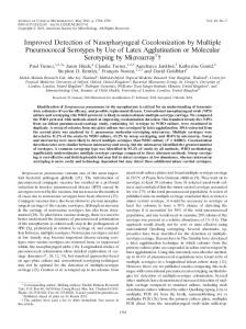
Clinical features Autosomal dominant inheritance is one of the features of Lynch syndrome (Table 1 and Figure 1). In contrast with familial adenomatous polyposis in which approximately one-fourth of cases
Table 1. Features of the Lynch syndrome Autosomal dominant inheritance Associated cancers: cancer of colorectum, stomach, small bowel, biliary tract, uroepithelial tract, ovary, endometrium, brain, skin (sebaceous adenoma) Development of cancer at an early age Development of multiple cancers Features of colorectal cancer: predeliction for proximal location, improved survival, multiple colorectal cancers, poorly differentiated tumors and Crohn’s-like infiltration of lymphocytes Features of adenomas: the numbers vary from one to a few, increased proportion of adenomas with a villous growth pattern, high degree of dysplasia, rapid progression from adenoma to carcinoma High frequency of microsatellite instability Immunohistochemistry: loss of MLH1, MSH2, MSH6 and PMS2 protein expression
14
General introduction
CRC 38 CRC 53
SmB 47
CRC 60
EC 44
CRC 47
CRC 29
CRC 33
EC 31
Figure 1. Pedigree of family with classical Lynch syndrome CRC = colorectal cancer EC = endometrial cancer SmB = small bowel cancer
is caused by a de novo APC-gene mutation, Lynch syndrome based on a de novo mismatch repair gene mutation has been rarely reported.26-28
As mentioned before, predisposed individuals from Lynch syndrome families have an in-
creased risk of developing CRC.29-31 The main precursors of CRC are adenomatous polyps.32 In
predisposed individuals to Lynch syndrome adenomas appear to develop at the same rate as in
individuals in the general population, but these adenomas develop at an earlier age, have more villous components and are more dysplastic than adenomas detected in the general population. Furthermore, the adenomas in predisposed individuals seem to progress more rapidly (2 to 3
years) to invasive colorectal cancer compared to those in the general population (8 to 10 years).33
Therefore, colorectal cancer in Lynch syndrome mutation carriers is often diagnosed at an early age, and synchronous and metachronous CRC are more common. Also, unlike in the general
population, the majority of these CRC is located in the proximal colon.34, 35 Specific pathological
characteristics of Lynch syndrome colorectal tumors have been identified but none of them are pathognomic. These features include poor differentiation, presence of mucinous and signet
cells, medullary features, peritumoral lymphocytic infiltration, Crohn’s like reaction, and tumor infiltrating lymphocytes mixed with tumor cells.36-38
Lynch syndrome mutation carriers are also at higher risk for other tumors including endome-
trial cancer (after CRC the most common cancer in Lynch syndrome patients) and to a lesser extent other cancers such as tumors of the stomach (particularly in Asian countries such as Japan 15
Chapter 2
CRC 56
Chapter 2
and Korea), small bowel, ovary, upper uroepithelial tract, biliary tract, skin and brain.29-31, 39 In
the presence of skin or brain tumors the phenotype is also addressed to as Muire-Torre syndrome and Turcot syndrome respectively.
Lynch syndrome associated colorectal tumors are adenocarcinomas. The endometrial cancers
seen in Lynch syndrome are mostly of the endometroid subtype 40, whereas ovarian cancers are serous or mucinous.30, 41 Gastric cancers are generally of the intestinal type.36, 42, 43 With respect
to tumors of the urinary tract, transitional cell carcinomas are associated with Lynch syndrome, localized in the ureter and renal pelvis but surprisingly not in the bladder.44, 45 The skin tumors in
Lynch syndrome are mostly sebaceous adenomas and adenocarcinomas, while the brain tumors are predominantly glioblastomas.46
Cancer risks in Lynch syndrome Lynch syndrome has a variable phenotype with respect to the tumor site, age of onset and the penetrance. Several studies have evaluated the cancer risks in Lynch syndrome. A summary of these studies is presented in Table 2.30, 39, 47-56 The cancer risks in Lynch syndrome are associated
with the affected MMR gene. In MLH1 and MSH2 mutation carriers the described CRC risk at
the age of 70 yrs ranges between 28-75% in males and 24-52% in females. Table 2. Life time cancer risks in Lynch syndrome Affected Mismatch repair gene Cancer type
MLH1+MSH2
MSH6
PMS2
Colorectal cancer (male)
28-75%
60-70-%
15-20%
Colorectal cancer (female)
24-52%
30-40%
15-20%
Endometrial cancer
27-60%
60-70%
15%
Small bowel cancer
4-7%
n.a.
Gastric cancer
2-13%
n.a.
Ovarian cancer
3-13%
n.a.
4%
n.a.
Upper urothelial tract
1-12%
n.a.
Brain
1-4%
n.a.
Biliary tract
25-32%*
n.a. = not available * all other Lynch syndrome-associated cancers Average life time risk based on the studies of Aarnio et al.30, Dunlop et al.47, Hampel et al.48, Hendriks et al.49, Plaschke et al.50, Quehenberger et al.51, Vasen et al.39, 52-54, 57, Barrow et al.55, 56
The mean age of CRC onset in MLH1 and MSH2 mutation carriers is approximately 45 years 30, 39, 47-56
Only few studies report the cumulative risk of CRC in MSH6 mutation carriers.50, 55 The
risk of CRC at the age of 70 years in male and female MSH6 mutation carriers is respectively
60-70% and 30-40%. The age of diagnosis of CRC in MSH6 mutation carriers is an average 5-10
years delayed compared to MLH1 or MSH2 carriers. The risk for developing endometrial cancer
in female MSH6 mutation carriers is 60-70%, while this is lower in MLH1 and MSH2 mutation 16
General introduction
carriers (27-60%). Furthermore, the mean age of onset of endometrial cancer seams slightly
lower in female MSH6 mutation carriers compared to MLH1 or MSH2 mutation carriers (54
years vs. 59 years). The risk of other Lynch syndrome associated tumors usually does not exceed
15% in MLH1 and MSH2 mutation carriers. In MSH6 mutation carriers these risks are at present
mutation carriers.57 The reported risk for developing colorectal cancer was 15-20%, while the risk of endometrial cancer was 15%. The risk for other Lynch syndrome associated cancer was 25-32%, which is much higher than in MLH1, MSH2 or MSH6 mutation carriers. More studies
evaluating the cancer risks in MSH6 and PMS2 mutation carriers are needed.
It should be noted that these risks could possibly be underestimated because some studies
evaluated families that were selected by using the Amsterdam criteria 52, or by including both
carriers as well as non carriers.51 On the other hand, the risks might also be biased because high risk families are being referred to a clinical genetic department, so called referral bias, while families without an apparent clustering of colorectal cancer are less frequently referred.
Diagnosis of Lynch syndrome Clinical diagnostic criteria Identification of subjects carrying a MMR mutation is important as surveillance can be restricted
to these individuals, while those without a gene defect can be reassured and spared from sur-
veillance. Surveillance in Lynch syndrome is important because it reduces the incidence and mortality of colorectal cancer.58-61 Currently, the Amsterdam criteria II and revised Bethesda criteria are used in clinical practice to select individuals for further analysis.
The Amsterdam criteria were formulated in 1990 by the International Collaborative Group
on HNPCC.15 However, in the following years various studies provided evidence that Lynch
syndrome was also associated with extracolonic tumors. This was the reason to propose a new set of criteria that include various extracolonic cancers, the Amsterdam criteria II (Table 3).62
These criteria are used in clinical practice as a selection criterion for mutation analysis in the MMR genes, however these criteria are too stringent to identify all Lynch syndrome families. Families suspected of Lynch syndrome but not fulfilling these criteria should not be falsely reassured and excluded from genetic counseling, DNA testing or surveillance.
The Bethesda criteria were formulated in 1996 and updated to the Revised Bethesda criteria in
2004 (table 2).25, 35, 63 The Bethesda criteria were formulated for the identification of tumors that
should be tested for MSI in order to select patients for subsequent MMR gene mutation analysis. Like the Amsterdam criteria II, the revised Bethesda criteria do not exclude a hereditary factor
for CRC. Whether the Amsterdam II and revised Bethesda criteria are adequate to identify Lynch syndrome patients is established by determining the proportion of cases with a MMR gene muta-
17
Chapter 2
largely unknown. So far, only one study has reported on data regarding cancer risk in PMS2
Chapter 2
Table 3. The Amsterdam Criteria II 62 and revised Bethesda criteria 35 Amsterdam criteria II There should be at least three relatives with a Lynch syndrome associated tumor (CRC, endometrial, small bowel, ureter/renal pelvis cancer); all of the following criteria should be present: •
one should be a first degree relative of the other two
•
at least two successive generations should be affected
•
at least one should be diagnosed before age 50 years
•
all tumors should be verified by pathological examination
•
FAP should be excluded in the CRC case
Revised Bethesda criteria •
CRC < age 50 years
•
presence of synchronous / metachronous Lynch syndrome-related cancer*, regardless of age
•
CRC with specific pathological features < 60 years**
• �CRC diagnosed in one or more 1st degree relatives with an Lynch syndrome- related cancer, with 1 of the diagnosis under age 50 years •
CRC in two or more 1st or 2nd degree relatives with an Lynch syndrome related cancer, regardless of age
* Lynch syndrome-related cancer: CRC, endometrial, stomach, ovarian, pancreas, ureter and renal pelvis, biliary tract, brain, sebaceous gland and small bowel carcinoma. * *tumor infiltrating lymphocytes, Crohn’s-like lymphocyte reaction, mucinous/signet ring differentiation or medullary growth pattern.
tion that would be missed using these criteria in a series of unselected CRC cases. One study 64
evaluated six studies 5, 65-69 in which either MSI or IHC analysis or both tests were performed as the primary screening tool in prospective, unselected series of colorectal cancer patients. The six studies showed that the sensitivity of the Amsterdam criteria II for detection of Lynch syndrome
MLH1 and MSH2 mutation carriers was approximately 40%, while the sensitivity of the revised Bethesda criteria was approximately 90%. This means that especially the Amsterdam criteria II miss a large proportion of MLH1 and MSH2 mutation carriers.70 These studies and our own
data (this thesis) indicate that the Amsterdam II and revised Bethesda criteria are suboptimal for the identification of Lynch syndrome mutation carriers, especially in families with a milder phenotype such as MSH6 and probably also PMS2 families.
Mutation prediction models In recent years several models to predict the likelihood of carrying a germline mutation have been developed.71-75 Mutation prediction models predict the probability of a mutation using
logistic regression or Bayesian methods. These models use information based on personal and family history as input to predict the probability of mutation carriership.
A major advantage of prediction models is that these models give a quantitative estimation
of the likelihood of mutation carriership instead of a bivariate (yes/no) assessment as provided
by the clinical diagnostic criteria. Mutation prediction models are thus potentially useful in clinical practice to optimize the identification of Lynch syndrome. The key issues involving
these models are their performance in clinical practice and their applicability in specific patient 18
General introduction
groups. However, most of these models are based on samples of Caucasian populations with European ancestry and further validation in other ethnic groups is necessary. Furthermore, the performance of these models has been evaluated in the same study setting which formed the basis
for their development. External validation is necessary to study generalizability of these models. study has evaluated the Premm1,2 model in a population based cohort of 1222 CRC patients
(the EPICOLON cohort). In this study cohort, the Premm1,2 model identified all the MLH1 and
MSH2 mutation carriers, indicating a good performance of this model.76 However, the number
of identified mutations (n=8) was very low, limiting the reliability of this study. Furthermore, the Premm1,2 model is not able to identify MSH6 and PMS2 mutation carriers. Another study used
the same EPICOLON cohort to compare the Premm1,2 model with the Edinburgh model reported a similar performance of both models.77
71
and
Microsatellite instability analysis (MSI) & immunohistochemistry (IHC) As mentioned before MSI is a hallmark of Lynch syndrome. Using the international set of recommended markers (D2S123, D5S346, D17S250, BAT25 and BAT26) more than 90% of
Lynch syndrome-associated CRC and about 15% of sporadic CRC exhibit microsatellite instability.22, 78-83 Comparing the marker size in normal and tumor DNA from the same individual
tumors is scored as MSI-high if at least two of the markers show instability, MSI low if one
marker shows instability, or MSI stable if none of the markers shows instability. The current
standard method for MSI analysis is relatively time consuming, laborious and expensive, due to the need to compare allelic profiles between tumor and matching germline DNA. However, new MSI analysis methods such as the fluorescent multiplex PCR of mononucleotide repeats and the
computerized fragment analysis method are promising. This alternative simple and straightfor-
ward MSI analysis system of mononucleotide microsatellite repeats can identify MSI-H tumors with a high sensitivity and specificity. An advantage of these systems is that comparison of tumor with matching germline DNA is unnecessary. Studies evaluating these new MSI analysis system are promising, but further validation is needed.84, 85
MSI analysis can be performed in DNA extracted from paraffin-wax embedded tumors. To
optimize MSI analyses good quality of tumor tissue is warranted and an experienced molecular laboratory/pathologist is needed. It should also be noted that an MSI-stable phenotype does not exclude Lynch syndrome, because of the possibility of a phenocopy. In proven Lynch syndrome
families, frequently tumors are encountered with no indication of MSI. These individuals could
have developed, for example, a CRC or endometrial carcinoma because these tumors have a relatively high prevalence in the population.86, 87 When a strong suspicion for Lynch syndrome
remains in spite of a MSI stable tumor, MSI analysis on a second tumor in the family may be considered.
19
Chapter 2
Data concerning the performance of these models in a population-based cohort are sparse. One
Chapter 2
MSI analysis is not suitable to predict which of the MMR genes is affected, but this can be
demonstrated by further IHC analysis. For this analysis, specific antibodies are used to visualize
the presence or absence of MLH1, MSH2, MSH6 and PMS2 proteins in tumor cells compared to normal cells. An IHC pattern with absent staining for MLH1 and PMS2 and positive staining
for MSH2 and MSH6 is indicative for a mutation in MLH1 (Table 4). This pattern is explained
by the fact that the MLH1 protein forms a heterodimer with the PMS2 protein. In the absence of MLH1 protein, the heterodimer will not be formed and the PMS2 protein will degrade resulting in the absence of staining of both proteins. Because the MSH2 protein forms a heterodimer
with MSH6, the specific immunohistochemical pattern observed in tumors of MSH2 mutation
carriers, comprises absence of staining of MSH2 and MSH6 with normal staining of MLH1 and PMS2. In tumors from MSH6 mutation carriers generally only absence of staining of the MSH6
protein is observed whereas in tumors from carriers of a PMS2 mutation absence of the PMS2 protein is found.
Table 4. IHC patterns associated with MLH1, MSH2, MSH6 and PMS2 mutations21, 105 MMR gene mutation IHC staining
MLH1
MSH2
MSH6
PMS2
MLH1
-
+
+
+
MSH2
+
-
+
+
MSH6
+
-
-
+
PMS2
-
+
+
-
However, a tumor with an MSI-high phenotype and absent staining of MLH1 can also be
caused by hypermethylation of the promotor region of the MLH1 gene, which is found in spo-
radic MSI-high CRC. In 50% of the sporadic CRC with MLH1 promotor hypermethylation
specific mutations in the BRAF gene are found in the tumor tissue. Therefore, additional BRAF
analysis and MLH1 promotor methylation analysis can differentiate between a somatic sporadic MSI-high CRC and a Lynch syndrome related cancer.88-91
IHC is especially indicative for MMR mutations that result in truncation of the protein, such
as nonsense, frameshift, splice site mutations and large genomic rearrangements. In case of missense mutations, IHC is not always diagnostic as the protein may be (partly) expressed and therefore still detected by IHC. Also, the value of IHC largely depends on the quality of the nuclear staining and the experience of the pathologist.92-94
Several studies have prospectively evaluated the results of MSI and IHC analysis in CRC tis-
sue for the identification of MMR mutations.67-69, 71, 95-97 The reported sensitivity and specificity of
MSI in these studies varied between 80%-100% and 70-95% respectively. For IHC, the reported
sensitivity and specificity were 85-95% and 80-95%, respectively. However, only two of these studies evaluated all the known MMR proteins.68, 96
MSI and IHC analysis is optimally performed on colorectal tumor tissue of the youngest
patient in the family. Other tumor tissues can be analyzed, however, the value of MSI/IHC in 20
General introduction
other Lynch syndrome related tumors is largely unknown. An American study evaluated the use of MSI and IHC analysis in 543 endometrial tumors in an unselected population.98 Of these 543 tumors, 98 (18%) were MSI-high and 20 (4%) were MSI-low. An abnormal IHC staining was
found in 90 (92%) of the MSI-high tumors. In 10 (8%) MSI/IHC positive tumors a MMR gene cancer. However, this study did not fully address the sensitivity and specificity of MSI and IHC in endometrial cancer since mutation testing in the MSI negative tumors was not performed. Nevertheless, other studies, although smaller, also have shown that MSI and IHC is feasible in endometrial cancer.99-101
Colorectal adenomas can also be used for MSI and IHC analysis, however previous studies
have shown that not all the colorectal adenomas found in mutation carriers exhibit an MSI-high phenotype. This is due to the fact that a MSI-high phenotype is not yet fully manifested in early or low grade colorectal adenomas.102-104
A combination of MSI and IHC provides the most optimal selection for mutation analysis, however in view of the costs some advocate IHC as the first step.95, 105 As mentioned before
the revised Bethesda criteria were designed to select CRC patients for MSI / IHC analysis.
Another strategy would be to perform MSI and IHC on all newly diagnosed colorectal cancer and or endometrial cancer cases. The advantage of such a strategy may be that families with
a mild phenotype would also be identified. Studies evaluating this strategy show promising results
106, 107
, but more studies are needed to evaluate the feasibility, diagnostic yield and cost
effectiveness of this strategy.
Mutation analysis To confirm the presence of a mutation in a MMR gene, mutation analysis is performed in DNA from blood derived lymphocytes. At this moment there are several techniques available for mu-
tation analysis including direct sequencing, denaturating gradient gel electrophoresis (DGGE) with sequencing of aberrant fragments and multiplex ligand dependent probe amplification (MLPA) for the detection of large genomic deletions, which occur most frequently in MSH2.
Mutation analysis is expensive and time consuming. Therefore it is generally performed when MSI and IHC analysis are indicative for a germline mutation or if there is a very high suspicion of a mutation based on the family history.
All genomic coding changes are potentially deleterious. However, as opposed to nonsense muta-
tions (which create a stop codon or lead to a frame shift) or those that cause abnormal splicing, missense mutations (which lead to the substitution of an amino acid) are usually not considered a priori pathogenic. Of all mutations identified in MLH1 and MSH2, 29% and 16%, respectively,
are missense mutations. A functional test to reliably assess the competence of the mismatch 21
Chapter 2
mutation was found. The authors concluded that MSI and IHC analysis is feasible in endometrial
Chapter 2
repair proteins is currently not available. Therefore, most missense mutations are designated as unclassified variants (UV). These variants cannot be used for diagnostic purposes.
Genetic testing and counseling The diagnosis of a germline mutation confirming a predisposition to cancer can be complex and may have considerable medical and psychosocial consequences.108 Individuals who opt for genetic testing should receive genetic counseling and psychosocial guidance.
Once a mutation has been detected in an affected individual, healthy family members can be
offered mutation analysis, so called presymptomatic diagnostic testing. A negative genetic test may result in emotional relief regarding personal and/or offspring cancer risk, and avoidance of unnecessary surveillance. However, feelings of guilt towards affected relatives, so called
survivor-guilt, may seriously harm inter-familial relations. A positive genetic test may lead to
emotional distress regarding personal cancer risk and frequent surveillance or considerations for prophylactic management. Furthermore, it may have consequences for offspring or the desire to have children. Also, a positive result can have financial consequences such as increased
mortgage and life-insurance costs. The protocol for genetic testing of the American Society of Clinical Oncology recommends three sessions with a clinical geneticist.108 During the first
session the discussed issues include the reasons for testing, the clinical features of the hereditary
colorectal cancer syndrome, the mode of inheritance, the consequences of the test results, the
options for surveillance or prophylactic procedures in case of a positive result and the DNA testing procedure. In the second session blood samples are taken and during the third session the test results are disclosed and if necessary further surveillance strategy discussed. In the
Netherlands genetic counseling for CRC is offered by the department of Clinical Genetics of the University Medical Centers. In clinical practice in the Netherlands blood samples are generally taken directly after the first counseling session.
The uptake for genetic testing in Lynch syndrome families varies. A Finnish study reported an
uptake rate of 75%, while an American study reported a rate of 43%.109, 110 However, both studies
were performed in a research setting and therefore these studies do not reflect clinical practice. We evaluated the uptake in clinical practice and reported uptake rates of 43% and 50%.111, 112 In
view of the preventative options in risk carriers for Lynch syndrome a higher uptake for genetic testing is desirable. The more since a previous study reported that genetic testing improves the compliance of colonoscopy surveillance from 19% to 88%.113 and mutation carriers are able to
cope well with their cancer susceptibility on the short as well on the long term.113-115
Studies at the reasons for risk carriers not to be tested and a better implementation of genetic
testing in clinical practice are desirable.
22
General introduction
Diagnostic approach in patients suspected of Lynch syndrome A detailed family history in all patients with cancer is the simplest and most cost-effective way to identify hereditary colorectal cancer. Characteristics of hereditary forms of colorectal cancer early age of onset, the presence of multiple tumors and the combined occurrence of colorectal cancer with endometrial cancer or another Lynch syndrome associated cancer.
In patients who comply to the revised Bethesda criteria MSI and/or IHC analysis should
be performed on the available tumor tissue.116 However, carcinomas in MSH6 mutation car-
riers, particularly endometrial carcinomas, have been shown to be present with a MSI-stable phenotype in a minority that cannot be neglected.86, 117(this thesis) Therefore, if an MSI-stable
phenotype is found in a family with clustering of endometrial carcinoma, IHC of MSH6 is the next step. If IHC is negative for MSH6, mutation analysis follows. If staining is present, MSI analysis of a second tumor can be considered. With respect to PMS2 mutations further studies are required.
In MSI-H cases with absent staining of MLH1 promotor hypermethylation analysis should be
performed before germline mutation analysis to exclude sporadic MSI-H cases.
In cases with a strong positive family history but an MSI stable tumor, MSI analysis on a second
tumor from the same family is recommended to exclude the possibility of phenocopies. When MSI and IHC analysis do not show abnormalities, germline mutation analysis is not useful.118
Surveillance in Lynch Syndrome Surveillance of the colon The surveillance program of Lynch syndrome includes colorectal surveillance by biennial colonoscopy starting from the age of 20-25 years (Table 5). The rationale for biennial colonos-
copy screening is that the risk for developing an invasive CRC within this two year period is
small.119, 120 There is evidence that colonoscopy surveillance is effective in reducing the incidence
and mortality of CRC.58-61 A Finnish study followed 22 Lynch syndrome families during a period
of 15 years.61 Colonoscopy surveillance at 3-year interval resulted in a 65% decrease in mortality
and an increased detection of early stage CRC. A decrease in mortality was also found in a
Table 5. Surveillance guidelines in Lynch syndrome 64 Surveillance
Examination
Start at age
Interval
Colon
Colonoscopy
20-25 years
1-2 year
Endometrium
Gynaecological examination, transvaginal US
30-35 years
1-2 year
Stomach*
Gastroscopy
30-35 years
1-2 year
Urinary tract*
Urine cytology
30-35 years
1-2 year
* if stomach or urinary tract cancer runs in the family (more than one case) 23
Chapter 2
that might be helpful in the differential diagnosis for non hereditary cases include an unusual
Chapter 2
Dutch study that evaluated the Dutch surveillance program for Lynch syndrome.58 This study reported a 70% decrease in the standardized mortality ratio for colorectal cancer when one or
more surveillance colonoscopies were performed. However, interval cancers have been reported despite surveillance colonoscopy.121
Currently, colonoscopy surveillance is performed by white light endoscopy, but a recent meta
analysis including 6 studies with tandem colonoscopies showed an adenoma miss rate of 2% for adenomas > 10 mm and even a 26% miss rate for adenomas < 5 mm.122 In Lynch syndrome,
flat adenomas are particularly prone to malignant transformation compared to adenomas in the general population.33, 34 Other endoscopic modalities such as high magnification chromo endos-
copy, narrow band imaging or autofluorescence endoscopy could decrease the adenoma miss
rate. Chromo endoscopy is a colonoscopic technique in which the colonic surface is sprayed
with a dye, such as indigo carmine, resulting in an enhanced view of the epithelial surface. Two small studies have evaluated the use of high magnification chromo-endoscopy in Lynch syn-
drome patients. A French study evaluated chromo-endoscopy in 36 consecutive asymptomatic
patients belonging to Lynch syndrome families.123 During white light colonoscopy 7 adenomas were detected in five patients and chromo endoscopy detected an additional 11 adenomas in
eight patients. The adenoma detection rate of chromo-endoscopy was significantly higher (p
=0.045) than white light colonoscopy. These results were confirmed by a British study, in which 25 asymptomatic patients fulfilling the modified Amsterdam criteria underwent both conven-
tional and chromo-endoscopy.124 White light colonoscopy detected 11 adenomas and chromo-
endoscopy detected an additional 32 adenomas (p < 0.01). However, chromo-endoscopy is time consuming because of the dye spraying. Both narrow band imaging and autofluorescence do
not need additional dye spraying thus these techniques might be less time consuming. Narrow band imaging is another endoscopic modality, in which superficial capillaries in the mucosa are
highlighted. Neoplasia in the mucosa has an increased vascular density and thus can be easily detected by narrow band imaging. So far, only one study has evaluated narrow band imaging for
colonoscopic surveillance in Lynch syndrome.125 In total 62 patients were evaluated and with
the use of narrow band imaging the number of patients with adenomas increased with 15% to a total of 42%.
Autofluorescence endoscopy also is another technique which may be used in the colono-
scopic surveillance of Lynch syndrome. The polyp detection rate with autofluorescence may be
higher 126 than conventional white light colonoscopy. More studies evaluating autofluorescence
endoscopy and narrow band imaging are needed. Until then, biennial colonoscopy screening by conventional high quality magnification white light colonoscopy remains the gold standard.
Surveillance of the endometrium Endometrial cancer is the second most common malignancy in Lynch syndrome and therefore female mutation carriers are offered endometrial cancer surveillance. The endometrial surveil24
General introduction
lance program includes biennial gynaecological examination and transvaginal US examination, starting at the age of 30-35 years. However, the effect of endometrial cancer surveillance is disputable.127-129 Because of the higher risk of developing endometrial carcinoma in MSH6 mutation carriers, hysterectomy can be suggested in these women after menopause.
may be considered in families with an excess of ovarian carcinoma. Also an ovariectomy can be considered in females with a prophylactic hysterectomy.130, 131 The value of surveillance for
endometrial cancer is not known and further studies need to prove that biennial surveillance
leads to the detection of premalignant lesions and early cancers. Accurate treatment of women with postmenopausal blood loss is probably the most important.
Surveillance of gastric carcinoma and upper urothelial cell carcinoma is recommended if two or more tumor cases occur within the family.116 However, the value of this strategy still remains
unclear. Surveillance guidelines for other Lynch syndrome associated tumors (small bowel, ovary, biliary tract, skin and brain) are lacking.
Therapy of colorectal carcinoma in Lynch syndrome Surgical treatment for colorectal carcinoma Previous studies have raised the question whether a subtotal colectomy instead of a segmental resection might be the preferred treatment in Lynch syndrome patients with a primary CRC.
A Finnish study reported that 15/37 Lynch syndrome patients who underwent a segmental
colon resection developed a metachronous CRC, compared to 4/17 Lynch syndrome patients who underwent a subtotal colectomy.132 An American study reported a metachronous CRC in
16/70 Lynch syndrome patients who underwent a segmental colon resection versus 0/23 Lynch syndrome patients who underwent a subtotal colectomy.133 The results of these studies sug-
gest that a subtotal colectomy is the preferred treatment in Lynch syndrome related CRC. In a Dutch study a decision analysis was performed to compare the life expectancy for patients who underwent either a subtotal colectomy or a segmental colon resection.134 The authors concluded
that a subtotal colectomy performed at a young age (< 47 years) would result in an increased
life expectancy of up to 2.3 years. A subtotal colectomy in a 67 years old patient resulted in an
increased life expectancy up to only 0.3 years. Based on these findings a subtotal colectomy with
an ileorectal anastomosis should be the treatment of first choice in young patients (< 60 years) presenting with CRC, while in older patients a segmental colon resection might be appropriate. Of course, surveillance of the residual colon remains important.
25
Chapter 2
In view of the risk of ovarian carcinoma, the failure of early cancer detection with trans-
vaginal US and determination of the tumor marker CA125, bilateral salpingo-oophorectomy
Chapter 2
Chemotherapy Currently, chemotherapeutic regimes for colorectal cancer include 5FU with or without
leucovorin, oxaliplatin and irinotecan. The effect of chemotherapy on MSI-high tumors have been reported in a few studies.135-139 Most of these studies reported that there was no benefit of
5FU-based chemotherapy and questioned the benefit of such therapy for patients with MSI-high
tumors. However, one study was a prospective non randomized study in 244 patients, including 52 with a MSI-high tumor.135 The authors concluded that patients with an MSI-high tumor that
received 5FU-based chemotherapy had a better survival. Prospective clinical trials are needed to evaluate the effect of different chemotherapeutic regimes in MSI-high tumors.
Chemoprevention in Lynch syndrome The use of aspirin has been associated with a moderate reduction in the risk of colonic adenomas and colorectal cancer in the general population, while resistant starch (an isomer of starch) has been associated with an antineoplastic effect.140-143 Therefore, aspirin and resistant starch would
be a candidate for chemoprevention in Lynch syndrome. Recently, a randomized, placebo con-
trolled trial evaluated the use of aspirin and resistant starch in proven mutation carriers during a
follow up period of 4 years. Of 693 participants randomly assigned to receive aspirin or placebo,
neoplasia developed in 66 (18.9%) participants receiving aspirin versus 65 (19%) participants
receiving placebo. The trial did not report a significant difference between the two groups with respect to the development of advanced colorectal neoplasia (7.4% vs. 9.9%, p =0.33). Of the
727 participants receiving resistant starch or placebo, also no significant difference was reported (18.7% vs. 18.4%). Furthermore, the distribution of advanced adenoma and colorectal cancer were evenly distributed in both groups. Although the concept of chemoprevention is promising,
there are currently no chemopreventive agents available for the prevention of colorectal cancer in Lynch syndrome.
26
General introduction
1. Knudsen AL, Bisgaard ML, Bulow S. Attenuated familial adenomatous polyposis (AFAP). A review of the literature. Fam Cancer 2003;2:43-55. 2. Sampson JR, Dolwani S, Jones S, Eccles D, Ellis A, et al. Autosomal recessive colorectal adenomatous polyposis due to inherited mutations of MYH. Lancet 2003;362:39-41. 3. Al-Tassan N, Chmiel NH, Maynard J, Fleming N, Livingston AL, et al. Inherited variants of MYH associated with somatic G:C-->T:A mutations in colorectal tumors. Nat Genet 2002;30:227-232. 4. Bisgaard ML, Fenger K, Bulow S, Niebuhr E, Mohr J. Familial adenomatous polyposis (FAP): frequency, penetrance, and mutation rate. Hum Mutat 1994;3:121-125. 5. Aaltonen LA, Salovaara R, Kristo P, Canzian F, Hemminki A, et al. Incidence of hereditary nonpolyposis colorectal cancer and the feasibility of molecular screening for the disease. N Engl J Med 1998;338:1481-1487. 6. Warthin AS. Heredity with reference to carcinoma; as shown by the study of cases examined in the pathological laboratory of the University of Michigan 1895-1913. 12 ed. 1913:546-555. 7. Warthin AS. The further study of a cancer family. 117 ed. 1925:206-212. 8. Hauser IJ, Weller CV. A further report on the cancer family of Warthin. 27 ed. 1936:434-449. 9. Lynch HT, Shaw MW, Magnuson CW, Larsen AL, Krush AJ. Hereditary factors in cancer. Study of two large midwestern kindreds. Arch Intern Med 1966;117:206-212. 10. Lynch HT, Krush AJ. Cancer family “G” revisited: 1895-1970. Cancer 1971;27:1505-1511. 11. Mecklin JP, Jarvinen HJ, Peltokallio P. Cancer family syndrome. Genetic analysis of 22 Finnish kindreds. Gastroenterology 1986;90:328-333. 12. Vasen HF, den Hartog Jager FC, Menko FH, Nagengast FM. Screening for hereditary non-polyposis colorectal cancer: a study of 22 kindreds in The Netherlands. Am J Med 1989;86:278-281. 13. Ponz De LM, Sassatelli R, Sacchetti C, Zanghieri G, Scalmati A, et al. Familial aggregation of tumors in the three-year experience of a population-based colorectal cancer registry. Cancer Res 1989;49:4344-4348. 14. Lynch HT, Cristofaro G, Rozen P, Vasen H, Lynch P, et al. History of the International Collaborative Group on Hereditary Non Polyposis Colorectal Cancer. Fam Cancer 2003;2:3-5. 15. Vasen HF, Mecklin JP, Khan PM, Lynch HT. The International Collaborative Group on Hereditary Non-Polyposis Colorectal Cancer (ICG-HNPCC). Dis Colon Rectum 1991;34:424-425. 16. Fishel R, Lescoe MK, Rao MR, Copeland NG, Jenkins NA, et al. The human mutator gene homolog MSH2 and its association with hereditary nonpolyposis colon cancer. Cell 1993;75:1027-1038. 17. Bronner CE, Baker SM, Morrison PT, Warren G, Smith LG, et al. Mutation in the DNA mismatch repair gene homologue hMLH1 is associated with hereditary non-polyposis colon cancer. Nature 1994;368:258-261. 18. Papadopoulos N, Nicolaides NC, Wei YF, Ruben SM, Carter KC, et al. Mutation of a mutL homolog in hereditary colon cancer. Science 1994;263:1625-1629. 19. Miyaki M, Konishi M, Tanaka K, Kikuchi-Yanoshita R, Muraoka M, et al. Germline mutation of MSH6 as the cause of hereditary nonpolyposis colorectal cancer. Nat Genet 1997;17:271-272. 20. Akiyama Y, Sato H, Yamada T, Nagasaki H, Tsuchiya A, et al. Germ-line mutation of the hMSH6/ GTBP gene in an atypical hereditary nonpolyposis colorectal cancer kindred. Cancer Res 1997;57:3920-3923. 21. de Jong AE, van Puijenbroek M, Hendriks Y, Tops C, Wijnen J, et al. Microsatellite instability, immunohistochemistry, and additional PMS2 staining in suspected hereditary nonpolyposis colorectal cancer. Clin Cancer Res 2004;10:972-980. 27
Chapter 2
References
Chapter 2
22. Kunkel TA. Nucleotide repeats. Slippery DNA and diseases. Nature 1993;365:207-208. 23. Jiricny J. Mediating mismatch repair. Nat Genet 2000;24:6-8. 24. Lipkin SM, Wang V, Jacoby R, Banerjee-Basu S, Baxevanis AD, et al. MLH3: a DNA mismatch repair gene associated with mammalian microsatellite instability. Nat Genet 2000;24:27-35. 25. Boland CR, Thibodeau SN, Hamilton SR, Sidransky D, Eshleman JR, et al. A National Cancer Institute Workshop on Microsatellite Instability for cancer detection and familial predisposition: development of international criteria for the determination of microsatellite instability in colorectal cancer. Cancer Res 1998;58:5248-5257. 26. Ripa R, Bisgaard ML, Bulow S, Nielsen FC. De novo mutations in familial adenomatous polyposis (FAP). Eur J Hum Genet 2002;10:631-637. 27. Desai DC, Lockman JC, Chadwick RB, Gao X, Percesepe A, et al. Recurrent germline mutation in MSH2 arises frequently de novo. J Med Genet 2000;37:646-652. 28. Kraus C, Kastl S, Gunther K, Klessinger S, Hohenberger W, et al. A proven de novo germline mutation in HNPCC. J Med Genet 1999;36:919-921. 29. Watson P, Lynch HT. The tumor spectrum in HNPCC. Anticancer Res 1994;14:1635-1639. 30. Aarnio M, Sankila R, Pukkala E, Salovaara R, Aaltonen LA, et al. Cancer risk in mutation carriers of DNA-mismatch-repair genes. Int J Cancer 1999;81:214-218. 31. Park YJ, Shin KH, Park JG. Risk of gastric cancer in hereditary nonpolyposis colorectal cancer in Korea. Clin Cancer Res 2000;6:2994-2998. 32. Watanabe T, Muto T, Sawada T, Miyaki M. Flat adenoma as a precursor of colorectal carcinoma in hereditary nonpolyposis colorectal carcinoma. Cancer 1996;77:627-634. 33. Jass JR, Stewart SM. Evolution of hereditary non-polyposis colorectal cancer. Gut 1992;33:783-786. 34. Lynch HT, de la Chapelle A. Hereditary colorectal cancer. N Engl J Med 2003;348:919-932. 35. Umar A, Boland CR, Terdiman JP, Syngal S, de la CA, et al. Revised Bethesda Guidelines for hereditary nonpolyposis colorectal cancer (Lynch syndrome) and microsatellite instability. J Natl Cancer Inst 2004;96:261-268. 36. Lynch HT, Lanspa S, Smyrk T, Boman B, Watson P, et al. Hereditary nonpolyposis colorectal cancer (Lynch syndromes I & II). Genetics, pathology, natural history, and cancer control, Part I. Cancer Genet Cytogenet 1991;53:143-160. 37. Mecklin JP, Jarvinen HJ. Clinical features of colorectal carcinoma in cancer family syndrome. Dis Colon Rectum 1986;29:160-164. 38. Young J, Simms LA, Biden KG, Wynter C, Whitehall V, et al. Features of colorectal cancers with high-level microsatellite instability occurring in familial and sporadic settings: parallel pathways of tumorigenesis. Am J Pathol 2001;159:2107-2116. 39. Ten Kate GL, Kleibeuker JH, Nagengast FM, Craanen M, Cats A, et al. Is surveillance of the small bowel indicated for Lynch syndrome families? Gut 2007. 40. de Leeuw WJ, Dierssen J, Vasen HF, Wijnen JT, Kenter GG, et al. Prediction of a mismatch repair gene defect by microsatellite instability and immunohistochemical analysis in endometrial tumours from HNPCC patients. J Pathol 2000;192:328-335. 41. Lynch HT, Smyrk TC, Watson P, Lanspa SJ, Lynch JF, et al. Genetics, natural history, tumor spectrum, and pathology of hereditary nonpolyposis colorectal cancer: an updated review. Gastroenterology 1993;104:1535-1549. 42. Aarnio M, Salovaara R, Aaltonen LA, Mecklin JP, Jarvinen HJ. Features of gastric cancer in hereditary non-polyposis colorectal cancer syndrome. Int J Cancer 1997;74:551-555.
28
43. Rodriguez-Bigas MA, Vasen HF, Lynch HT, Watson P, Myrhoj T, et al. Characteristics of small bowel carcinoma in hereditary nonpolyposis colorectal carcinoma. International Collaborative Group on HNPCC. Cancer 1998;83:240-244. 44. Sijmons RH, Kiemeney LA, Witjes JA, Vasen HF. Urinary tract cancer and hereditary nonpolyposis colorectal cancer: risks and screening options. J Urol 1998;160:466-470. 45. Watson P, Lynch HT. Extracolonic cancer in hereditary nonpolyposis colorectal cancer. Cancer 1993;71:677-685. 46. Hamilton SR, Liu B, Parsons RE, Papadopoulos N, Jen J, et al. The molecular basis of Turcot’s syndrome. N Engl J Med 1995;332:839-847. 47. Dunlop MG, Farrington SM, Carothers AD, Wyllie AH, Sharp L, et al. Cancer risk associated with germline DNA mismatch repair gene mutations. Hum Mol Genet 1997;6:105-110. 48. Hampel H, Stephens JA, Pukkala E, Sankila R, Aaltonen LA, et al. Cancer risk in hereditary nonpolyposis colorectal cancer syndrome: later age of onset. Gastroenterology 2005;129:415-421. 49. Hendriks YM, Wagner A, Morreau H, Menko F, Stormorken A, et al. Cancer risk in hereditary nonpolyposis colorectal cancer due to MSH6 mutations: impact on counseling and surveillance. Gastroenterology 2004;127:17-25. 50. Plaschke J, Engel C, Kruger S, Holinski-Feder E, Pagenstecher C, et al. Lower incidence of colorectal cancer and later age of disease onset in 27 families with pathogenic MSH6 germline mutations compared with families with MLH1 or MSH2 mutations: the German Hereditary Nonpolyposis Colorectal Cancer Consortium. J Clin Oncol 2004;22:4486-4494. 51. Quehenberger F, Vasen HF, van Houwelingen HC. Risk of colorectal and endometrial cancer for carriers of mutations of the hMLH1 and hMSH2 gene: correction for ascertainment. J Med Genet 2005;42:491-496. 52. Vasen HF, Stormorken A, Menko FH, Nagengast FM, Kleibeuker JH, et al. MSH2 mutation carriers are at higher risk of cancer than MLH1 mutation carriers: a study of hereditary nonpolyposis colorectal cancer families. J Clin Oncol 2001;19:4074-4080. 53. Vasen HF, Wijnen JT, Menko FH, Kleibeuker JH, Taal BG, et al. Cancer risk in families with hereditary nonpolyposis colorectal cancer diagnosed by mutation analysis. Gastroenterology 1996;110:1020-1027. 54. Watson P, Vasen HF, Mecklin JP, Bernstein I, Aarnio M, et al. The risk of extra-colonic, extraendometrial cancer in the Lynch syndrome. Int J Cancer 2008;123:444-449. 55. Barrow E, Alduaij W, Robinson L, Shenton A, Clancy T, et al. Colorectal cancer in HNPCC: cumulative lifetime incidence, survival and tumour distribution. A report of 121 families with proven mutations. Clin Genet 2008;74:233-242. 56. Barrow E, Robinson L, Alduaij W, Shenton A, Clancy T, et al. Cumulative lifetime incidence of extracolonic cancers in Lynch syndrome: a report of 121 families with proven mutations. Clin Genet 2009;75:141-149. 57. Senter L, Clendenning M, Sotamaa K, Hampel H, Green J, et al. The clinical phenotype of Lynch syndrome due to germ-line PMS2 mutations. Gastroenterology 2008;135:419-428. 58. de Jong AE, Hendriks YM, Kleibeuker JH, de Boer SY, Cats A, et al. Decrease in mortality in Lynch syndrome families because of surveillance. Gastroenterology 2006;130:665-671. 59. Jablonska M, Reznikova L, Kotrlik J, Svitavsky M, Mikova M, et al. Clinical implications of recognition of the hereditary non-polyposis colon cancer syndrome (HNPCC) for the early detection of colorectal cancer. Sb Lek 1995;96:275-282. 60. Vasen HF, Taal BG, Nagengast FM, Griffioen G, Menko FH, et al. Hereditary nonpolyposis colorectal cancer: results of long-term surveillance in 50 families. Eur J Cancer 1995;31A:1145-1148.
29
Chapter 2
General introduction
Chapter 2
61. Jarvinen HJ, Aarnio M, Mustonen H, Aktan-Collan K, Aaltonen LA, et al. Controlled 15-year trial on screening for colorectal cancer in families with hereditary nonpolyposis colorectal cancer. Gastroenterology 2000;118:829-834. 62. Vasen HF, Watson P, Mecklin JP, Lynch HT. New clinical criteria for hereditary nonpolyposis colorectal cancer (HNPCC, Lynch syndrome) proposed by the International Collaborative group on HNPCC. Gastroenterology 1999;116:1453-1456. 63. Rodriguez-Bigas MA, Boland CR, Hamilton SR, Henson DE, Jass JR, et al. A National Cancer Institute Workshop on Hereditary Nonpolyposis Colorectal Cancer Syndrome: meeting highlights and Bethesda guidelines. J Natl Cancer Inst 1997;89:1758-1762. 64. Vasen HF, Moslein G, Alonso A, Bernstein I, Bertario L, et al. Guidelines for the clinical management of Lynch syndrome (HNPCC). J Med Genet 2007. 65. Debniak T, Kurzawski G, Gorski B, Kladny J, Domagala W, et al. Value of pedigree/clinical data, immunohistochemistry and microsatellite instability analyses in reducing the cost of determining hMLH1 and hMSH2 gene mutations in patients with colorectal cancer. Eur J Cancer 2000;36:49-54. 66. Salovaara R, Loukola A, Kristo P, Kaariainen H, Ahtola H, et al. Population-based molecular detection of hereditary nonpolyposis colorectal cancer. J Clin Oncol 2000;18:2193-2200. 67. Cunningham JM, Kim CY, Christensen ER, Tester DJ, Parc Y, et al. The frequency of hereditary defective mismatch repair in a prospective series of unselected colorectal carcinomas. Am J Hum Genet 2001;69:780-790. 68. Hampel H, Frankel WL, Martin E, Arnold M, Khanduja K, et al. Screening for the Lynch syndrome (hereditary nonpolyposis colorectal cancer). N Engl J Med 2005;352:1851-1860. 69. Pinol V, Castells A, Andreu M, Castellvi-Bel S, Alenda C, et al. Accuracy of revised Bethesda guidelines, microsatellite instability, and immunohistochemistry for the identification of patients with hereditary nonpolyposis colorectal cancer. JAMA 2005;293:1986-1994. 70. Ramsoekh D, Wagner A, van Leerdam ME, Dinjens WN, Steyerberg EW, et al. A high incidence of MSH6 mutations in Amsterdam criteria II-negative families tested in a diagnostic setting. Gut 2008;57:1539-1544. 71. Barnetson RA, Tenesa A, Farrington SM, Nicholl ID, Cetnarskyj R, et al. Identification and survival of carriers of mutations in DNA mismatch-repair genes in colon cancer. N Engl J Med 2006;354:2751-2763. 72. Balmana J, Stockwell DH, Steyerberg EW, Stoffel EM, Deffenbaugh AM, et al. Prediction of MLH1 and MSH2 mutations in Lynch syndrome. JAMA 2006;296:1469-1478. 73. Chen S, Wang W, Lee S, Nafa K, Lee J, et al. Prediction of germline mutations and cancer risk in the Lynch syndrome. JAMA 2006;296:1479-1487. 74. Wijnen JT, Vasen HF, Khan PM, Zwinderman AH, van der KH, et al. Clinical findings with implications for genetic testing in families with clustering of colorectal cancer. N Engl J Med 1998;339:511-518. 75. Lipton LR, Johnson V, Cummings C, Fisher S, Risby P, et al. Refining the Amsterdam Criteria and Bethesda Guidelines: testing algorithms for the prediction of mismatch repair mutation status in the familial cancer clinic. J Clin Oncol 2004;22:4934-4943. 76. Balaguer F, Balmana J, Castellvi-Bel S, Steyerberg EW, Andreu M, et al. Validation and extension of the PREMM1,2 model in a population-based cohort of colorectal cancer patients. Gastroenterology 2008;134:39-46. 77. Balmana J, Balaguer F, Castellvi-Bel S, Steyerberg EW, Andreu M, et al. Comparison of predictive models, clinical criteria and molecular tumour screening for the identification of patients with Lynch syndrome in a population-based cohort of colorectal cancer patients. J Med Genet 2008;45:557-563.
30
78. Thibodeau SN, Bren G, Schaid D. Microsatellite instability in cancer of the proximal colon. Science 1993;260:816-819. 79. Jiricny J. Replication errors: cha(lle)nging the genome. EMBO J 1998;17:6427-6436. 80. Lothe RA, Peltomaki P, Meling GI, Aaltonen LA, Nystrom-Lahti M, et al. Genomic instability in colorectal cancer: relationship to clinicopathological variables and family history. Cancer Res 1993;53:5849-5852. 81. Moslein G, Tester DJ, Lindor NM, Honchel R, Cunningham JM, et al. Microsatellite instability and mutation analysis of hMSH2 and hMLH1 in patients with sporadic, familial and hereditary colorectal cancer. Hum Mol Genet 1996;5:1245-1252. 82. Aaltonen LA, Peltomaki P, Mecklin JP, Jarvinen H, Jass JR, et al. Replication errors in benign and malignant tumors from hereditary nonpolyposis colorectal cancer patients. Cancer Res 1994;54:1645-1648. 83. Lipkin SM, Wang V, Stoler DL, Anderson GR, Kirsch I, et al. Germline and somatic mutation analyses in the DNA mismatch repair gene MLH3: Evidence for somatic mutation in colorectal cancers. Hum Mutat 2001;17:389-396. 84. Deschoolmeester V, Baay M, Wuyts W, van Marck E, van Damme N, et al. Detection of microsatellite instability in colorectal cancer using an alternative multiplex assay of quasi-monomorphic mononucleotide markers. J Mol Diagn 2008;10:154-159. 85. Berginc G, Glavac D. Rapid and accurate approach for screening of microsatellite unstable tumours using quasimonomorphic mononucleotide repeats and denaturating high performance liquid chromatography (DHPLC). Dis Markers 2009;26:19-26. 86. Hendriks YM, Wagner A, Morreau H, Menko F, Stormorken A, et al. Cancer risk in hereditary nonpolyposis colorectal cancer due to MSH6 mutations: impact on counseling and surveillance. Gastroenterology 2004;127:17-25. 87. Hendriks YM, Jagmohan-Changur S, van der Klift HM, Morreau H, van Puijenbroek M, et al. Heterozygous mutations in PMS2 cause hereditary nonpolyposis colorectal carcinoma (Lynch syndrome). Gastroenterology 2006;130:312-322. 88. McGivern A, Wynter CV, Whitehall VL, Kambara T, Spring KJ, et al. Promoter hypermethylation frequency and BRAF mutations distinguish hereditary non-polyposis colon cancer from sporadic MSI-H colon cancer. Fam Cancer 2004;3:101-107. 89. Domingo E, Niessen RC, Oliveira C, Alhopuro P, Moutinho C, et al. BRAF-V600E is not involved in the colorectal tumorigenesis of HNPCC in patients with functional MLH1 and MSH2 genes. Oncogene 2005;24:3995-3998. 90. Raedle J, Trojan J, Brieger A, Weber N, Schafer D, et al. Bethesda guidelines: relation to microsatellite instability and MLH1 promoter methylation in patients with colorectal cancer. Ann Intern Med 2001;135:566-576. 91. Cunningham JM, Christensen ER, Tester DJ, Kim CY, Roche PC, et al. Hypermethylation of the hMLH1 promoter in colon cancer with microsatellite instability. Cancer Res 1998;58:3455-3460. 92. Muller W, Burgart LJ, Krause-Paulus R, Thibodeau SN, Almeida M, et al. The reliability of immunohistochemistry as a prescreening method for the diagnosis of hereditary nonpolyposis colorectal cancer (HNPCC)--results of an international collaborative study. Fam Cancer 2001;1:87-92. 93. Overbeek LI, Ligtenberg MJ, Willems RW, Hermens RP, Blokx WA, et al. Interpretation of immunohistochemistry for mismatch repair proteins is only reliable in a specialized setting. Am J Surg Pathol 2008;32:1246-1251.
31
Chapter 2
General introduction
Chapter 2
94. Mangold E, Pagenstecher C, Friedl W, Fischer HP, Merkelbach-Bruse S, et al. Tumours from MSH2 mutation carriers show loss of MSH2 expression but many tumours from MLH1 mutation carriers exhibit weak positive MLH1 staining. J Pathol 2005;207:385-395. 95. Engel C, Forberg J, Holinski-Feder E, Pagenstecher C, Plaschke J, et al. Novel strategy for optimal sequential application of clinical criteria, immunohistochemistry and microsatellite analysis in the diagnosis of hereditary nonpolyposis colorectal cancer. Int J Cancer 2006;118:115-122. 96. Southey MC, Jenkins MA, Mead L, Whitty J, Trivett M, et al. Use of molecular tumor characteristics to prioritize mismatch repair gene testing in early-onset colorectal cancer. J Clin Oncol 2005;23:6524-6532. 97. Niessen RC, Berends MJ, Wu Y, Sijmons RH, Hollema H, et al. Identification of mismatch repair gene mutations in young patients with colorectal cancer and in patients with multiple tumours associated with hereditary non-polyposis colorectal cancer. Gut 2006;55:1781-1788. 98. Hampel H, Frankel W, Panescu J, Lockman J, Sotamaa K, et al. Screening for Lynch syndrome (hereditary nonpolyposis colorectal cancer) among endometrial cancer patients. Cancer Res 2006;66:7810-7817. 99. Yoon SN, Ku JL, Shin YK, Kim KH, Choi JS, et al. Hereditary nonpolyposis colorectal cancer in endometrial cancer patients. Int J Cancer 2008;122:1077-1081. 100. Lu KH, Schorge JO, Rodabaugh KJ, Daniels MS, Sun CC, et al. Prospective determination of prevalence of lynch syndrome in young women with endometrial cancer. J Clin Oncol 2007;25:5158-5164. 101. Walsh MD, Cummings MC, Buchanan DD, Dambacher WM, Arnold S, et al. Molecular, pathologic, and clinical features of early-onset endometrial cancer: identifying presumptive lynch syndrome patients. Clin Cancer Res 2008;14:1692-1700. 102. Muller A, Beckmann C, Westphal G, Bocker ET, Friedrichs N, et al. Prevalence of the mismatchrepair-deficient phenotype in colonic adenomas arising in HNPCC patients: results of a 5-year follow-up study. Int J Colorectal Dis 2006;21:632-641. 103. Giuffre G, Muller A, Brodegger T, Bocker-Edmonston T, Gebert J, et al. Microsatellite analysis of hereditary nonpolyposis colorectal cancer-associated colorectal adenomas by laser-assisted microdissection: correlation with mismatch repair protein expression provides new insights in early steps of tumorigenesis. J Mol Diagn 2005;7:160-170. 104. Iino H, Simms L, Young J, Arnold J, Winship IM, et al. DNA microsatellite instability and mismatch repair protein loss in adenomas presenting in hereditary non-polyposis colorectal cancer. Gut 2000;47:37-42. 105. Hendriks YM, de Jong AE, Morreau H, Tops CM, Vasen HF, et al. Diagnostic approach and management of Lynch syndrome (hereditary nonpolyposis colorectal carcinoma): a guide for clinicians. CA Cancer J Clin 2006;56:213-225. 106. Julie C, Tresallet C, Brouquet A, Vallot C, Zimmermann U, et al. Identification in Daily Practice of Patients With Lynch Syndrome (Hereditary Nonpolyposis Colorectal Cancer): Revised Bethesda Guidelines-Based Approach Versus Molecular Screening. Am J Gastroenterol 2008. 107. Hampel H, Frankel WL, Martin E, Arnold M, Khanduja K, et al. Feasibility of Screening for Lynch Syndrome Among Patients With Colorectal Cancer. J Clin Oncol 2008. 108. American Society of Clinical Oncology policy statement update: genetic testing for cancer susceptibility. J Clin Oncol 2003;21:2397-2406. 109. Aktan-Collan K, Mecklin JP, Jarvinen H, Nystrom-Lahti M, Peltomaki P, et al. Predictive genetic testing for hereditary non-polyposis colorectal cancer: uptake and long-term satisfaction. Int J Cancer 2000;89:44-50.
32
110. Lerman C, Hughes C, Trock BJ, Myers RE, Main D, et al. Genetic testing in families with hereditary nonpolyposis colon cancer. JAMA 1999;281:1618-1622. 111. Wagner A, Tops C, Wijnen JT, Zwinderman K, van der Meer C, et al. Genetic testing in hereditary non-polyposis colorectal cancer families with a MSH2, MLH1, or MSH6 mutation. J Med Genet 2002;39:833-837. 112. Ramsoekh D, van Leerdam ME, Tops CM, Dooijes D, Steyerberg EW, et al. The use of genetic testing in hereditary colorectal cancer syndromes: genetic testing in HNPCC, (A)FAP and MAP. Clin Genet 2007;72:562-567. 113. Wagner A, van K, I, Kriege MG, Tops CM, Wijnen JT, et al. Long term follow-up of HNPCC gene mutation carriers: compliance with screening and satisfaction with counseling and screening procedures. Fam Cancer 2005;4:295-300. 114. van Oosten I, Meijers-Heijboer H, Duivenvoorden HJ, Brocker-Vriends AH, van Asperen CJ, et al. Comparison of individuals opting for BRCA1/2 or HNPCC genetic susceptibility testing with regard to coping, illness perceptions, illness experiences, family system characteristics and hereditary cancer distress. Patient Educ Couns 2007;65:58-68. 115. Aktan-Collan K, Haukkala A, Mecklin JP, Uutela A, Kaariainen H. Psychological consequences of predictive genetic testing for hereditary non-polyposis colorectal cancer (HNPCC): a prospective follow-up study. Int J Cancer 2001;93:608-611. 116. Richtlijn Erfelijke Darmkanker 2008. Vereniging Klinische Genetica Nederland; Kwaliteitsinstituut voor de gezondheidszorg CBO, 2008. 117. Wu Y, Berends MJ, Mensink RG, Kempinga C, Sijmons RH, et al. Association of hereditary nonpolyposis colorectal cancer-related tumors displaying low microsatellite instability with MSH6 germline mutations. Am J Hum Genet 1999;65:1291-1298. 118. Kets CM, van Krieken JH, Hebeda KM, Wezenberg SJ, Goossens M, et al. Very low prevalence of germline MSH6 mutations in hereditary non-polyposis colorectal cancer suspected patients with colorectal cancer without microsatellite instability. Br J Cancer 2006;95:1678-1682. 119. Lanspa SJ, Jenkins JX, Cavalieri RJ, Smyrk TC, Watson P, et al. Surveillance in Lynch syndrome: how aggressive? Am J Gastroenterol 1994;89:1978-1980. 120. de Vos tot Nederveen Cappel WH, Nagengast FM, Griffioen G, Menko FH, Taal BG, et al. Surveillance for hereditary nonpolyposis colorectal cancer: a long-term study on 114 families. Dis Colon Rectum 2002;45:1588-1594. 121. Vasen HF, Nagengast FM, Khan PM. Interval cancers in hereditary non-polyposis colorectal cancer (Lynch syndrome). Lancet 1995;345:1183-1184. 122. van Rijn JC, Reitsma JB, Stoker J, Bossuyt PM, van Deventer SJ, et al. Polyp miss rate determined by tandem colonoscopy: a systematic review. Am J Gastroenterol 2006;101:343-350. 123. Lecomte T, Cellier C, Meatchi T, Barbier JP, Cugnenc PH, et al. Chromoendoscopic colonoscopy for detecting preneoplastic lesions in hereditary nonpolyposis colorectal cancer syndrome. Clin Gastroenterol Hepatol 2005;3:897-902. 124. Hurlstone DP, Karajeh M, Cross SS, McAlindon ME, Brown S, et al. The role of high-magnificationchromoscopic colonoscopy in hereditary nonpolyposis colorectal cancer screening: a prospective “back-to-back” endoscopic study. Am J Gastroenterol 2005;100:2167-2173. 125. East JE, Suzuki N, Stavrinidis M, Guenther T, Thomas HJ, et al. Narrow band imaging for colonoscopic surveillance in hereditary non-polyposis colorectal cancer. Gut 2008;57:65-70. 126. Matsuda T, Saito Y, Fu KI, Uraoka T, Kobayashi N, et al. Does autofluorescence imaging videoendoscopy system improve the colonoscopic polyp detection rate?--a pilot study. Am J Gastroenterol 2008;103:1926-1932.
33
Chapter 2
General introduction
Chapter 2
127. Dove-Edwin I, Boks D, Goff S, Kenter GG, Carpenter R, et al. The outcome of endometrial carcinoma surveillance by ultrasound scan in women at risk of hereditary nonpolyposis colorectal carcinoma and familial colorectal carcinoma. Cancer 2002;94:1708-1712. 128. Rijcken FE, Mourits MJ, Kleibeuker JH, Hollema H, van der Zee AG. Gynecologic screening in hereditary nonpolyposis colorectal cancer. Gynecol Oncol 2003;91:74-80. 129. Renkonen-Sinisalo L, Butzow R, Leminen A, Lehtovirta P, Mecklin JP, et al. Surveillance for endometrial cancer in hereditary nonpolyposis colorectal cancer syndrome. Int J Cancer 2007;120:821-824. 130. Schmeler KM, Lu KH. Gynecologic cancers associated with Lynch syndrome/HNPCC. Clin Transl Oncol 2008;10:313-317. 131. Kwon JS, Sun CC, Peterson SK, White KG, Daniels MS, et al. Cost-effectiveness analysis of prevention strategies for gynecologic cancers in Lynch syndrome. Cancer 2008;113:326-335. 132. Mecklin JP, Jarvinen H. Treatment and follow-up strategies in hereditary nonpolyposis colorectal carcinoma. Dis Colon Rectum 1993;36:927-929. 133. van Dalen R, Church J, McGannon E, Fay S, Burke C, et al. Patterns of surgery in patients belonging to amsterdam-positive families. Dis Colon Rectum 2003;46:617-620. 134. de Vos tot Nederveen Cappel WH, Buskens E, van Duijvendijk P, Cats A, Menko FH, et al. Decision analysis in the surgical treatment of colorectal cancer due to a mismatch repair gene defect. Gut 2003;52:1752-1755. 135. Liang JT, Huang KC, Lai HS, Lee PH, Cheng YM, et al. High-frequency microsatellite instability predicts better chemosensitivity to high-dose 5-fluorouracil plus leucovorin chemotherapy for stage IV sporadic colorectal cancer after palliative bowel resection. Int J Cancer 2002;101:519-525. 136. Ribic CM, Sargent DJ, Moore MJ, Thibodeau SN, French AJ, et al. Tumor microsatellite-instability status as a predictor of benefit from fluorouracil-based adjuvant chemotherapy for colon cancer. N Engl J Med 2003;349:247-257. 137. Carethers JM, Smith EJ, Behling CA, Nguyen L, Tajima A, et al. Use of 5-fluorouracil and survival in patients with microsatellite-unstable colorectal cancer. Gastroenterology 2004;126:394-401. 138. Fallik D, Borrini F, Boige V, Viguier J, Jacob S, et al. Microsatellite instability is a predictive factor of the tumor response to irinotecan in patients with advanced colorectal cancer. Cancer Res 2003;63:5738-5744. 139. de Vos tot Nederveen Cappel WH, Meulenbeld HJ, Kleibeuker JH, Nagengast FM, Menko FH, et al. Survival after adjuvant 5-FU treatment for stage III colon cancer in hereditary nonpolyposis colorectal cancer. Int J Cancer 2004;109:468-471. 140. Sandler RS, Halabi S, Baron JA, Budinger S, Paskett E, et al. A randomized trial of aspirin to prevent colorectal adenomas in patients with previous colorectal cancer. N Engl J Med 2003;348:883-890. 141. Baron JA, Cole BF, Sandler RS, Haile RW, Ahnen D, et al. A randomized trial of aspirin to prevent colorectal adenomas. N Engl J Med 2003;348:891-899. 142. Benamouzig R, Deyra J, Martin A, Girard B, Jullian E, et al. Daily soluble aspirin and prevention of colorectal adenoma recurrence: one-year results of the APACC trial. Gastroenterology 2003;125:328-336. 143. Logan RF, Grainge MJ, Shepherd VC, Armitage NC, Muir KR. Aspirin and folic acid for the prevention of recurrent colorectal adenomas. Gastroenterology 2008;134:29-38.
34
Chapter 3 Cancer risk in MLH1, MSH2 and MSH6 mutation carriers; different risk profiles may influence clinical management
Dewkoemar Ramsoekh1,2, Anja Wagner3, Monique E. van Leerdam1, Dennis Dooijes3, Carli Tops4, Ewout W. Steyerberg2 and Ernst J. Kuipers1,5
Departments of 1Gastroenterology and Hepatology, 2Public Health, 3Clinical Genetics and 5Internal Medicine, Erasmus MC University Medical Center, Rotterdam, the Netherlands Department of Human and Clinical Genetics, Leiden University Medical Center, Leiden, the Netherlands 4
Submitted for publication
Chapter 3
Abstract Background & Aims: Lynch syndrome (LS) is associated with a high risk for colorectal cancer (CRC) and extracolonic malignancies, such as endometrial carcinoma (EC). The risk is depen-
dent of the affected mismatch repair gene. The aim of the present study was to calculate the cumulative risk of LS related cancers in proven MLH1, MSH2 and MSH6 mutation carriers.
Methods: The study population consisted out of 67 proven LS families. Clinical information
including mutation status and tumour diagnosis was collected. Cumulative risks were calculated and compared using Kaplan Meier survival analysis.
Results: MSH6 mutation carriers, both males and females had the lowest risk for developing
CRC at age 70 years, 54% and 30% respectively and the age of onset was delayed by 3-5 years in males. With respect to endometrial carcinoma, female MSH6 mutation carriers had the highest
risk at age 70 years (61%) compared to MLH1 (25%) and MSH2 (49%). Also, the age of EC
onset was delayed by 5-10 years in comparison with MLH1 and MSH2.
Conclusions: Although the cumulative lifetime risk of LS related cancer is similar, MLH1,
MSH2 and MSH6 mutations seem to cause distinguishable cancer risk profiles. MSH6 mutation
carriers have a low CRC risk under the age of 30. As a possible consequence, surveillance colonoscopy starting at a higher age is more suitable.
36
Cancer risk in MLH1, MSH2 and MSH6 mutation carriers; different risk profiles may influence clinical management
Introduction Lynch syndrome (LS), also known as hereditary non-polyposis colorectal cancer, is the most
common hereditary colorectal cancer (CRC) syndrome and accounts for 2-5% of all colorectal cancer cases.1 Germline mutations in any of the four mismatch repair (MMR) genes, MLH12,
MSH23, MSH64 and PMS25, are the underlying cause of LS. Subjects carrying a mutation in one
of the MMR genes have a higher risk for developing colorectal cancer, but also endometrial carcinoma and to a lesser extent malignancies of the stomach, small bowel, ovaries, upper uroepithelial tract, biliary tract, skin and brain are more often seen in these subjects.6-9
lifetime risk for colorectal cancer in the literature varies from 28-100% in males and 25-83% in females.7, 10-18 The risk of developing endometrial carcinoma ranges from 30-71% and the risk of
other LS-associated cancers is less than 10-15%.9 Furthermore, some studies have suggested that extracolonic cancers are more often observed in MSH2 mutation families compared to MLH1
mutation families.13, 19 MSH6 mutation families probably have a milder clinical phenotype with
a later onset of both CRC an EC and clustering of endometrial carcinoma.17 The risks in PMS2
mutation families are largely unknown. One study reported that PMS mutation families have a milder phenotype compared to MLH1 or MSH2 mutation families.20
Unfortunately, the precise lifetime risk for CRC and endometrial carcinoma may be biased
because the families selected in previous studies were mainly selected on basis of clustering of CRC or fulfilment of clinical criteria (Amsterdam II criteria). Furthermore, it was not always
clear whether the affected subjects were proven mutation carriers. In addition, most studies have only evaluated lifetime risks for MLH1 and MSH2 mutations, while studies evaluating MSH6
mutation families are sparse. The most efficient way to calculate the lifetime risks of CRC and EC in Lynch syndrome would be to calculate these risks based on a cohort of proven mutation carriers. Therefore, the aim of the present study was to calculate the cumulative lifetime risks for CRC and EC in Lynch syndrome using a cohort of proven MLH1, MSH2 and MSH6 mutation
carriers.
Materials and Methods Study population During the period 1994-2007, an MMR gene mutation was detected in 67 families who were counselled at the Department of Clinical Genetics of the Erasmus MC University Medical Center, because of a clinical suspicion for Lynch syndrome. Clinical data of family members including sex, age, mutation status, age at diagnosis of both LS-associated and other cancers
were collected. LS-associated cancer included colorectal, endometrial, stomach, ovaries, up37
Chapter 3
The colorectal cancer risk in LS is dependent on sex and the MMR gene involved. The reported
Chapter 3
per uroepithelial tract, biliary tract, skin and brain cancer. Also, the site of the tumour, age at death and cause of death were collected. The cancer diagnosis was confirmed by pathology and
medical reports. In addition, data regarding colonoscopic surveillance of affected and unaffected family members were collected.
Only subjects with a proven MMR gene mutation were included in this study.
Mutation analysis Mutation analysis was performed by denaturing gradient gel electrophoresis, sequencing and multiplex ligation-dependent probe amplification (MRC-Holland kits P003 and P008). Mutation
nomenclature was used according to international guidelines (www.hgvs.org). A variant was
considered a mutation when leading to a predicted truncated protein or based on previously published data. Silent or missense variants which were previously unreported or of unclear status were labelled unclassified variants (UV) and not considered as an MMR gene mutation.
Statistical analysis Data were submitted for statistical testing using the Statistical Package for the Social Sciences (SPSS Inc, Chicago, IL), version 12.0.1. Data are given as median and range or as mean with
standard deviation when appropriate. The chi square test, Student’s t test and log rank test were used to compare differences between MLH1, MSH2 and MSH6 mutation carriers. Penetrance for
age was calculated using the Kaplan Meier survival analysis method and included the 67 index
cases. In case of multiple or recurrent colorectal carcinoma or endometrial adenocarcinoma,
only the first diagnosis of either cancer was included in the analysis. The observation time for the different cancers was from birth until the date of first cancer diagnosis, death, date of hysterectomy (only for the observation time of endometrial carcinoma) or the end of the study (31 December 2007). A p value below .05 was considered statistically significant.
Results Study population In the 67 families with an MMR gene mutation, 26 (39%) were detected with an MLH1 mutation,
20 (30%) with an MSH2 mutation and 21 (31%) with an MSH6 mutation. Of the 67 families, 46
(69%) met the Amsterdam II criteria. Mutation analysis was performed in 725 subjects (296 men and 429 women) and a mutation was identified in 246 subjects (92 men, 154 women) (Table 1).
At the time of mutation analysis the mean age of the 246 mutation carriers was 49 (± 16) years.
38
Cancer risk in MLH1, MSH2 and MSH6 mutation carriers; different risk profiles may influence clinical management
Table 1. Study population characteristics MLH1
MSH2
MSH6
Families
26
20
21
Total 67
Mutation carriers
70
67
109
246
Males (%)
28 (40)
28 (42)
36 (33)
92 (37)
Subjects with colorectal cancer (%)
36 (51)
21 (31)
26 (24)
83 (34)
Subjects with endometrial carcinoma
7 (10)
9 (13)
21 (19)
37 (15)
10 (4)
Ovarian carcinoma
1 (1)
3 (4)
6 (6)
Small bowel cancer
1 (1)
2 (3)
0 (0)
3 (1)
Transitional cell carcinoma
0 (0)
3 (4)
3 (3)
6 (2)
* No histological proven stomach cancers were reported.
Of the 246 mutation carriers, 115 (47%) were diagnosed with a Lynch syndrome associated
tumour. One hundred and four (42%) mutation carriers already had been diagnosed with a Lynch syndrome associated tumour before mutation analysis was performed. Colorectal cancer was
diagnosed in 83 (34%) mutation carriers, including 17 (7%) mutation carriers who developed 2 or more CRCs during their lifetime. Endometrial carcinoma was diagnosed in 37 (24%) of the
154 female mutation carriers, including 13 mutation carriers who also developed CRC during
their life. Of the six families with a strong family history of endometrial carcinoma (two or more cases within the family), five (83%) were diagnosed with an MSH6 mutation. With respect to the
other LS-associated cancers, 19 (8%) mutation carriers developed another LS-associated cancer during their life (Table 1). Seven of these nineteen mutation carriers were also diagnosed with CRC, one mutation carrier also with endometrial carcinoma and four mutation carriers with both CRC and EC.
In total, 194 mutation carriers were under colonoscopic surveillance, including 69 subjects
who had already been diagnosed with colorectal cancer before mutational testing was performed. One of these 69 mutation carriers was already diagnosed with endometrial carcinoma at the time of mutation analysis and developed colorectal cancer shortly after being one year under colono-
scopic surveillance. The other 68 mutation carriers were included in a colonoscopic surveillance
program after being diagnosed with colorectal cancer. These 68 subjects were treated surgically
(partial colectomy) for colorectal cancer and colonoscopic surveillance of the remaining colon was performed. Of the remaining 125 mutation carriers none developed colorectal cancer and in 23 (18%) adenomatous polyps had been detected and removed. The person-years of follow up was 1414 years and the mean follow up time of the subjects under colonoscopic surveillance was 7 ± 4 years.
39
Chapter 3
Subjects with other Lynch associated cancer (%)*
Chapter 3
Lifetime risks The respective lifetime risks curves are shown in figures 1-4. For all LS-associated tumours, the
cumulative risks in both male and female mutation carriers at 70 years was 71% for MLH1, 77%
for MSH2 and 75% for MSH6 mutation carriers (Figure 1). Although the cumulative risks at age 70 years were similar for the three different MMR genes, the log rank test showed a significant
difference for developing any Lynch syndrome associated cancer between MSH6, MLH1 and
MSH2 mutation carriers (p = 0.01). This was due to the fact that before the age of 70 years the
risk of developing any Lynch syndrome associated cancer in MSH6 carriers was lower compared to MLH1 or MSH2 mutation carriers (Figure 1).
Figure 1. All Lynch associated tumours: cumulative risks for MLH1, MSH2 and MSH6 mutation carriers.
In Figure 2, the age related cumulative risk for CRC is shown for male MLH1, MSH2 and
MSH6 mutation carriers. At age 70 years, the cumulative risk was the highest for MLH1 muta-
tion carriers, 78%, while the cumulative risks for MSH2 and MSH6 mutation carriers were 57% and 54% respectively. There was no significant difference in age related cumulative risk between MSH6 mutation carriers (p = 0.05) compared to MLH1 and MSH2 mutation carriers. However,
the highest increase in risk in male MLH1 and MSH2 mutation carriers was observed between
the ages of 40 to 50 years, while the risk in male MSH6 mutation carriers mostly increased 40
Chapter 3
Cancer risk in MLH1, MSH2 and MSH6 mutation carriers; different risk profiles may influence clinical management
Figure 2. Colorectal cancer in males; cumulative risks for MLH1, MSH2 and MSH6 mutation carriers.
between the ages of 50 to 60 years. Although the age related risks were not significant different between the three different MMR genes, there was a trend in male MLH1 and MSH2 mutation
carriers to develop CRC at an earlier age than male MSH6 mutation carriers. The cumulative
risks for CRC in females were lower compared to males, 57% for MLH1, 52% for MSH2 and
30% for MSH6 mutation carriers (Figure 3), with a significantly lower age related cumulative risk in MSH6 mutation carriers (p = 0.001) compared to MLH1 and MSH2 mutation carriers.
For endometrial carcinoma, the highest cumulative risk was observed in the MSH6 mutation
carriers (61%), while the cumulative risks for MLH1 and MSH2 mutation carriers were 25% and 49% respectively. However, the log rank test showed no significant difference in age related
cumulative risk (p = 0.58) between MSH6 mutation carriers compared to MLH1 and MSH2 mutation carriers.
Median age of onset The median age of CRC onset in males was significantly higher in MSH6 mutation carriers (48
years; range 32-84 years) compared to MSH2 mutation carriers (43 years; range 20-51 years,
p = 0.03), but not significantly higher compared to MLH1 mutation carriers (45 years; range
33-63 years, p = 0.07). For female mutation carriers, no significant differences in the median age of CRC onset were found when comparing MSH6 (53 years; range 34-61 years) with MLH1
(50 years; range 25-79 years, p = 0.88) and MSH2 (44 years; range 29-82 years, p = 0.28). The 41
Chapter 3
Figure 3. Colorectal cancer in females, cumulative risks for MLH1, MSH2 and MSH6 mutation carriers.
median age of EC onset was significant higher in MSH6 mutation carriers (56 years; 47-67
years) compared to MLH1 mutation carriers (51 years; 46-54 years, p = 0.02) and MSH2 muta-
tion carriers (46 years; 36-55 years, p = 0.001). There were no significant differences in the age of onset of other LS-associated cancers between MLH1 (53 years; range 52-54 years), MSH2
(42 years; range 23-59 years) and MSH6 (50 years; range: 35-76) mutation carriers (MLH1 vs.
MSH2, p = 0.41; MLH1 vs. MSH6, p =0.76 and MSH2 vs. MSH6, p = 0.41).
Discussion In this study, we evaluated 246 individuals from 67 families with a proven mismatch repair gene
mutation to determine the cumulative lifetime risk of developing cancer. Previous studies have shown different lifetime risks for developing CRC in Lynch patients.
One of the first studies evaluating the lifetime risk reported a lifetime risk for CRC at age 75
years of 92% in males and 83% in females.10 Most later studies reported somewhat similar risks
for CRC ranging from 65-100% in males and 30- 63% risk in females.7, 11-13 A more recently published study reported the lowest lifetime risk for CRC so far, 27% for males and 22% for
females at age 70 years.15 All these studies only evaluated the risks associated with MLH1 and MSH2 mutations. Thirty one percent of the families included in our study carried an MSH6
42
Chapter 3
Cancer risk in MLH1, MSH2 and MSH6 mutation carriers; different risk profiles may influence clinical management
Figure 4. Endometrial carcinoma in females, cumulative risks for MLH1, MSH2 and MSH6 mutation carriers.
mutation. This frequency is higher than previously reported.4, 21-23 Studies evaluating the lifetime
risks of cancer amongst MLH1, MSH2 and MSH6 families are sparse. A study evaluating the risk
in 20 MSH6 families showed that colorectal cancer was less frequent and developed 10 years
later in MSH6 compared to MLH1 and MSH2. In addition a significant higher lifetime risk of
endometrial carcinoma of 71% in MSH6 mutation carriers with a later age of onset (54 years
vs. 48 and 49 years for MLH1 and MSH2) was reported.17 A German study comparing 27 MSH6
mutation families with 156 MLH1 and MSH2 mutation families confirmed the lower risk and
later age of onset of CRC in MSH6 families.24 These results were also confirmed by a recently
published British study, but this study only included 11 proven MSH6 mutation carriers.18
Our study indicates that, however the cumulative risks of cancer at age 70 years in MLH1,
MSH2 and MSH6 mutation carriers is similar, each mutated gene has a distinguishable cancer
risk profile. In MSH6 mutation carriers the risk at age 70 years for developing CRC was the lowest in both male (54%) and female (30%) when compared to carriers of MLH1 and MSH2
mutations. Between male MSH6 and MSH2 mutation carriers also a significant difference in the
age of CRC onset (48 vs. 43 years, p = 0.03) was found and there was a trend in higher age of
CRC onset between male MSH6 and MLH1 mutation carriers. For female mutation carriers, no
significant differences were found in the mean age of onset of CRC. This can be explained by the fact that female MLH1 and MSH2 mutation carriers still developed CRC at an older age. The
43
Chapter 3
lower risk of CRC onset in female MSH6 mutation carriers under the age of 50 years raises the
question whether colonoscopic surveillance guidelines in these subjects can be changed. Current guidelines advise to start with biennial colonoscopy surveillance from the age of 20-25 years.25
In our study population, the youngest affected female MSH6 mutation carrier with CRC was 34
years. Our data and the data from previous studies support that colonoscopic surveillance can be started at an age of 30 years in female MSH6 mutation carriers.17
However our numbers are too small to draw definite conclusions, CRC seems to be the pre-
dominant cancer in MLH1 mutation carriers. In MSH2 and MSH6 mutation carriers extracolonic
cancers appear to contribute more to the similar cumulative lifetime risk of cancer in MLH1,
MSH2 and MSH6 mutation carriers. A higher risk of extracolonic-LS-associated cancer was previously reported in MSH2 mutation carriers compared to MLH1 mutation carriers.13, 19 Unfor-
tunately, the number of extracolonic-LS associated cancer (excluding endometrial carcinoma) in our study population was too low to calculate accurate risk estimates for these cancers. In concordance with other studies 17, 26 our study indicates that MSH6 carriers have the highest en-
dometrial cancer risk followed by MSH2 and MLH1 mutation carriers. Also, this risk increases
sharply after the age of 50 years. In view of the disputable effect of endometrial carcinoma surveillance
27, 28
, in female MSH6 carriers aged 45 years or above prophylactic hysterectomy
may be suggested in order to decrease the risk for developing endometrial carcinoma.29 In MSH2
and MLH1 female mutation carriers this option may be more questionable. In MSH2 muta-
tion carriers the risk of other extracolonic and extraendometrial cancers may reduce faith in and benefit of risk reducing surgery. In MLH1 mutation carriers the risk of endometrial cancer
may not outweigh the disadvantages of surgery. In case of surgery for another cause, additional hysterectomy should be considered also in MLH1 en MSH2 mutation carriers.
A strength of the present study was that the age related risks where calculated using proven
mutation carriers. However, the age related risks might be somewhat lower since not all the unaffected individuals from proven mutation families opted for genetic testing and thus the total number of unaffected mutation carriers in the mutation families may be underestimated.
In addition, individuals with a higher risk for mutation carriership, i.e. with an affected first
degree relative, more often opt for genetic testing.30 This may also have introduced some bias
with respect to the age related risks. Also, we included the index cases in our study population. Index cases give rise to the suspicion of Lynch syndrome and they always have cancer. This may also have resulted in a slightly higher age related risk. On the other hand, the majority (77%) of
not affected mutation carriers was under colonoscopy surveillance, which likely has influenced
the age related risks for developing invasive CRC, since colonoscopy surveillance in Lynch syndrome patients is effective in reducing the incidence and mortality of CRC.31 A limitation of our study was that our study population was not very large (n=246), and the number of male carriers was 92. This could explain why we did not find a significant difference in both the mean age
of CRC onset and the age related risk between male MLH1, MSH2 and MSH6 mutation carriers.
44
Cancer risk in MLH1, MSH2 and MSH6 mutation carriers; different risk profiles may influence clinical management
In conclusion, the present study indicates that, although the cumulative risks at age 70 years of LS related cancer in MLH1, MSH2 and MSH6 mutation carriers are similar, each mutated
gene has a distinguishable cancer risk profile. It underlines that MSH6 mutation carriers have a
distinct clinical phenotype with a lower CRC risk and a higher risk for developing endometrial
carcinoma. Starting with biennial colonoscopic surveillance at an age of 30 years instead of an age of 20-25 years in female MSH6 mutation carriers is more suitable. Moreover, in female
Chapter 3
MSH6 mutation carriers prophylactic hysterectomy may be considered from an age of 45 years.
45
Chapter 3
References 1. Aaltonen LA, Salovaara R, Kristo P, Canzian F, Hemminki A, et al. Incidence of hereditary nonpolyposis colorectal cancer and the feasibility of molecular screening for the disease. N Engl J Med 1998;338:1481-1487. 2. Bronner CE, Baker SM, Morrison PT, Warren G, Smith LG, et al. Mutation in the DNA mismatch repair gene homologue hMLH1 is associated with hereditary non-polyposis colon cancer. Nature 1994;368:258-261. 3. Fishel R, Lescoe MK, Rao MR, Copeland NG, Jenkins NA, et al. The human mutator gene homolog MSH2 and its association with hereditary nonpolyposis colon cancer. Cell 1993;75:1027-1038. 4. Miyaki M, Konishi M, Tanaka K, Kikuchi-Yanoshita R, Muraoka M, et al. Germline mutation of MSH6 as the cause of hereditary nonpolyposis colorectal cancer. Nat Genet 1997;17:271-272. 5. Nicolaides NC, Papadopoulos N, Liu B, Wei YF, Carter KC, et al. Mutations of two PMS homologues in hereditary nonpolyposis colon cancer. Nature 1994;371:75-80. 6. Rodriguez-Bigas MA, Vasen HF, Lynch HT, Watson P, Myrhoj T, et al. Characteristics of small bowel carcinoma in hereditary nonpolyposis colorectal carcinoma. International Collaborative Group on HNPCC. Cancer 1998;83:240-244. 7. Aarnio M, Sankila R, Pukkala E, Salovaara R, Aaltonen LA, et al. Cancer risk in mutation carriers of DNA-mismatch-repair genes. Int J Cancer 1999;81:214-218. 8. Park YJ, Shin KH, Park JG. Risk of gastric cancer in hereditary nonpolyposis colorectal cancer in Korea. Clin Cancer Res 2000;6:2994-2998. 9. Watson P, Vasen HF, Mecklin JP, Bernstein I, Aarnio M, et al. The risk of extra-colonic, extraendometrial cancer in the Lynch syndrome. Int J Cancer 2008;123:444-449. 10. Vasen HF, Wijnen JT, Menko FH, Kleibeuker JH, Taal BG, et al. Cancer risk in families with hereditary nonpolyposis colorectal cancer diagnosed by mutation analysis. Gastroenterology 1996;110:1020-1027. 11. Dunlop MG, Farrington SM, Carothers AD, Wyllie AH, Sharp L, et al. Cancer risk associated with germline DNA mismatch repair gene mutations. Hum Mol Genet 1997;6:105-110. 12. Lin KM, Shashidharan M, Thorson AG, Ternent CA, Blatchford GJ, et al. Cumulative incidence of colorectal and extracolonic cancers in MLH1 and MSH2 mutation carriers of hereditary nonpolyposis colorectal cancer. J Gastrointest Surg 1998;2:67-71. 13. Vasen HF, Stormorken A, Menko FH, Nagengast FM, Kleibeuker JH, et al. MSH2 mutation carriers are at higher risk of cancer than MLH1 mutation carriers: a study of hereditary nonpolyposis colorectal cancer families. J Clin Oncol 2001;19:4074-4080. 14. Wagner A, Hendriks Y, Meijers-Heijboer EJ, de Leeuw WJ, Morreau H, et al. Atypical HNPCC owing to MSH6 germline mutations: analysis of a large Dutch pedigree. J Med Genet 2001;38:318-322. 15. Quehenberger F, Vasen HF, van Houwelingen HC. Risk of colorectal and endometrial cancer for carriers of mutations of the hMLH1 and hMSH2 gene: correction for ascertainment. J Med Genet 2005;42:491-496. 16. Hampel H, Stephens JA, Pukkala E, Sankila R, Aaltonen LA, et al. Cancer risk in hereditary nonpolyposis colorectal cancer syndrome: later age of onset. Gastroenterology 2005;129:415-421. 17. Hendriks YM, Wagner A, Morreau H, Menko F, Stormorken A, et al. Cancer risk in hereditary nonpolyposis colorectal cancer due to MSH6 mutations: impact on counseling and surveillance. Gastroenterology 2004;127:17-25.
46
18. Barrow E, Alduaij W, Robinson L, Shenton A, Clancy T, et al. Colorectal cancer in HNPCC: cumulative lifetime incidence, survival and tumour distribution. A report of 121 families with proven mutations. Clin Genet 2008;74:233-242. 19. Lin KM, Shashidharan M, Ternent CA, Thorson AG, Blatchford GJ, et al. Colorectal and extracolonic cancer variations in MLH1/MSH2 hereditary nonpolyposis colorectal cancer kindreds and the general population. Dis Colon Rectum 1998;41:428-433. 20. Hendriks YM, Jagmohan-Changur S, van der Klift HM, Morreau H, van Puijenbroek M, et al. Heterozygous mutations in PMS2 cause hereditary nonpolyposis colorectal carcinoma (Lynch syndrome). Gastroenterology 2006;130:312-322. 21. Peltomaki P, Vasen H. Mutations associated with HNPCC predisposition -- Update of ICG-HNPCC/ INSiGHT mutation database. Dis Markers 2004;20:269-276. 22. Peterlongo P, Nafa K, Lerman GS, Glogowski E, Shia J, et al. MSH6 germline mutations are rare in colorectal cancer families. Int J Cancer 2003;107:571-579. 23. Kolodner RD, Tytell JD, Schmeits JL, Kane MF, Gupta RD, et al. Germ-line msh6 mutations in colorectal cancer families. Cancer Res 1999;59:5068-5074. 24. Plaschke J, Engel C, Kruger S, Holinski-Feder E, Pagenstecher C, et al. Lower incidence of colorectal cancer and later age of disease onset in 27 families with pathogenic MSH6 germline mutations compared with families with MLH1 or MSH2 mutations: the German Hereditary Nonpolyposis Colorectal Cancer Consortium. J Clin Oncol 2004;22:4486-4494. 25. Vasen HF, Moslein G, Alonso A, Bernstein I, Bertario L, et al. Guidelines for the clinical management of Lynch syndrome (hereditary non-polyposis cancer). J Med Genet 2007;44:353-362. 26. Barrow E, Robinson L, Alduaij W, Shenton A, Clancy T, et al. Cumulative lifetime incidence of extracolonic cancers in Lynch syndrome: a report of 121 families with proven mutations. Clin Genet 2009;75:141-149. 27. Dove-Edwin I, Boks D, Goff S, Kenter GG, Carpenter R, et al. The outcome of endometrial carcinoma surveillance by ultrasound scan in women at risk of hereditary nonpolyposis colorectal carcinoma and familial colorectal carcinoma. Cancer 2002;94:1708-1712. 28. Renkonen-Sinisalo L, Butzow R, Leminen A, Lehtovirta P, Mecklin JP, et al. Surveillance for endometrial cancer in hereditary nonpolyposis colorectal cancer syndrome. Int J Cancer 2007;120:821824. 29. Schmeler KM, Lynch HT, Chen LM, Munsell MF, Soliman PT, et al. Prophylactic surgery to reduce the risk of gynecologic cancers in the Lynch syndrome. N Engl J Med 2006;354:261-269. 30. Ramsoekh D, van Leerdam ME, Tops CM, Dooijes D, Steyerberg EW, et al. The use of genetic testing in hereditary colorectal cancer syndromes: genetic testing in HNPCC, (A)FAP and MAP. Clin Genet 2007;72:562-567. 31. de Jong AE, Hendriks YM, Kleibeuker JH, de Boer SY, Cats A, et al. Decrease in mortality in Lynch syndrome families because of surveillance. Gastroenterology 2006;130:665-671.
47
Chapter 3
Cancer risk in MLH1, MSH2 and MSH6 mutation carriers; different risk profiles may influence clinical management
Chapter 4 A high incidence of MSH6 mutations in Amsterdam Criteria II negative families tested in a diagnostic setting
Dewkoemar Ramsoekh1,2, Anja Wagner3, Monique E. van Leerdam1, Winand N.M. Dinjens4, Ewout W. Steyerberg2, Dicky J.J. Halley3, Ernst J. Kuipers1,5 and Dennis Dooijes3 Departments of 1Gastroenterology and Hepatology, 2Public Health, 3Clinical Genetics, 4Pathology and 5Internal Medicine, Erasmus MC University Medical Center, Rotterdam, The Netherlands Gut 2008;57(11):1539-44
Chapter 4
Abstract Background/aims: In Lynch syndrome, the clinical phenotype in MSH6 mutation families dif-
fers from that in MLH1 and MSH2 families. Therefore, MSH6 mutation families are less likely
to fulfil diagnostic criteria such as the Amsterdam II criteria (AC II) and the revised Bethesda
guidelines (rBG), and will be underdiagnosed. The aim of the present study was to evaluate the contribution of MSH6 gene mutations in families that were analysed for Lynch syndrome in a
diagnostic setting.
Methods: Families that had molecular analysis for Lynch syndrome were included in this study. Complete molecular screening of the MLH1, MSH2 and MSH6 genes was performed in all fami-
lies. Microsatellite instability (MSI) and immunohistochemical (IHC) analysis was performed in almost all families. Clinical data were collected from medical records and family pedigrees.
Results: A total of 108 families were included. MSI and IHC analysis was performed in 97
families and in 40 an MSI-high phenotype with absent protein expression was found. Germline
mutation analysis detected mutations in 23 families (7 MLH1, 4 MSH2 and 12 MSH6). The
majority of MSH6 families were AC II negative, but fulfilled the rBG.
Conclusions: There is a high incidence of MSH6 mutations in families tested for Lynch syn-
drome in a diagnostic setting. Many of these families remain underdiagnosed using the AC II. The rBG are more useful to select these families for further analysis. However, to optimize the
detection of MSH6 families, MSI and IHC analysis should also be performed in families with
clustering of late onset endometrial carcinoma.
50
A high incidence of MSH6 mutations in Amsterdam Criteria II negative families tested in a diagnostic setting
Introduction Lynch syndrome (LS), also known as hereditary non-polyposis colorectal cancer (HNPCC),
is the most common form of hereditary colorectal cancer (CRC) and accounts for 2-5% of all CRC.1, 2 This syndrome is mainly characterized by early onset of CRC and endometrial carci-
noma. In addition, tumours of the stomach, small bowel, urinary tract, ovaries, brain and skin occur at higher frequencies in Lynch syndrome families compared to the general population.3
Traditionally, LS is clinically defined by the diagnostic Amsterdam criteria, which were initially introduced in 1990 (AC-I).4 In 1999 these criteria were modified with the inclusion of the LS
associated extracolonic tumours (AC-II, Table 1).5 In order to increase the identification of LS
carriers the Bethesda guidelines were formulated and subsequently revised in 2004 into the
revised Bethesda guidelines (rBG).6, 7 The rBG are used to identify tumours that should be tested
for microsatellite instability (MSI) in order to select patients for further analysis of mismatch repair (MMR) gene mutations. Mutations in these genes (MLH1, MSH2, MSH6 and PMS2)
clinical phenotype of MSH6 mutation carriers seems to differ from MLH1 and MSH2 mutation
carriers. These differences include a later age of onset of the tumours, a lower risk of CRC and a higher risk of endometrial carcinoma in female MSH6 mutation carriers.8, 9 Several reports showed that mutations in the MLH1 and MSH2 genes account for almost 90% of Lynch syn-
drome cases, while mutations in the MSH6 gene account for approximately 10% of cases. PMS2
mutations have been reported in only a few families thus far.10-13 The aim of the present study was to evaluate the contribution of mutations in the MSH6 gene, in comparison to MLH1 and Table 1. The Amsterdam Criteria II and the revised Bethesda guidelines Amsterdam Criteria II There should be at least three relatives with a LS associated tumour (CRC, endometrial, small bowel, ureter/renal pelvis cancer); all of the following criteria should be present: •
one should be a first degree relative of the other two
•
at least two successive generations should be affected
•
at least one should be diagnosed before age 50 years
•
all tumours should be verified by pathological examination
•
FAP should be excluded in the CRC case
Revised Bethesda guidelines •
CRC diagnosed before age 50 years
•
presence of synchronous / metachronous LS-related cancer*, regardless of age
•
CRC with specific pathological features < 60 years**
•
Patient with CRC with one or more 1st degree relatives with an LS related cancer, with 1 of the diagnosis under age 50 years
•
Patient with CRC with two or more 1st or 2nd degree relatives with an LS related cancer, regardless of age
* LS related cancer: CRC, endometrial, stomach, ovarian, pancreas, ureter and renal pelvis, biliary tract, brain, sebaceous gland and small bowel carcinoma. * *tumour infiltrating lymphocytes, Crohn’s-like lymphocyte reaction, mucinous/signet ring differentiation or medullary growth pattern. 51
Chapter 4
underlie LS, but the phenotype may differ depending on the affected gene. In particular, the
Chapter 4
MSH2 mutations, in families that presented in a diagnostic setting with a familial clustering of CRC and other LS associated tumours.
Methods Study population Following genetic counselling at the department of Clinical Genetics of the Erasmus MC Uni-
versity Medical Center, a total of 108 families were tested for germline mutations of the MMR genes during the period 2000-2006 because of a clinical suspicion of LS.
Microsatellite instability (MSI) analysis and/or immunohistochemistry (IHC) analysis was
performed in tumours originating from one or more index cases of these families. Mutation analysis of the MLH1, MSH2 and MSH6 genes was mostly performed in the youngest colorectal
cancer or endometrial carcinoma patient in the family. The family pedigrees of the 108 families were evaluated for compliance to the AC II and rBG (Table 1).
Late onset colorectal cancer or endometrial cancer was defined as cancer diagnosed above the
age of 50 years. Clinical data were collected from family pedigrees and medical records. The
data collection included the diagnosis of cancer, the age at diagnosis, the presence of multiple (synchronous or metachronous) tumours, results of MSI and immunohistochemical (IHC) analysis and the results of DNA mutation analysis.
Molecular analyses Microsatellite instability analysis MSI analysis was performed on paired tumour and normal tissue DNA using a panel of 5 mic-
rosatellite markers (BAT 25, BAT 26, BAT 40, D2S123 and D5S346), as described previously.14
Tumours were regarded MSI-high if at least two of the five markers showed instability, MSI-low if one of the five markers showed instability and MSI-stable if all markers were stable.
Immunohistochemical analysis IHC analysis for MLH1, MSH2 and MSH6 was carried out by using the primary antibodies anti-MLH1, anti-MSH2 and anti-MSH6, as described previously.14 The sections were scored
as either negative (i.e. the absence of detectable nuclear staining of cancer cells) or positive for MLH1, MSH2 and MSH6 staining.
52
A high incidence of MSH6 mutations in Amsterdam Criteria II negative families tested in a diagnostic setting
BRAF mutation analysis BRAF mutation analysis for the p.Val600Glu mutation (p.V600E) was performed to determine
the presence of a sporadic CRC and to rule out Lynch syndrome in patients with MSI-positive
tumours and absent MLH1 staining. Mutation analysis was performed by direct sequencing of tumour DNA, as described previously.15
Germline mutation analysis Genomic DNA from the index patients was isolated from peripheral blood samples. The analysed
families were predominantly of Dutch ethnic background. All coding regions and intron-exon boundaries of the MLH1, MSH2 and MSH6 genes were completely analysed using direct se-
quence analysis (PCR primers and reaction conditions available on request). Reaction products were analysed using an ABI 3730xl capillary automated sequencer. In addition, all three genes
cation (MLPA kits P003 & P008, MRC Holland). Mutation nomenclature was used according to international guidelines (http://www.hgvs.org/mutnomen). In case of novel DNA variants, 200
Dutch anonymous control chromosomes were screened to identify common polymorphisms. A variant was considered a mutation when leading to a predicted truncated protein or based on
previously published data. Silent or missense variants, which were previously unreported or of unclear status were labelled unclassified variants (UVs).
Haplotype analysis in MSH6 mutation families To identify possible founder effects underlying the presence of the c.1614_1615delCTinsAG
and p.Ser156X mutations in the population extended haplotype analysis was performed. Several intragenic polymorphisms and extragenic polymorphic markers flanking the MSH6 gene (D2S2298, D2S2240, D2S2378, D2S391, D2S288, D2S2227, AFM196XF6, AFM073WF3,
AFM079XG9, D2S1248, D2S2156, D2S2251, D2S2153 and D2S378) were used to construct
h aplotypes segregating with the c.1614_1615delCTinsAG mutation and the p.Ser156X muta-
tion in affected families. Intra familial segregation analysis was performed where possible, to confirm the validity of the constructed haplotypes.
Statistical analysis Data were submitted for statistical testing by using the Statistical Package for the Social Sci-
ences (SPSS Inc, Chicago, IL), version 12.0.1. Data are given as median and range or as mean with standard deviation when appropriate. Chi square and student’s t test analysis was carried
53
Chapter 4
were analysed for genomic rearrangements using Multiplex Ligation-dependent Probe Amplifi-
Chapter 4
out to evaluate differences between families. Sensitivity and specificity of the AC II and rBG were calculated. A two-sided p value less than 0.05 was considered significant.
Results Molecular analyses and germline mutation analysis MSI and/or IHC analysis were performed in tumour tissue of 97/108 (90%) families (Figure 1)
and an MSI-high phenotype was found in 40 (41%) of these families. Consecutive IHC analysis
showed an absence of expression of at least one of the three MMR proteins in 37 of these 40 families, including 22 families with an absence of MLH1 expression. BRAF p.V600E mutation
analysis in these 22 families showed the presence of this mutation in 6 (27%) families.
Figure 1. Molecular screening and germline mutation analysis in 97 families.
Germline mutation analysis for the MLH1, MSH2 and MSH6 genes was performed in all 108
families. We detected 17 MMR mutations (5 MLH1, 4 MSH2 and 8 MSH6) in 94 families, in
which both MSI and IHC analysis was performed. This included one MSH2 mutation in a family
with an MSI-stable tumour and normal protein expression of the three MMR proteins. In two families, MSI analysis alone was performed and in both an MSI-stable phenotype was found.
Germline mutation analysis did not detect a pathogenic mutation in these two families. In one family, MSI analysis failed, but consecutive IHC analysis showed an absence of MSH6 protein expression. Germline mutation analysis in this family resulted in the identification of an MSH6 mutation.
In 11 families only germline mutation analysis was performed and in 5 of these families a
mutation was identified (2 MLH1 and 3 MSH6). Taken together, a pathogenic MMR mutation
was found in 23/108 families (7 MLH1, 4 MSH2 and 12 MSH6) (Tables 2 and 3). 54
A high incidence of MSH6 mutations in Amsterdam Criteria II negative families tested in a diagnostic setting
Table 2. Family characteristics of the 108 families Families
No. Families
AC II +
rBG +
No. CRC
Mean age of CRC onset (yrs)
No. EC
Mean age of EC onset (yrs)
Mutation MLH1
7
4 (57%)
6 (86%)
17
56 ± 11
4
45 ± 5
MSH2
4
2 (50%)
4 (100%)
11
49 ± 21
2
52 ± 5
MSH6
12
3 (25%)
10 (83%)
27
60 ± 14
19
57 ± 5
No Mutation
85
8 (9%)
50 (59%)
157
59 ± 14
9
62 ± 13
Total
108
17 (16%)
70 (65%)
212
58 ± 15
34
57 + 10
AC II = Amsterdam II criteria rBG = revised Bethesda guidelines CRC = colorectal carcinoma EC = endometrial carcinoma
Seventeen (16%) of the 108 families fulfilled the AC II criteria, including 9 (39%) with an MMR mutation (4 MLH1, 2 MSH2 and 3 MSH6)(Table 2). The sensitivity and specificity of the AC II
for the detection of mutation families in this high-risk group was 39% and 91%, respectively. Seventy (65%) families, including 20 MMR mutation families, fulfilled the rBG, corresponding with a sensitivity and specificity of 87% and 41%, respectively, for the detection of mutation carriers. In three families that did not fulfil the rBG a pathogenic MMR mutation was identified.
These families showed clustering of late onset CRC (one MLH1 family) or clustering of late onset endometrial carcinoma (2 MSH6 families). Of the 35 families that did not fulfil the rBG
and in which no mutation was identified, 17 families showed clustering of late onset CRC and 9 families showed clustering of other Lynch associated tumours. In the remaining 9 families
there was one family member affected with late onset CRC in combination with the presence of adenomatous polyps in other family members. Combining the rBG with the presence of cluster-
ing of late onset CRC in a family would increase the sensitivity for detecting mutation carriers
to 91%, but decrease the specificity to 21%. Combining the rBG with the presence of clustering of late onset endometrial carcinoma in the family would result in an increase of the sensitivity to 96% and a small increase of specificity to 42%.
Characteristics of the MSH6 mutation families Nine (75%) of the twelve MSH6 mutation families did not fulfil the AC II criteria, while 2 (17%)
MSH6 mutation families did not fulfill the rBG. In these two families there was a clustering of
late onset endometrial carcinoma. Seven (58%) of the 12 index cases from the MSH6 mutation
families were diagnosed with endometrial carcinoma and three (25%) were diagnosed with colorectal cancer. Of the remaining two index cases, one was diagnosed with ovarian carcinoma
55
Chapter 4
Performance of the Amsterdam II criteria and revised Bethesda guidelines
Chapter 4
and the other with uroepithelial carcinoma. In contrast to other reports, the mean age of CRC
onset in the MSH6 families (60 ± 14 years) was not higher compared to the MLH1 mutation
families (56 ± 11 years, p=0.3), the MSH2 mutation families (49 ± 21 years, p=0.07) or the non mutation families (59 ± 21 years, p=0.7). However, due to small numbers, this conclusion was
not statistically significant. In our study, the mean age of endometrial carcinoma onset in the MSH6 families (57 ± 7 years) was significantly higher compared to the MLH1 mutation families
(45 ± 5 years, p = 0.004), but not compared to the MSH2 mutation families (52 ± 5 years, p = 0.29) or the non mutation families (62 ± 13 years, p=0.3).
The MSH6 mutations p.Ser156X and c.1614_1615delCTinsAG are founder mutations In the families with a mutation in the MSH6 gene, two mutations were seen in more than one family
(table 3). Analyses were performed to test whether the p.Ser156X and c.1614_1615delCTinsAG mutations showed a founder effect. Extended haplotype analysis was performed in probands of seven MSH6 mutation families with the p.Ser156X mutation. Five of these seven probands had been diagnosed with this MSH6 mutation elsewhere (Bonn, Germany and Leiden, the Nether-
lands). All seven p.Ser156X mutation carriers showed shared alleles from marker D2S391 to Table 3. Mutations in the MLH1, MSH2 and MSH6 genes that were identified during the study Gene MLH1
Exon
Nucl. change
Amino Acid
Frequency
Consequence
No. of mutation carriers
4
c.380G>A
(p.Arg127Lys)
6
deletion exon 6
1
Splice donor site
1
1
Truncated/no protein
10
c.806C>G
1
p.Ser269X
1
Nonsense
1
12
c.1039-2A>T
12
c.1225C>T
1
Splice acceptor site
1
p.Gln409X
1
Nonsense
3
16 16
deletion exon 16
1
Truncated/no protein
2
c.1896G>A
1
Splice donor site
3
2
c.212-1G>A
3
c.551delT
1
Splice acceptor site
1
1
Frameshift
5
c.942+3A>T
1
1
Splice donor site
13
c.2020G>C
6
p.Gly674Arg
1
Missense
4
3
c.476C>G
p.Ser156X
4
4
c.651dupT
Nonsense
20
1
Frameshift
4
c.1614_1615delTCinsAG
2
3
Nonsense
17
7 MSH2
4 MSH6
56
p.Tyr538X
4
c.1784delT
1
Frameshift
12
4
c.2719_2720delGT
1
Frameshift
9
6
c.3519_3522dup4
1
Frameshift
4
9
c.3932_3935dup4
1
Frameshift
2
A high incidence of MSH6 mutations in Amsterdam Criteria II negative families tested in a diagnostic setting
marker AFM079XG9, with a large number of recombination events in the regions outside the shared haplotype, indicating that the p.Ser156X is a founder mutation of ancient origin (Figure
Figure 2a. The haplotype associated with the p.Ser156X mutation as determined in 7 probands from independent families. The MSH6 gene is between the dotted lines. Alleles of intragenic polymorphisms in the MSH6 gene are shown between the dotted lines. A schematic representation of alleles of extragenic polymorphic markers that were analysed are shown outside the dotted lines. All carriers of the p.Ser156X mutation shared alleles from marker D2S391 to marker AFM079XG9 (dark grey area). A large number of recombination events in the regions outside the shared haplotype (light gray) indicate an ancient origin of the p.Ser156X mutation. n.d.: not determined.
mutation showed shared alleles from marker D2S2240 to marker D2S2153, demonstrating a founder effect for this MSH6 mutation too.
Comparison of the c.1614_1615delCTinsAG haplotype with the haplotype of the similar
c.1614_1615delCTinsG mutation showed that the c.1614_1615delCTinsAG mutation is on a different chromosomal background than the very similar c.1614_1615delCTinsG mutation (Fig-
ure 2b) and identifies the c. 1614_1615 dinucleotide in the MSH6 gene as a mutational hotspot.
Discussion In this study we have evaluated the contribution of mutations in the MSH6 gene in families ana-
lysed for Lynch syndrome in a diagnostic setting and compared this with contributions of MLH1
and MSH2 mutations. Twenty-three (21%) of the 108 analysed families carried a mutation in one of the mismatch repair genes, in particular in the MSH6 gene (52%). This finding is in contrast
with previous studies, which reported that MSH6 gene mutations account for approximately
10% of LS cases.10-13 There are a number of possible explanations for the high number of MSH6 mutation families in our study population.
57
Chapter 4
2a). Haplotype analysis in the probands of the three families with the c.1614_1615delCTinsAG
Chapter 4
Figure 2b. The haplotype associated with the c.1614_1615delTCinsAG mutation as determined in probands from 3 different families. The MSH6 gene is between the dotted lines. Alleles of intragenic polymorphisms in the MSH6 gene are shown between the dotted lines. A schematic representation of alleles of extragenic polymorphic markers that were analysed are shown outside the dotted lines. All carriers of the c.1614_1615delTCinsAG mutation shared alleles from marker D2S2240 to marker D2S2153 (dark grey area). The c.1614_1615delTCinsAG mutation is on a different chromosomal background than the similar c.1614_1615delTCinsG mutation (see haplotype 4), indicating a different origin and defining the c.1614_1615 position in the MSH6 gene as a mutational “hotspot”
Firstly, the MSH6 gene has been recognized as a cause of Lynch syndrome a few years later
than the MLH1 and MSH2 genes.10, 16, 17 As a result, diagnostic MSH6 testing was implemented
in laboratories a few years later and is still not routine analysis in all LS screening programs, causing an underdiagnosis of MSH6 families.
Secondly, previous studies may have underestimated the presence of MSH6 mutations due to
the more atypical presentation of disease in MSH6 families 10, 18. As reported previously, MSH6 mutations are associated with a lower colorectal cancer risk in combination with a later age of
onset. Female MSH6 mutations carriers have a higher risk of developing endometrial carcinoma, but at a later age of onset than female MLH1 and MSH2 mutation carriers.8, 19 Clinicians may
fail to recognize a positive family history of endometrial carcinoma as Lynch syndrome. Of
all the endometrial carcinomas (n = 34) diagnosed in our study cohort, more than half were diagnosed in the twelve MSH6 families (n = 19). This supports previous findings suggesting a 5-fold increased likelihood of finding a deleterious mutation in MSH6 among endometrial car-
cinoma patients compared to colorectal cancer patients.20, 21 Awareness of the high prevalence of
endometrial carcinoma combined with the later onset of both CRC and endometrial carcinoma in MSH6 mutation families could result in an improved detection of MSH6 mutation families.
Thirdly, mutation analysis was performed in families presenting with clustering of Lynch
syndrome associated tumours and not only restricted to AC II or rBG positive families. This may
have resulted in a higher detection rate of MSH6 families. In our study population, the sensitivity
of the AC II criteria was 39%. These findings are in line with previous studies which reported a sensitivity of 40% of the AC II criteria for the detection of MMR mutations.22-28 58
A high incidence of MSH6 mutations in Amsterdam Criteria II negative families tested in a diagnostic setting
The reported sensitivity of the rBG is 90% and a similar sensitivity of 87% was found in
our study.22-28 The MSH6 mutation families missed by the rBG showed only clustering of late
onset endometrial carcinoma.29 Combining the rBG with the presence of clustering of late onset CRC showed an increase in the sensitivity to 91%. Also, to increase the detection of mutation
families, combining the rBG with the clustering of late onset endometrial carcinoma might be an
option. In our study, we found that this combination resulted in an increase of sensitivity to 96%. In seven of the twelve (58%) MSH6 mutation families a pathogenic founder mutation (p.Ser156X or c.1614_1615delCTinsAG ) was detected. A founder mutation arises in the DNA of an indi-
vidual who is a founder of a distinct population. Such a mutation can subsequently get passed on to next generations if it does not pose a great burden on reproductive fitness. Typically, the
incidence of founder mutations can be increased at population bottlenecks. The identification of two founder mutations in the MSH6 gene during the present study suggests that the MSH6
gene may accommodate founder mutations easily, possibly due to the fact that mutations in this
suggest that likely more MSH6 founder effects are out in the population waiting to be identified.
Theoretically, the presence of founder mutations may have influenced the high number of
MSH6 families in our study population. In relatively small, homogenous and isolated popula-
tions the incidence of founder mutations may increase or at least remain stable because of the small effects of genetic drift. However, the Dutch population is not known to be a genetic isolate.
Furthermore, data on the incidence of the contribution of MSH6 mutations in similar clinical
patient groups in other populations is lacking, making this possibility difficult to address. Thus, it is presently not clear whether the high incidence of MSH6 mutations in our region can partially
be explained by the presence of two founder mutations or whether it reflects the fact that the use of very stringent inclusion criteria for MSH6 mutation analysis in other studies (AC II) resulted in a failure to detect the majority of MSH6 families.
A limitation of our study was the use of a selected clinical population consisting of families
referred for genetic counselling of Lynch syndrome. All families were counselled and mutation
analysis was performed at our clinical genetics department during a period of 7 years. Using such a clinical population will likely have introduced some selection bias. However, based on the large difference in the incidence of MSH6 mutations, we think that the previously reported incidence of 10% MSH6 mutations is an underestimation.
In conclusion, the present study reports a high number of MSH6 mutations, in families tested for Lynch syndrome in a diagnostic setting. The revised Bethesda guidelines identify the majority of
MSH6 families. However, also in Bethesda negative families presenting with clustering of late onset endometrial carcinoma, MSI and IHC analysis should be performed.
59
Chapter 4
gene cause a later onset of disease and thus form a smaller reproductive burden. This would also
Chapter 4
References 1. de la Chapelle A. The incidence of Lynch syndrome. Fam Cancer 2005;4:233-237. 2. Lynch HT, de la Chapelle A. Genetic susceptibility to non-polyposis colorectal cancer. J Med Genet 1999;36:801-818. 3. Aarnio M, Sankila R, Pukkala E, Salovaara R, Aaltonen LA, et al. Cancer risk in mutation carriers of DNA-mismatch-repair genes. Int J Cancer 1999;81:214-218. 4. Vasen HF, Mecklin JP, Khan PM, Lynch HT. The International Collaborative Group on Hereditary Non-Polyposis Colorectal Cancer (ICG-HNPCC). Dis Colon Rectum 1991;34:424-425. 5. Vasen HF, Watson P, Mecklin JP, Lynch HT. New clinical criteria for hereditary nonpolyposis colorectal cancer (HNPCC, Lynch syndrome) proposed by the International Collaborative group on HNPCC. Gastroenterology 1999;116:1453-1456. 6. Boland CR, Thibodeau SN, Hamilton SR, Sidransky D, Eshleman JR, et al. A National Cancer Institute Workshop on Microsatellite Instability for cancer detection and familial predisposition: development of international criteria for the determination of microsatellite instability in colorectal cancer. Cancer Res 1998;58:5248-5257. 7. Umar A, Boland CR, Terdiman JP, Syngal S, de la CA, et al. Revised Bethesda Guidelines for hereditary nonpolyposis colorectal cancer (Lynch syndrome) and microsatellite instability. J Natl Cancer Inst 2004;96:261-268. 8. Hendriks YM, Wagner A, Morreau H, Menko F, Stormorken A, et al. Cancer risk in hereditary nonpolyposis colorectal cancer due to MSH6 mutations: impact on counseling and surveillance. Gastroenterology 2004;127:17-25. 9. Plaschke J, Engel C, Kruger S, Holinski-Feder E, Pagenstecher C, et al. Lower incidence of colorectal cancer and later age of disease onset in 27 families with pathogenic MSH6 germline mutations compared with families with MLH1 or MSH2 mutations: the German Hereditary Nonpolyposis Colorectal Cancer Consortium. J Clin Oncol 2004;22:4486-4494. 10. Miyaki M, Konishi M, Tanaka K, Kikuchi-Yanoshita R, Muraoka M, et al. Germline mutation of MSH6 as the cause of hereditary nonpolyposis colorectal cancer. Nat Genet 1997;17:271-272. 11. Peltomaki P, Vasen H. Mutations associated with HNPCC predisposition -- Update of ICG-HNPCC/ INSiGHT mutation database. Dis Markers 2004;20:269-276. 12. Kolodner RD, Tytell JD, Schmeits JL, Kane MF, Gupta RD, et al. Germ-line msh6 mutations in colorectal cancer families. Cancer Res 1999;59:5068-5074. 13. Peterlongo P, Nafa K, Lerman GS, Glogowski E, Shia J, et al. MSH6 germline mutations are rare in colorectal cancer families. Int J Cancer 2003;107:571-579. 14. Poley JW, Wagner A, Hoogmans MM, Menko FH, Tops C, et al. Biallelic germline mutations of mismatch-repair genes: a possible cause for multiple pediatric malignancies. Cancer 2007;109:23492356. 15. Benlloch S, Paya A, Alenda C, Bessa X, Andreu M, et al. Detection of BRAF V600E mutation in colorectal cancer: comparison of automatic sequencing and real-time chemistry methodology. J Mol Diagn 2006;8:540-543. 16. Bronner CE, Baker SM, Morrison PT, Warren G, Smith LG, et al. Mutation in the DNA mismatch repair gene homologue hMLH1 is associated with hereditary non-polyposis colon cancer. Nature 1994;368:258-261. 17. Fishel R, Lescoe MK, Rao MR, Copeland NG, Jenkins NA, et al. The human mutator gene homolog MSH2 and its association with hereditary nonpolyposis colon cancer. Cell 1993;75:1027-1038.
60
18. Wagner A, Hendriks Y, Meijers-Heijboer EJ, de Leeuw WJ, Morreau H, et al. Atypical HNPCC owing to MSH6 germline mutations: analysis of a large Dutch pedigree. J Med Genet 2001;38:318-322. 19. Wijnen J, de Leeuw W, Vasen H, van der KH, Moller P, et al. Familial endometrial cancer in female carriers of MSH6 germline mutations. Nat Genet 1999;23:142-144. 20. Hampel H, Frankel W, Panescu J, Lockman J, Sotamaa K, et al. Screening for Lynch syndrome (hereditary nonpolyposis colorectal cancer) among endometrial cancer patients. Cancer Res 2006;66:7810-7817. 21. Hampel H, Panescu J, Lockman J, Sotamaa K, Fix D, et al. Comment on: Screening for Lynch Syndrome (Hereditary Nonpolyposis Colorectal Cancer) among Endometrial Cancer Patients. Cancer Res 2007;67:9603. 22. Aaltonen LA, Salovaara R, Kristo P, Canzian F, Hemminki A, et al. Incidence of hereditary nonpolyposis colorectal cancer and the feasibility of molecular screening for the disease. N Engl J Med 1998;338:1481-1487. 23. Debniak T, Kurzawski G, Gorski B, Kladny J, Domagala W, et al. Value of pedigree/clinical data, immunohistochemistry and microsatellite instability analyses in reducing the cost of determining hMLH1 and hMSH2 gene mutations in patients with colorectal cancer. Eur J Cancer 2000;36:49-54. 24. Salovaara R, Loukola A, Kristo P, Kaariainen H, Ahtola H, et al. Population-based molecular detection of hereditary nonpolyposis colorectal cancer. J Clin Oncol 2000;18:2193-2200. 25. Cunningham JM, Kim CY, Christensen ER, Tester DJ, Parc Y, et al. The frequency of hereditary defective mismatch repair in a prospective series of unselected colorectal carcinomas. Am J Hum Genet 2001;69:780-790. 26. Hampel H, Frankel WL, Martin E, Arnold M, Khanduja K, et al. Screening for the Lynch syndrome (hereditary nonpolyposis colorectal cancer). N Engl J Med 2005;352:1851-1860. 27. Pinol V, Castells A, Andreu M, Castellvi-Bel S, Alenda C, et al. Accuracy of revised Bethesda guidelines, microsatellite instability, and immunohistochemistry for the identification of patients with hereditary nonpolyposis colorectal cancer. JAMA 2005;293:1986-1994. 28. Wijnen JT, Vasen HF, Khan PM, Zwinderman AH, van der KH, et al. Clinical findings with implications for genetic testing in families with clustering of colorectal cancer. N Engl J Med 1998;339:511-518. 29. Vasen HF, Moslein G, Alonso A, Bernstein I, Bertario L, et al. Guidelines for the clinical management of Lynch syndrome (HNPCC). J Med Genet 2007.
61
Chapter 4
A high incidence of MSH6 mutations in Amsterdam Criteria II negative families tested in a diagnostic setting
Chapter 5 Mutation prediction models in Lynch syndrome: evaluation in a clinical genetic setting
Dewkoemar Ramsoekh1,2, Monique E. van Leerdam1, Anja Wagner3, Ernst J. Kuipers1,4 and Ewout W. Steyerberg2 Departments of 1Gastroenterology and Hepatology, 2Public Health, 3Clinical Genetics and 4Internal Medicine, Erasmus MC University Medical Center, Rotterdam, The Netherlands Accepted for publication in the Journal of Medical Genetics
Chapter 5
Abstract Background/aims: The identification of Lynch syndrome is hampered by the absence of
specific diagnostic features and underutilization of genetic testing. Prediction models have
therefore been developed, but they have not been validated for a clinical genetic setting. The aim of the present study was to evaluate the usefulness of currently available prediction models. Methods: We collected data of 321 index probands who were referred to the department of Clinical Genetics of the Erasmus Medical Center because of a family history of colorectal cancer. These data were used as input for five previously published models. External validity was
assessed by discriminative ability (AUC: area under the receiver operating characteristic curve)
and calibration. For further insight, predicted probabilities were categorized with cut-offs of 5%,
10%, 20% and 40%. Furthermore, costs of different testing strategies were related to the number of extra detected mutation carriers.
Results: Of the 321 index probands, 66 harboured a germline mutation. All models discrimi-
nated well between high risk and low risk index probands (AUC: 0.82-0.84). Calibration was well for the Premm1,2 and Edinburgh model, but poor for the other models. Cut-offs could be found for the prediction models where costs could be saved while missing only few mutations.
Conclusions: The Edinburgh and Premm1,2 model were the models with the best performance
for an intermediate to high-risk setting. These models may well be of use in clinical practice to select patients for further testing of mismatch repair gene mutations.
64
Mutation prediction models in Lynch syndrome: evaluation in a clinical genetic setting
Introduction Lynch syndrome, also known as hereditary non-polyposis colorectal cancer, is the most common form of hereditary colorectal cancer (CRC). This syndrome is characterized by early onset of
CRC and endometrial cancer. In addition, tumours of the stomach, small bowel, urinary tract,
ovaries, brain and skin occur at higher frequencies in Lynch syndrome families compared to the general population.1, 2 Lynch syndrome is caused by mutations in the mismatch repair (MMR)
genes (MLH1, MSH2, MSH6 and PMS2). As a consequence, Lynch syndrome-associated tu-
mours exhibit microsatellite instability (MSI) and absent protein expression of the affected gene. Early identification of mutation carriers is important because of the high risk for colorectal cancer and other malignancies.
Identification of mutation carriers can be based on a combination of clinical diagnostic
criteria, the Amsterdam II criteria (AC II) and the revised Bethesda guidelines (BG).3, 4 The
AC II, however, have a limited sensitivity for the detection of mutation carriers and therefore it is suboptimal for identification of mutation carriers. The BG were therefore developed to identify individuals eligible for molecular screening by microsatellite instability (MSI) and im-
munohistochemical (IHC) staining of MMR proteins. However, some components of the BG are
rather complex. Molecular screening by MSI testing and IHC staining of proteins has a higher sensitivity compared to the AC II and BG 5-11 but MSI with absent staining of MLH1 and PMS2
be performed when tumour tissue is available. The optimal diagnostic strategy for Lynch syn-
drome is still under debate. Performing MSI and IHC in CRC cases selected by the pathologists has been proposed as a diagnostic strategy by some authors9, 15, 16, while others propose fulfilment
of BG followed by MSI and IHC analysis as a more effective strategy.10 These different views
emphasize the need for other diagnostic tools in the identification of Lynch syndrome. In recent years, several models have been developed to predict the likelihood of carrying a germline muta-
tion.11, 17-20 These models use information from personal and family history as input to predict the probability of mutation carriership. A major advantage of prediction models is that these models
give a quantitative estimate instead of a binary (yes/no) assessment as provided by the clinical
diagnostic criteria. Mutation prediction models are thus potentially useful in clinical practice to
optimise the identification of Lynch syndrome carriers, but the performances of these models have not been evaluated other than in similar settings as where they were developed. External
validation is necessary to study generalizability of these models. Moreover, we need to consider
the cut-off that should be used to select patients for further evaluation for Lynch syndrome. For example, a cut-off of 5% may be preferable to 10% if the additional costs of testing patients with risks between 5% and 10% are reasonable compared to the detection of some extra mutation carriers in this group. The aim of the present study was to evaluate the usefulness of five easily applicable mutation prediction models in a clinical population of familial CRC.
65
Chapter 5
is also present in 15% of sporadic CRC cases.12-14 Furthermore, MSI and IHC analysis can only
Chapter 5
Methods Study population Since 1992 high-risk families for Lynch syndrome have been referred by general practitioners and medical specialists to the department of Clinical Genetics of the Erasmus MC University
Medical Center for oncogenetic counselling. We included families with one or more subjects who had undergone cancer risk assessment for Lynch syndrome. From every family one or more family members underwent tumour analysis by MSI and IHC analysis, and /or underwent
germline mutation analysis for mutations in the MLH1, MSH2 and MSH6 genes. In total, 321
unrelated families with 17552 members were included in this study. In every family, an index
proband was identified, defined as the youngest family member diagnosed with CRC and in which MSI, IHC analysis and/or germline mutation analysis had been performed. Demographic,
clinical and tumour related characteristics of the index proband and a detailed family history were obtained from medical records and family pedigrees. Only findings that were confirmed
by pathology or medical records were included. Family pedigrees were traced backwards and
laterally as far as possible, at least to second-degree relatives. For all the affected individuals the age of cancer onset, type, location, tumour stage of the malignancy, the presence and number of colorectal adenomas and results of MSI, IHC and/or germline mutation analysis were recorded. Furthermore, the presence and number of colorectal adenomas and the results of germline mutation analysis in healthy individuals was recorded.
Molecular screening MSI analysis was performed on paired tumour and normal tissue DNA using a panel of 5 vali-
dated microsatellite markers, as described previously.21 Tumours were regarded as MSI-high if at least two of the five markers showed instability, MSI-low if one of the five markers showed instability and MSI-stable if none of the markers showed instability.
Immunostaining for MLH1, MSH2 and MSH6 was carried out as described previously.21 The
slides were scored as either negative (i.e. the absence of detectable nuclear staining of cancer cells), or positive for MLH1, MSH2 and MSH6 staining. Individuals found to have tumours with either an MSI-high phenotype or lack of MLH1, MSH2 or MSH6 protein expression were
considered MMR deficient, while individuals with a tumour exhibiting an MSI-stable phenotype with normal MLH1, MSH2 or MSH6 protein expression were considered MMR proficient.
66
Mutation prediction models in Lynch syndrome: evaluation in a clinical genetic setting
Germline mutation analysis All individuals with a MMR deficient tumour underwent germline mutation analysis of the
MLH1, MSH2 and MSH6 gene. Germline mutation analysis was performed by sequencing and
Multiplex Ligation-dependent Probe Amplification (MRC Holland).
Classification of index probands Index probands were considered positive if germline mutation analysis revealed a mutation in one of the mismatch repair genes. Index probands in whom germline mutation analysis detected
an unclassified variant or no mutation were classified as non-mutation carrier. Index probands in which only MSI and IHC analysis had been performed and the analysis showed a MMR proficient tumour were also classified as non-mutation carrier.
Prediction models Mutation probability estimates for MLH1, MSH2 and MSH6 were calculated using five, previ-
ously published prediction models, i.e. the Leiden11, PREMM1,219, Edinburgh18, UK-Amsterdam
Plus (UK-Ams)17, and UK-Alternative (UK-Alt) model.17 The Leiden and PREMM1,2 model preand UK-Alt model predict the likelihood of finding a MLH1, MSH2 or MSH6 gene mutation in
high risk individuals (Table 1).
Cost effectiveness analysis We estimated the costs and effects (number of detected mutations) of different diagnostic strategies. The costs of tumour MMR deficiency (MSI and IHC) testing were estimated as € 590,
the tariff recommended by the Dutch Pathology association (www.pathology.nl). The costs of
germline mutation analysis were assumed to be € 620 for each MMR gene.22 In case of direct
germline mutation analysis, three MMR genes have to be analysed, thus the costs associated with this strategy was € 1,860. Tumour MMR deficiency analysis is mostly indicative for the mutated MMR gene and therefore the costs of subsequent germline mutation analysis was cal-
culated for a single MMR gene (€ 620). Costs were related to the number of mutations detected in MLH1, MSH2, or MSH6 genes for all prediction models. The diagnostic strategies included:
i) direct germline mutation analysis in all index probands, ii) tumour MMR deficiency analysis in all index probands followed by germline mutation analysis in those with MMR deficient
tumours and iii) a strategy in which prediction models were used to select cases eligible for ad-
ditional tumour MMR deficiency analysis and subsequent germline mutation analysis. Strategy
iii considered consecutive cut-off values for the predicted probability of a mutation of each 67
Chapter 5
dict the likelihood of finding an MLH1 or MSH2 gene mutation, while the Edinburgh, UK-Ams
Chapter 5
Table 1. Input variables and features of the five prediction models. Leiden
Premm1,2
CRC status counselee
x
x
CRC status relatives
x
x
Variable
Edinburgh
UK-Ams
UK-Alt
x
x
x
x
x
x
Model input Gender counselee
x
No. of CRC in counselee
x
x
x
No. of relatives with CRC
x
x
x
CRC age of onset counselee
x
x
x
x
x
CRC age of onset relatives
x
x
x
x
x
EC status counselee
x
x
x
x
x
EC status relatives
x
x
x
x
x
No. of relatives with EC
x
x
x x
EC age of onset counselee
x
x
EC age of onset relatives
x
x
x
Colonic adenoma status counselee
x
x†
x†
Colonic adenoma status relatives
x†
x†
No. of relatives with colonic adenoma
x†
Lynch syndrome associated cancer status counselee
x
Lynch syndrome associated cancer status relatives Fulfilment of Amsterdam II criteria
x†
x x
x
Presence of syn- / metachronous cancer
x
Proximal location CRC
x
x‡
x‡
x
x
Model output Predictions for carrying a MLH1 or MSH2 mutation
x
x
Predictions for carrying a MLH1, MSH2 or MSH6
x
CRC = colorectal cancer EC = endometrial carcinoma † For the UK-Ams and UK-Alt only counselees and relatives with > 5 colonic adenomas are included. ‡ For the UK-Ams and UK-Alt only individuals with syn- / metachronous CRC and EC are included.
prediction model, such that incremental cost-effectiveness could be calculated, expressed as extra costs (k€ = thousands of Euro) per extra detected mutation.
Statistical analysis Sensitivity and specificity were calculated for the five different models with respect to the presence
of a MLH1, MSH2, or MSH6 germline mutation, considering cut-offs for the predicted probability of a mutation of 5%, 10%, 20% and 40%. The predicted probabilities for each index proband
included in the study were calculated using an extensive coding program in the SPSS software
package (version 12.0.1, SPSS Inc, Chicago, IL), with verification in an independently developed 68
Mutation prediction models in Lynch syndrome: evaluation in a clinical genetic setting
Microsoft Excel spreadsheet (Microsoft Corporation, Redmond, WA). The performance of the
models was assessed with respect to discrimination and calibration. Discrimination is the model’s ability to separate patients with and without mutations. To quantify discrimination, the area under the receiver operating characteristic curve (AUC) was calculated. A model with an AUC of 0.50
has no discriminative power, while an AUC of 1.0 reflects perfect discrimination. Calibration reflects the ability of a model to produce unbiased estimates of the probability of an outcome. For
example, if patients with certain characteristics are predicted to have a 10% chance of carrying a mutation, the observed prevalence of mutation carriership should also be 10%. Calibration was
assessed graphically by plotting observed outcome against the predicted probability (0%-100%). Calibration was further tested with the Hosmer-Lemeshow goodness-of-fit test.23 For the five mod-
els we compared observed outcome vs. predicted risks for each decile. Discrimination and calibra-
tion were calculated using R software (Version 2.5.1, the R Foundation for Statistical Computing).
All p-values were two sided and a p-value of less than 0.05 was considered statistically significant.
Results Index proband characteristics
in this study (Table 2). Twenty-seven (8%) and 155 (48%) of them met the Amsterdam II criteria
and Bethesda guidelines respectively. Among the MLH1 and MSH2 mutation carriers, the BG
were positive in respectively 84% and 91%. Among the MSH6 mutation carriers, the BG were positive in only 50%. The index probands were primarily analysed by MSI and IHC (n=277;
86%) or by direct germline mutation analysis (n=44; 14%). Of the 277 (86%) index probands who underwent molecular analysis by MSI and IHC, 54 (19%) index probands had a tumour
demonstrating MMR deficiency. Germline mutation analysis was performed in 175 (55%) index
probands, 54 (31%) with an MMR deficient tumour, 77(44%) with an MMR proficient tumour and 44 (25%) who directly underwent germline mutation analysis. In total, 66 mutations in either MLH1 (n=25; 38%), MSH2 (n = 23; 35%) or MSH6 (n= 18; 27%) were identified. In
146 (45%) index probands only MSI and IHC analysis was performed which showed an MMR proficient tumour. These index probands were considered as non-mutation carriers.
Detection of mutations The AC II had a sensitivity for identification of mutation carriers of 30% (20/66) and a specificity of 89% (228/255). The BG missed 15 of the 66 mutation carriers, corresponding with a sensitivity of 77% and a specificity of 59% (151/255). Molecular analysis with MSI and IHC in tumours of 277 index probands identified 54 (19%) index probands with a MMR deficient
69
Chapter 5
A total of 321 unrelated index probands (155 male / 166 female) of 321 families were included
Chapter 5
Table 2. Family history and index probands characteristics (n=321) MLH1 mutation
MSH2 mutation
MSH6 mutation
No mutation
Total
n (%)
n (%)
n (%)
n (%)
n (%)
25
23
18
255
321
No. of CRC
65
64
41
426
596
No. of EC
13
13
23
5
54
No. of other LS-associated cancer
7
16
6
63
92
Characteristics
Mutation status Family history
Amsterdam II criteria +*
7 (28)
9 (39)
4 (22)
7 (3)
27 (8)
Revised Bethesda guidelines +*
21(84)
21 (91)
9 (50)
104 (41)
155 (48)
Proband characteristics Male gender
11 (44)
10 (43)
8 (44)
126 (49)
155 (48)
Mean age CRC onset (yrs ± SD )
44 (± 11)
30 (± 11)
52 (± 14)
54 (±13)
51 (± 13)
Mean age EC onset (yrs ± SD)
51 (± 4)
49 (± 4)
57 (± 5)
52 (± 5)
53 (± 6)
Proximal
22 (88)
16 (70)
10 (56)
66 (26)
114 (36)
Synchronous CRC
4 (16)
6 (26)
1 (6)
9 (4)
20 (6)
Site of tumour
First degree relative with CRC 0 relatives
11 (44)
11 (48)
10 (56)
161 (63)
193 (60)
1 relative
8 (32)
8 (35)
5 (28)
81 (32)
102 (32)
≥ 2 relatives
6 (24)
4 (17)
3 (16)
13 (5)
26 (8)
9/9
7/7
11/11
27/250
54/277
MMR deficient tumour†
CRC = colorectal cancer EC = endometrial cancer LS = Lynch syndrome SD = standard deviation * fulfilling the Amsterdam II criteria or the revised Bethesda guidelines † defined as MSI-high phenotype or loss of MLH1, MSH2 or MSH6 protein expression by immunohistochemistry
tumour, corresponding with a sensitivity of 100% and a specificity of 89% for detection of mutation carriers.
Performance of prediction models All prediction models discriminated well between high risk and low risk probands, with an AUC of 0.82 for the UK-Ams and UK-Alt model and an AUC of 0.84 for the Leiden, Premm1,2
and Edinburgh model. Calibration curves showed considerable differences between observed mutation frequency and predictions from the Leiden, UK-Ams and UK-Alt models (Figure 1,
Hosmer-Lemeshow goodness of fit test p < 0.0001). For example, the calibration curve in the
Leiden model was above the dotted ideal line. This implies that most predicted probabilities for mutation carriership were systematically too low; for example index probands with a predicted
probability of 40% actually had an observed probability for a mutation carriership around 70%.
70
Chapter 5
Mutation prediction models in Lynch syndrome: evaluation in a clinical genetic setting
Figure 1. Validation of the five mutation prediction models. The smooth curves reflect the relationship between observed fraction with a mutation and predictions from each model. Perfect calibration is represented by the straight dotted line through the origin. Triangles indicate the fraction of mutations in quintiles of patients with similar predictions, with 95% confidence intervals. Spikes at the bottom of each graph represent the distribution of predictions for those with and without mutations respectively. 71
Chapter 5
Using a probability cut-off value of ≥ 5%, the UK-Alt model identified all mutation carriers,
corresponding with a sensitivity of 100% (Table 3). The 100% sensitivity of the UK-Alt model
is explained by the fact that the predicted probabilities were systematically too high, as shown in Figure 1. The two well-calibrated models, the Premm1,2 and Edinburgh, both had a sensitiv-
ity of 98% at a 5% cut-off. The specificity of both models at a 5% cut-off was 22% and 9%,
respectively. The Leiden model had the lowest sensitivity at a 5% cut-off (73%) and the highest specificity (80%), which relates to systematic underestimation of actual frequencies of muta-
Table 3. Performance characteristics of the prediction models according to different strategies Model
Without MMR deficiency analysis
With MMR deficiency analysis*
Sensitivity (%)
Specificity (%)
Specificity (%)
≥ 5%
73
80
80
≥ 10%
58
90
89
≥ 20%
38
98
100
≥ 40%
27
99
100
≥ 5%
98
22
82
≥ 10%
88
67
87
≥ 20%
67
84
86
≥ 40%
29
94
87
Leiden model
Premm1,2 model
Edinburgh model ≥ 5%
98
9
90
≥ 10%
94
29
90
≥ 20%
83
69
90
≥ 40%
53
96
89
≥ 5%
82
55
89
≥ 10%
76
80
84
≥ 20%
56
95
93
≥ 40%
38
99
100
≥ 5%
100
13
90
≥ 10%
91
24
91
≥ 20%
77
60
89
≥ 40%
65
91
87
UK-Ams model
UK-Alt model
* The sensitivity was calculated for 66 mutation carriers; specificity without MMR deficiency analysis for 255 index probands without mutation. The specificity with MMR deficiency analysis was calculated for 250 index probands without mutation among the 277 index probands who underwent tumour MMR deficiency analysis. 72
Mutation prediction models in Lynch syndrome: evaluation in a clinical genetic setting
tions (Fig 1). Using cut-off values of 10% or above resulted in a progressive loss of sensitivity of all the models (Table 3).
As expected, the combination of prediction models with subsequent MMR deficiency analysis
before MMR mutation testing increased the specificity associated with each model. For example, at a 5% cut-off the specificity of the Premm1,2 model increased from 22% to 82%.
Cost effectiveness of different diagnostic strategies The most expensive diagnostic strategy was direct germline mutation analysis in all index probands (k€ 597 or k€ 9,1 per mutation). Performing tumour MMR deficiency analysis in
all index probands followed by germline mutation analysis in those with a MMR deficient tumour was less expensive (k€ 248, k€ 3,8 per mutation, Table 4). Using prediction models to select cases for additional tumour MMR deficiency analysis and subsequent germline mutation
analysis was associated with lower costs, at the expense of missing mutations. At a 5% cut-off, the Leiden model was associated with the lowest costs (k€ 84), but 23 mutations would be
missed. To detect all mutations, the extra costs per extra detected mutation were k€ 7. The Edinburgh and Premm1,2 models detected nearly all mutations at a 5% cut-off (65/66 and 64/66
respectively). To detect the one or two missed mutations would cost an additional k€ 16 or k€
20 respectively. With a cut-off of 10%, three (65-62) or 11 (64-53) more mutations would be
model respectively. A cut-off of 20% would lead to many more mutations missed (62-55 = 7 and 53-41 = 12 respectively), with costs of k€ 11 and k€ 4 per extra detected mutation. A cut-off
of 5% for the Premm1,2 model hence had a similar cost-effectiveness as a threshold of 10% for
the Edinburgh model, which is related to the slight difference in calibration at low predicted probabilities (Figure 1). Accepting a threshold for the costs per extra detected mutation of ap-
proximately k€ 10, would result in the use of different cut-off values. The Leiden and UK-Ams model should be used with a cut-off value less than 5%, the UK-Alt and Premm1,2 model with a ≥ 5% cut-off and the Edinburgh model with a cut-off value of ≥ 10%
73
Chapter 5
missed. The extra costs were k€ 37 and k€ 8 per extra mutation for the Edinburgh and Premm1,2
Chapter 5
Table 4. Cost effectiveness of the prediction models at different cut-off values.
MMR deficiency analysis
MMR deficiency analysis (n)
Germline mutation analysis (n)
Total costs
Mutations detected (n)
Costs per extra detected mutation
321
94
€ 247,670
66
NA
Leiden model ≥ 0%
321
94
€ 247,670
66
€ 7,112
≥ 5%
90
50
€ 84,100
43
€ 2,871
≥ 10%
55
37
€ 55,390
33
€ 2,351
≥ 20%
24
21
€ 27,180
21
€ 1,505
≥ 40%
15
15
€ 18,150
15
€ 1,210
≥ 0%
321
94
€ 247,670
66
€ 20,475
≥ 5%
260
86
€ 206,720
64
€ 8,331
≥ 10%
132
60
€ 115,080
53
€ 3,831
Premm1,2 model
≥ 20%
73
42
€ 69,110
41
€ 1,724
≥ 40%
31
18
€ 29,450
18
€ 1,636
≥ 0%
321
94
€ 247,670
66
€ 16,640
≥ 5%
297
90
€ 231,030
65
€ 37,410
≥ 10%
242
82
€ 193,620
62
€ 10,524
≥ 20%
135
65
€ 119,950
55
€ 3,554
≥ 40%
45
36
€ 48,870
35
€ 1,396
Edinburgh model
UK-Ams model ≥ 0%
321
94
€ 247,670
66
€ 8,819
≥ 5%
170
67
€ 141,840
54
€ 11,270
≥ 10%
102
59
€ 96,760
50
€ 3,316
≥ 20%
51
38
€ 53,650
37
€ 1,852
≥ 40%
27
25
€ 31,430
25
€ 1,257
UK-Alt model ≥ 0%
321
94
€ 247,670
66
∞
≥ 5%
288
89
€ 225,100
66
€ 4,372
≥ 10%
253
80
€ 198,870
60
€ 7,796
≥ 20%
153
62
€ 128,710
51
€ 7,656
≥ 40%
66
46
€ 67,460
43
€ 1,569
NA = not applicable
Discussion Identification of high-risk subjects at high risk for Lynch syndrome still remains difficult. In this
study we found adequate discriminative ability of five different mutation prediction models for Lynch syndrome using clinical data of 321 index probands with suspicion of Lynch syndrome. 74
Mutation prediction models in Lynch syndrome: evaluation in a clinical genetic setting
Among the five models considered, the Premm1,2 and Edinburgh model had the best performance
in predicting mutation carriership, because these two models had the highest discriminative
ability and were well calibrated. Combining prediction models with tumour MMR deficiency analysis resulted in a substantial increase of the specificity.
The calibration of the Leiden, UK-Ams and UK-Alt models was relatively poor, reflecting that
predicted probabilities were systematically too high or too low. Although the Edinburgh model was developed using population-based data, this model was well calibrated in our intermediate to high-risk population. The Premm1,2 model and the Leiden model were designed to predict only the presence of MLH1 and MSH2 mutations. Recently, the Premm1,2 model was evaluated in a population based cohort of 1222 colorectal cancer patients, and identified all MLH1 and MSH2
mutation carriers.24 However, the number of identified mutations (n=8) was very low, limiting
the strength of the conclusions from this study.
In subjects suspected of Lynch syndrome, high sensitivity is important to identify mutation car-
riers. Tumour MMR testing in all by definition has the highest sensitivity (100%), but the lowest
specificity (0%). This strategy was associated with high costs. Therefore, selection of high-risk individuals may be considered. The low sensitivity of the AC II and the complexity of the BG necessitate new diagnostic strategies to identify individuals at risk for Lynch syndrome. Predic-
tion models have a higher sensitivity compared to the AC II and BG if relatively low cut-offs well be useful in a clinical setting. External validation showed that especially the Premm1,2 and Edinburgh model were accurate in predicting mutation carriership, with a sensitivity of 98% at
a 5% cut-off. In clinical practice, these two models can be used to identify high-risk individuals who are eligible for tumour MMR deficiency analysis. Using prediction models led to lower
costs than tumour MMR deficiency analysis in all CRC cases at the expensive of missing no or only few mutation carriers. The cost-effectiveness is determined by the cut-off value used, with
lower cut-off values resulting in higher costs per extra detected mutation. The optimal threshold in terms of cost-effectiveness is not known and may depend on the specific health care setting.
One might accept high costs per extra detected mutations when proven mutation carriers will
follow colonoscopic surveillance. Such surveillance is highly effective in Lynch syndrome. A 25-year-old person would gain 13.5 years of life expectance compared to no surveillance.25, 26
The reported compliance in mutation carriers is approximately 90%, thus the expected gain in life expectancy may be only slightly less.27 Further establishment of the optimal threshold
in terms of cost effectiveness and life years gained is needed. We note that the availability of user-friendly formats, such as the web-based Premm1,2 model (http://www.dana-farber.org/
pat/cancer/gastrointestinal/crc-calculator/), enables easy calculation of predicted probabilities in clinical practice.
75
Chapter 5
are used for the predicted probability of mutation. With cut-offs of 5% to 10% such models can
Chapter 5
Our study population consisted of families that had been counselled at the department of clini-
cal genetics. This enabled verification of the family history by medical records and pathology reports. The family history was as complete as possible which can be considered as strength of our study. But the results of this study may not apply to the performance of mutation prediction models in the general population, because we used an intermediate to high-risk study popula-
tion. Another limitation of our study was that not all probands underwent germline mutation analysis. We classified the probands with normal tumor MMR results and in whom no germline
mutation analysis was performed as non-mutation carriers. Theoretically, these probands could harbour a mutation. However, the reported sensitivity of both MSI (80%-100%) and IHC analy-
sis (85-95%) are high.8-10, 18, 28-30 Finally, we only evaluated logistic regression based prediction
models and therefore excluded the MMRpro model. MMRpro is a Mendelian model, which
uses Bayesian calculations considering information from the full pedigree, including unaffected relatives. The performance of MMRpro may be similar to the Edinburgh and Premm1,2 models
in the clinical setting, but further evaluation is needed.
In conclusion, we have evaluated five easily applicable mutation prediction models for Lynch
syndrome in a selected clinical population. The models have a high accuracy and cost-effective-
ness for detecting germline mutations in the mismatch repair genes. The Edinburgh and Premm1,2 model had the best performance in an intermediate to high-risk setting and these models may
well be of use in clinical practice. The poor calibration of the Leiden, UK-Ams and UK-Alt model hampers direct application of these mutation prediction models. Further evaluation of mutation prediction models across different settings is needed.
76
Mutation prediction models in Lynch syndrome: evaluation in a clinical genetic setting
1. Watson P, Lynch HT. The tumor spectrum in HNPCC. Anticancer Res 1994;14:1635-1639. 2. Aarnio M, Sankila R, Pukkala E, Salovaara R, Aaltonen LA, et al. Cancer risk in mutation carriers of DNA-mismatch-repair genes. Int J Cancer 1999;81:214-218. 3. Vasen HF, Watson P, Mecklin JP, Lynch HT. New clinical criteria for hereditary nonpolyposis colorectal cancer (HNPCC, Lynch syndrome) proposed by the International Collaborative group on HNPCC. Gastroenterology 1999;116:1453-1456. 4. Umar A, Boland CR, Terdiman JP, Syngal S, de la Chapelle A, et al. Revised Bethesda Guidelines for hereditary nonpolyposis colorectal cancer (Lynch syndrome) and microsatellite instability. J Natl Cancer Inst 2004;96:261-268. 5. Aaltonen LA, Salovaara R, Kristo P, Canzian F, Hemminki A, et al. Incidence of hereditary nonpolyposis colorectal cancer and the feasibility of molecular screening for the disease. N Engl J Med 1998;338:1481-1487. 6. Debniak T, Kurzawski G, Gorski B, Kladny J, Domagala W, et al. Value of pedigree/clinical data, immunohistochemistry and microsatellite instability analyses in reducing the cost of determining hMLH1 and hMSH2 gene mutations in patients with colorectal cancer. Eur J Cancer 2000;36:49-54. 7. Salovaara R, Loukola A, Kristo P, Kaariainen H, Ahtola H, et al. Population-based molecular detection of hereditary nonpolyposis colorectal cancer. J Clin Oncol 2000;18:2193-2200. 8. Cunningham JM, Kim CY, Christensen ER, Tester DJ, Parc Y, et al. The frequency of hereditary defective mismatch repair in a prospective series of unselected colorectal carcinomas. Am J Hum Genet 2001;69:780-790. 9. Hampel H, Frankel WL, Martin E, Arnold M, Khanduja K, et al. Screening for the Lynch syndrome (hereditary nonpolyposis colorectal cancer). N Engl J Med 2005;352:1851-1860. 10. Pinol V, Castells A, Andreu M, Castellvi-Bel S, Alenda C, et al. Accuracy of revised Bethesda guidelines, microsatellite instability, and immunohistochemistry for the identification of patients with hereditary nonpolyposis colorectal cancer. JAMA 2005;293:1986-1994. 11. Wijnen JT, Vasen HF, Khan PM, Zwinderman AH, van der Klift H, et al. Clinical findings with implications for genetic testing in families with clustering of colorectal cancer. N Engl J Med 1998;339:511-518. 12. Aaltonen LA, Peltomaki P, Mecklin JP, Jarvinen H, Jass JR, et al. Replication errors in benign and malignant tumors from hereditary nonpolyposis colorectal cancer patients. Cancer Res 1994;54:1645-1648. 13. Ionov Y, Peinado MA, Malkhosyan S, Shibata D, Perucho M. Ubiquitous somatic mutations in simple repeated sequences reveal a new mechanism for colonic carcinogenesis. Nature 1993;363:558-561. 14. Muller W, Burgart LJ, Krause-Paulus R, Thibodeau SN, Almeida M, et al. The reliability of immunohistochemistry as a prescreening method for the diagnosis of hereditary nonpolyposis colorectal cancer (HNPCC)--results of an international collaborative study. Fam Cancer 2001;1:87-92. 15. de Bruin JH, Kievit W, Ligtenberg MJ, Nagengast FM, Adang EM, et al. More hereditary intestinal cancer can be detected if patients with colorectal carcinoma that are selected by the pathologist are examined for microsatellite instability. Ned Tijdschr Geneeskd 2005;149:1792-1798. 16. Kievit W, de Bruin JH, Adang EM, Severens JL, Kleibeuker JH, et al. Cost effectiveness of a new strategy to identify HNPCC patients. Gut 2005;54:97-102. 17. Lipton LR, Johnson V, Cummings C, Fisher S, Risby P, et al. Refining the Amsterdam Criteria and Bethesda Guidelines: testing algorithms for the prediction of mismatch repair mutation status in the familial cancer clinic. J Clin Oncol 2004;22:4934-4943. 77
Chapter 5
References
Chapter 5
18. Barnetson RA, Tenesa A, Farrington SM, Nicholl ID, Cetnarskyj R, et al. Identification and survival of carriers of mutations in DNA mismatch-repair genes in colon cancer. N Engl J Med 2006;354:2751-2763. 19. Balmana J, Stockwell DH, Steyerberg EW, Stoffel EM, Deffenbaugh AM, et al. Prediction of MLH1 and MSH2 mutations in Lynch syndrome. JAMA 2006;296:1469-1478. 20. Chen S, Wang W, Lee S, Nafa K, Lee J, et al. Prediction of germline mutations and cancer risk in the Lynch syndrome. JAMA 2006;296:1479-1487. 21. Poley JW, Wagner A, Hoogmans MM, Menko FH, Tops C, et al. Biallelic germline mutations of mismatch-repair genes: a possible cause for multiple pediatric malignancies. Cancer 2007;109:23492356. 22. Vasen HF, Hendriks Y, de Jong AE, Van PM, Tops C, et al. Identification of HNPCC by molecular analysis of colorectal and endometrial tumors. Dis Markers 2004;20:207-213. 23. Hosmer DW, Lemeshow S. Applied logistic regression. New York: Wiley and Sons, 2000. 24. Balaguer F, Balmana J, Castellvi-Bel S, Steyerberg EW, Andreu M, et al. Validation and extension of the PREMM1,2 model in a population-based cohort of colorectal cancer patients. Gastroenterology 2008;134:39-46. 25. Jarvinen HJ, Aarnio M, Mustonen H, Aktan-Collan K, Aaltonen LA, et al. Controlled 15-year trial on screening for colorectal cancer in families with hereditary nonpolyposis colorectal cancer. Gastroenterology 2000;118:829-834. 26. Syngal S, Weeks JC, Schrag D, Garber JE, Kuntz KM. Benefits of colonoscopic surveillance and prophylactic colectomy in patients with hereditary nonpolyposis colorectal cancer mutations. Ann Intern Med 1998;129:787-796. 27. Wagner A, van Kessel, I, Kriege MG, Tops CM, Wijnen JT, et al. Long term follow-up of HNPCC gene mutation carriers: compliance with screening and satisfaction with counseling and screening procedures. Fam Cancer 2005;4:295-300. 28. Engel C, Forberg J, Holinski-Feder E, Pagenstecher C, Plaschke J, et al. Novel strategy for optimal sequential application of clinical criteria, immunohistochemistry and microsatellite analysis in the diagnosis of hereditary nonpolyposis colorectal cancer. Int J Cancer 2006;118:115-122. 29. Southey MC, Jenkins MA, Mead L, Whitty J, Trivett M, et al. Use of molecular tumor characteristics to prioritize mismatch repair gene testing in early-onset colorectal cancer. J Clin Oncol 2005;23:6524-6532. 30. Niessen RC, Berends MJ, Wu Y, Sijmons RH, Hollema H, et al. Identification of mismatch repair gene mutations in young patients with colorectal cancer and in patients with multiple tumours associated with hereditary non-polyposis colorectal cancer. Gut 2006;55:1781-1788.
78
Chapter 6 The use of genetic testing in hereditary colorectal cancer syndromes; Genetic testing in HNPCC, (A)FAP and MAP
D Ramsoekh1, ME van Leerdam1, CMJ Tops2, D Dooijes3, EW Steyerberg4, EJ Kuipers1 and A Wagner3 Department of 1Gastroenterology and Hepatology, 3Clinical Genetics and 4 Public Health, Erasmus MC University Medical Center, Rotterdam, The Netherlands Department of 2Human and Clinical Genetics, Leiden University Medical Center, Leiden, The Netherlands Clinical Genetics 2007;72(6):562-7.
Chapter 6
Abstract This study evaluated the use of genetic testing and time trends in hereditary non polyposis colorectal cancer (HNPCC), (attenuated) familial adenomatous polyposis ((A)FAP) and MU-
TYH associated polyposis (MAP) families. Eighty-seven families, who were diagnosed with disease causing mutations between 1995-2006, were included in this study. The families con-
sisted of 1547 individuals at risk. Data of these individuals were collected from medical records and family pedigrees.
There was considerable interest in genetic testing with test rates of 41% in HNPCC families,
42% in (A)FAP families and 53% in MAP families. The use of genetic testing was associated
with age and parenthood. Despite the interest in genetic testing, many risk-carriers do not apply
for testing. Moreover, time trend analysis showed a decline in test rate in HNPCC families. Studies evaluating the reasons for not testing are needed. Furthermore, a better implementation of genetic testing in clinical practice is desirable.
Keywords: (A)FAP, genetic testing, HNPCC, MAP, mutation carrier.
80
The use of genetic testing in hereditary colorectal cancer syndromes; Genetic testing in HNPCC, (A)FAP and MAP
Introduction Hereditary Non Polyposis Colorectal Cancer (HNPCC) and Familial Adenomatous Polyposis (FAP) are the two most common hereditary colorectal cancer syndromes, accounting for 2-5% of
colorectal cancer cases.1, 2 These syndromes are caused respectively by germline mutations in the
MMR genes (MLH1, MSH2, MSH6 or PMS2) and in the APC gene.3-5 An attenuated form of FAP (AFAP) is characterized by the development of less polyps than in classical FAP (10-100 polyps vs. more that 100 polyps) at an older age. AFAP is associated with mutations in the APC gene
or mutations in the MUTYH gene.6, 7 The latter is also known as MUTYH Associated Polyposis
(MAP). HNPCC and (A)FAP are inherited in an autosomal dominant way, whereas MAP is recessively inherited. About 25% of the APC mutations are de novo.8, 9
Genetic testing in HNPCC, (A)FAP or MAP family members has a considerable medical and
psychological impact. Subjects with a known mutation can benefit from medical surveillance
programs, while subjects without a mutation are relieved from anxiety and can be dismissed
from surveillance. However, genetic testing can also be associated with negative effects such as
an increased anxiety about one’s health, uncertainty about the future, social discrimination and financial consequences, for instance related to health care and life-insurances.
Data on the use of genetic tests in clinical practice in HNPCC families are sparse. A few
studies have reported genetic test rates ranging from 43 to 75%.10-12 Data concerning the genetic
test rate in (A)FAP and MAP families are lacking. The aim of the present study was therefore to
evaluate the use of genetic testing and time trends in molecularly confirmed HNPCC, (A)FAP and MAP families in a clinical setting. Furthermore, factors associated with the use of genetic testing were evaluated.
Chapter 6
Material and Methods Subjects In total, a cohort of 87 families diagnosed with a hereditary colorectal cancer syndrome at the department of Clinical Genetics of the Erasmus MC University Medical Center between 1995
and 2006 was selected. Of these 87 families, 45 families were identified with a pathogenic germline mutation in the MLH1 (n=17), MSH2 (n=12), or MSH6 (n=16) gene. In 34 families a
pathogenic mutation in the APC gene was identified and in eight families pathogenic mutations in the MUTYH gene. DNA analysis was performed at the Department of Human and Clinical
Genetics of the Leiden University Medical Center or at the Department of Clinical Genetics of
the Erasmus MC University Medical Center in Rotterdam. Subjects originating from families with a germline HNPCC gene mutation or with two germline MUTYH mutations were included if they were aged 18 years or older and in case of an a priori risk of 100%, 50% or 25% for carry-
81
Chapter 6
ing the mutation(s) (Table 1). Subjects originating from families with a germline APC mutation were included if they were aged 10 years or older and in case of an a priori risk of 100%, 50% or 25% for carrying the mutation. Unaffected parents and unaffected siblings of apparently de novo APC mutation carriers were also included. Table 1. Definition of a priori risk groups. A priori risk HNPCC
(A)FAP
MAP
100%*
Person with a HNPCC related tumor or obligate carriers
Person with (attenuated) polyposis +/- CRC
Person with attenuated polyposis +/ - CRC
50%
1st degree relative to an affected person 1st degree relative to an affected person with an HNPCC related tumor or with (attenuated) polyposis +/- CRC or proven mutation proven mutation
25%
Person with an unaffected living or deceased parent with a 50% risk
Person with an unaffected living or deceased parent with a 50% risk
Parents
Parent of an apparently de novo mutation carrier
Siblings
Sibling of an apparently de novo mutation carrier
Person with a sibling with attenuated polyposis +/ - CRC or two proven mutations
* 100% a priori risk subjects may include phenocopies
Counseling procedure The first relatives seeking genetic advice for colorectal cancer in the family, the initial coun-
selees, were healthy risk carriers as well as relatives affected by an HNPCC, (A)FAP or MAP related tumor or polyposis. In general, the identification of a causative germline mutation in a family was performed on blood DNA of affected individuals. If the initial counselee was a
healthy risk carrier, affected relatives were approached to cooperate. After identification of a pathogenic mutation, the initial counselee was asked to inform all adult first- and second degree
relatives of patients with an HNPCC, (A)FAP, or MAP related tumor or polyposis about the possibility of genetic testing. Written information about the inheritance of familial cancer, risks
for developing cancer, the availability of diagnostic genetic testing and possibility of medical surveillance programs was available for relatives. Relatives who underwent genetic testing received one or more pre-test counseling sessions as recommended by the American Society
of Clinical Oncology.13 Psychological support was available for relatives throughout the testing
procedure.
82
The use of genetic testing in hereditary colorectal cancer syndromes; Genetic testing in HNPCC, (A)FAP and MAP
Data collection All data were collected from family pedigrees and medical records. Data collection included sex, presence of an HNPCC related tumor or A(FAP) and MAP related polyposis or tumors, age at tumor diagnosis, age at time of genetic testing, parenthood and a priori risk (Table 1).
The interval between the molecular diagnosis in the family and the individual genetic test was evaluated for HNPCC, (A)FAP and MAP families.
Time trend analysis regarding genetic testing was evaluated using data of genetic test use in 18 HNPCC families 12 and 13 (A)FAP families who were molecularly diagnosed in the period 1995-
2000. These data were compared with the data of 27 HNPCC families and 21 (A)FAP families
who were molecularly diagnosed in the period 2001-2006. All MAP families were molecularly diagnosed after 2001 and therefore not included in the time trend analysis.
Statistics Data were submitted for statistical testing using the Statistical Package for the Social Sciences (SPSS Inc, Chicago, IL), version 12.0.1. Data are given as median and range or as mean with
standard deviation when appropriate. Descriptive statistics were used to establish test rates of
genetic testing. Chi square analysis was performed to calculate differences between the different a priori risk groups. A two-sided p value < 0.05 was considered significant. Univariate analysis
was used to establish the influence of parenthood, gender and age on the use of genetic testing in 50% a priori risk subjects. Multivariate logistic regression analysis was used to establish the simultaneous influence of age, gender and parenthood on the use of genetic testing in 50%
a priori risk subjects. Age was categorized into subjects younger or older than 50 years for younger than 18 years, aged 18-40 and older than 40 years. Subjects younger than 18 years were not included in the univariate and multivariate analysis regarding the influence of parenthood.
83
Chapter 6
HNPCC families. For the (A)FAP families, subjects were categorized into three groups, subjects
Chapter 6
Results HNPCC families The 45 HNPCC families consisted of 1230 living subjects, including 112 subjects with a 100% a priori risk, 640 subjects with a 50% a priori risk and 478 subjects with a 25% a priori risk (Table
2). At the time of clinical ascertainment, 39 of the 45 families fulfilled the Amsterdam II criteria. The median follow-up time after identification of the family specific mutation was 82 months (10-140 months).
Table 2. Genetic testing and outcome in the 87 families A priori risk
100%
50%
25% Parent alive
Parent dead
Residual risk Total
Parent*
Sibling*
Total
HNPCC families (n=45) 112
640
279
199
478
1230
Tested
95 (85%)
330 (52%)
12 (4%)
62 (31%)
74 (15%)
499 (41%)
Mutation Carrier
91 (96%)
147 (45%)
1 (8%)
4 (6%)
5 (7%)
243 (49%)
Number
(A)FAP families (n=34) 46
102
31
26
57
34
48
287
Tested
43 (93%)
46 (45%)
0
3 (12%)
3 (5%)
15 (44%)
13 (27%)
120 (42%)
Mutation Carrier
41 (95%)
9 (20%)
0
0
0
0
0
50 (42%)
Number
MAP families (n=8) Number
8
22
30
Tested
8 (100%)
8 (36%)
16 (53%)
Mutation Carrier
8 (100%)
5 (63%)
13 (81%)
homozygous
6
0
heterozygous
0
5
c� ompound heterozygous
2
0
* of a subject with an apparently de novo APC mutation
Genetic testing was used in 499 subjects (41%). Of the subjects with a 100%, 50% and 25% a
priori risk for carrying the mutation, genetic testing was used in 85%, 52% and 15% respectively (p < 0.001 for 100% vs. 50% and p< 0.001 for 50% vs. 25%). Multivariate analysis showed
that gender and parenthood were significantly associated with the use of genetic testing in 50% a priori risk subjects (Table 3). The effect of age was masked by having children, but was significantly associated with genetic testing in the multivariate analysis. 84
The use of genetic testing in hereditary colorectal cancer syndromes; Genetic testing in HNPCC, (A)FAP and MAP
Table 3. Univariate and multivariate analysis of the 50% a priori risk subjects in the HNPCC and the APC mutation families. Total
Tested (%)
Univariate OR (95% CI)
p value
Adjusted OR (95% CI)
p value
< 50 year
455
244 (54%)
1.3 (0.9-1.9)
0.1
2.5 (1.7-3.8)
< 0.001
> 50 year
185
86 (46%)
1.8 (1.3-2.5)
< 0.001
1.7 (1.2-2.4)
< 0.01
3.0 (2.2-4.3)
< 0.001
4.1 (2.8-6.0)
< 0.001
HNPCC families (n=640) Age
Gender Female
333
196 (59%)
Male
307
134 (44%)
Children yes
301
199 (66%)
no
339
131 (39%)
(A)FAP families (n=102) Age (categorized) < 18 yr
22
10 (45%)
18- 40 yr
36
27 (75%)
0.5 (0.2-1.5)
0.04 0.6 (0.2-1.8)
0.005
> 40 yr
44
9 (20%)
2.5 (0.8-7.7)
8.1 (1.4-47.0)
1.5 (0.7-3.2)
0.3
1.3 (0.6-3.1)
0.5
1.1 (0.4-2.8)
0.8
5.3 (1.1-24.4)
0.04
Gender Female
50
25 (50%)
Male
52
21 (40%)
Children* yes
30
14 (47%)
no
50
22 (44%)
* only for the 50% a priori risk subjects aged 18 years or older
Twenty-eight percent of 50% a priori risk subjects decided to opt for genetic testing within one (Figure 1).
Comparison of the genetic test rate for 50% a priori risk subjects who were found to be eligible for genetic testing during the period 1995-2000 and during the period 2001-2006 showed a significant decline in genetic test rate from 57% to 46% (p = 0.03).
(A)FAP families The (A)FAP families consisted of 287 living subjects, including 46 subjects with a 100% a priori
risk, 102 subjects with a 50% a priori risk, 57 subjects with a 25% a priori risk, 34 unaffected
parents of an apparently de novo mutation carrier and 48 unaffected siblings of an apparently de novo mutation carrier (Table 2). The median follow up time after identification of the familyspecific mutation was 74 months (range 17-137 months).
Genetic testing was used in 120 subjects (42%). Of the subjects with an a priori risk of
100%, 50% and 25% for carrying the mutation, genetic testing was used in 93%, 45% and 5% 85
Chapter 6
year after the molecular diagnosis in the family, 46% after two years and 52% after three years
Chapter 6
1.0
Proportion tested
0.8
0.6
0.4
0.2
0.0
0
12
24
36
48
T (Months)
60
72
84
Figure 1. Proportion of 50% HNPCC and (A)FAP risk subjects and 25% MAP risk subjects tested since identification of the familial mutation.
respectively (p < 0.0001 for 100% vs. 50% and p < 0.0001 for 50% vs. 25%) (Table 2). Of the
parents and siblings of an apparently de novo mutation carrier respectively 44% and 27% used genetic testing. The test rate in 50% a priori risk subjects was significantly lower for subjects
older than 40 years of age (Table 3). Multivariate analysis showed that age and parenthood were significantly associated with the use of genetic testing in 50% a priori risk subjects (Table 3).
Thirty-one percent of 50% a priori risk subjects decided to opt for genetic testing within one
year after the molecular diagnosis in the family, 36% after two years and 39% after three years (Figure 1).
Comparison of the genetic test rate for 50% a priori risk subjects who were found to be
eligible for genetic testing in the period 1995-2000 and in the period 2001-2006 showed no significant decline in genetic test rate from 47% to 43% (p = 0.2).
MAP families The eight MAP families consisted of 30 living subjects, including eight subjects with a 100% a
priori risk and 22 subjects with a 25% a priori risk (Table 2). The median follow up time after
identification of the family specific mutations was 31 months (range 12-38 months). Genetic testing was used in eight 25% a priori risk subjects (36%) (Table 2).
86
The use of genetic testing in hereditary colorectal cancer syndromes; Genetic testing in HNPCC, (A)FAP and MAP
Discussion There is a clear interest in genetic testing for hereditary colorectal cancer syndromes. However, more than half of the subjects at risk do not opt for testing. A higher genetic test rate would be
desirable, since subjects without a mutation can be dismissed from burdensome surveillance.14-16
Furthermore, preventive options are available for identified mutation carriers. Besides, subjects with a known mutation who have knowledge of their mutation status comply better with colonoscopy surveillance.17
A reason for refraining from testing may be a lack of adequate information about the possibility
of testing. This may be due to the current procedure in the Netherlands. In The Netherlands, the initial counselee should inform all relatives. Studies on the efficiency of the current procedure and other possible procedures to inform risk carriers are needed.
It is also possible that well-informed risk carriers refrain from genetic testing because of pos-
sible psychosocial consequences or the fear of screening. However, changes in distress amongst
mutation carriers of cancer predisposition syndromes appear to be only temporary. Immediately
after test disclosure there is increase in general anxiety. Carriers are able to cope with having the predisposition on the short as well as the long term.17-21 With respect to the burden of screening, the majority of HNPCC carriers experiences colonoscopic surveillance as uncomfortable. Nev-
ertheless, 88% of proven mutation carriers comply with colonoscopic surveillance.17 Another
reason for declining genetic testing are the possible financial consequences such as higher costs for health insurances or a mortgage.22
The genetic test rate is correlated with the a priori risk for carrying a mutation. It should however genetic testing to identify the familial mutation. This probably contributed to the high test rate among 100% a priori risk subjects. The lower test rate in 25% a priori risk subjects may be due
to a lower perceived cancer risk in this group. Risk carriers may feel “more safe” with a parent between them and the family with the predisposition. Not willing to violate the parent’s “right not to know” may be another factor that contributes to the difference in test rate in 25% a priori risk subjects with and without a deceased parent.
In the HNPCC and (A)FAP families the uptake of the 50% a priori risk subjects was comparable
(52% vs. 45%) and associated with age. The influence of age may be due to a decreased risk to carry the familial mutation with increasing age. Furthermore, there may be a decrease of perceived risk of cancer being healthy at an older age.
Another factor associated with performing genetic testing among 50% a priori risk subjects is parenthood. In other cancer syndromes such as hereditary breast and ovary cancer the as-
87
Chapter 6
be taken into account that affected relatives (100% a priori risk) were actively recruited for
Chapter 6
sociation between parenthood and genetic testing has also been observed.23 Genetic testing
provides knowledge about the presence or absence of the cancer risks for offspring and parental responsibility is therefore a stimulation for testing.
In our study female 50% a priori risk subjects from HNPCC families tended to undergo genetic testing more often. This finding is in contrast with previous studies which did not report a gender
difference in the uptake of genetic testing in HNPCC families.10, 11, 24 However, female MMR
mutation carriers have an higher risk for developing endometrial cancer and this could possibly influence the uptake of genetic testing among females.25, 26 Besides, in general women are more
likely to use health care services compared to men.27, 28
Time trend analysis showed a significant decline in the performance of genetic testing by 50%
a priori risk subjects originating from HNPCC families. There were no major changes in the counseling procedure and staff at our department during the study period that can explain the
observed decline. The fear of financial consequences is a known factor to refrain from genetic
testing.22 The decline in genetic testing may therefore reflect a less favorable social situation for
testing in The Netherlands in the past years. Professionals in the field should be aware of this and actively contribute to a good social situation for genetic testing. Further studies into the reasons for refraining from testing should be performed.
Also, the awareness among health care providers should be improved. A previous study
reported that 95% of American gastroenterologists were aware of genetic counseling, but only 52% of the gastroenterologists were aware of genetic testing for FAP and 34% for HNPCC.29 Therefore further implementation of genetic testing in clinical practice is needed.
In conclusion, the present study shows a clear interest in genetic testing for hereditary colorectal cancer syndromes. However, more than half of the subjects at risk do not opt for testing and in
HNPCC families this proportion has increased over the past years. The reasons for refraining
from testing should be further studied. Also, in view of the considerable medical benefit of genetic testing, social discrimination of possible mutation carriers should be minimized and
the awareness of testing should be increased among at-risk individuals as well as health care providers. Further implementation of genetic testing in clinical practice is needed.
88
The use of genetic testing in hereditary colorectal cancer syndromes; Genetic testing in HNPCC, (A)FAP and MAP
1. Peel DJ, Ziogas A, Fox EA, Gildea M, Laham B, et al. Characterization of hereditary nonpolyposis colorectal cancer families from a population-based series of cases. J Natl Cancer Inst 2000;92:15171522. 2. Burt RW, Bishop DT, Lynch HT, Rozen P, Winawer SJ. Risk and surveillance of individuals with heritable factors for colorectal cancer. WHO Collaborating Centre for the Prevention of Colorectal Cancer. Bull World Health Organ 1990;68:655-665. 3. Hendriks YM, de Jong AE, Morreau H, Tops CM, Vasen HF, et al. Diagnostic approach and management of Lynch syndrome (hereditary nonpolyposis colorectal carcinoma): a guide for clinicians. CA Cancer J Clin 2006;56:213-225. 4. Groden J, Thliveris A, Samowitz W, Carlson M, Gelbert L, et al. Identification and characterization of the familial adenomatous polyposis coli gene. Cell 1991;66:589-600. 5. Nishisho I, Nakamura Y, Miyoshi Y, Miki Y, Ando H, et al. Mutations of chromosome 5q21 genes in FAP and colorectal cancer patients. Science 1991;253:665-669. 6. Spirio L, Olschwang S, Groden J, Robertson M, Samowitz W, et al. Alleles of the APC gene: an attenuated form of familial polyposis. Cell 1993;75:951-957. 7. Al-Tassan N, Chmiel NH, Maynard J, Fleming N, Livingston AL, et al. Inherited variants of MYH associated with somatic G:C-->T:A mutations in colorectal tumors. Nat Genet 2002;30:227-232. 8. Bisgaard ML, Fenger K, Bulow S, Niebuhr E, Mohr J. Familial adenomatous polyposis (FAP): frequency, penetrance, and mutation rate. Hum Mutat 1994;3:121-125. 9. Ripa R, Bisgaard ML, Bulow S, Nielsen FC. De novo mutations in familial adenomatous polyposis (FAP). Eur J Hum Genet 2002;10:631-637. 10. Lerman C, Hughes C, Trock BJ, Myers RE, Main D, et al. Genetic testing in families with hereditary nonpolyposis colon cancer. JAMA 1999;281:1618-1622. 11. Aktan-Collan K, Mecklin JP, Jarvinen H, Nystrom-Lahti M, Peltomaki P, et al. Predictive genetic testing for hereditary non-polyposis colorectal cancer: uptake and long-term satisfaction. Int J Cancer 2000;89:44-50. 12. Wagner A, Tops C, Wijnen JT, Zwinderman K, van der Meer C, et al. Genetic testing in hereditary non-polyposis colorectal cancer families with a MSH2, MLH1, or MSH6 mutation. J Med Genet 2002;39:833-837. 13. American Society of Clinical Oncology policy statement update: genetic testing for cancer susceptibility. J Clin Oncol 2003;21:2397-2406. 14. Jarvinen HJ, Aarnio M, Mustonen H, Aktan-Collan K, Aaltonen LA, et al. Controlled 15-year trial on screening for colorectal cancer in families with hereditary nonpolyposis colorectal cancer. Gastroenterology 2000;118:829-834. 15. Bulow S. Results of national registration of familial adenomatous polyposis. Gut 2003;52:742-746. 16. Heiskanen I, Luostarinen T, Jarvinen HJ. Impact of screening examinations on survival in familial adenomatous polyposis. Scand J Gastroenterol 2000;35:1284-1287. 17. Wagner A, van Kessel I, Kriege MG, Tops CM, Wijnen JT, et al. Long term follow-up of HNPCC gene mutation carriers: compliance with screening and satisfaction with counseling and screening procedures. Fam Cancer 2005;4:295-300. 18. Aktan-Collan K, Haukkala A, Mecklin JP, Uutela A, Kaariainen H. Psychological consequences of predictive genetic testing for hereditary non-polyposis colorectal cancer (HNPCC): a prospective follow-up study. Int J Cancer 2001;93:608-611.
89
Chapter 6
References
Chapter 6
19. Claes E, Denayer L, Evers-Kiebooms G, Boogaerts A, Legius E. Predictive testing for hereditary non-polyposis colorectal cancer: motivation, illness representations and short-term psychological impact. Patient Educ Couns 2004;55:265-274. 20. Meiser B, Collins V, Warren R, Gaff C, St John DJ, et al. Psychological impact of genetic testing for hereditary non-polyposis colorectal cancer. Clin Genet 2004;66:502-511. 21. Michie S, Bobrow M, Marteau TM. Predictive genetic testing in children and adults: a study of emotional impact. J Med Genet 2001;38:519-526. 22. Hadley DW, Jenkins J, Dimond E, Nakahara K, Grogan L, et al. Genetic counseling and testing in families with hereditary nonpolyposis colorectal cancer. Arch Intern Med 2003;163:573-582. 23. Meijers-Heijboer EJ, Verhoog LC, Brekelmans CT, Seynaeve C, Tilanus-Linthorst MM, et al. Presymptomatic DNA testing and prophylactic surgery in families with a BRCA1 or BRCA2 mutation. 335 ed. 2000:2015-2020. 24. Codori AM, Petersen GM, Miglioretti DL, Larkin EK, Bushey MT, et al. Attitudes toward colon cancer gene testing: factors predicting test uptake. Cancer Epidemiol Biomarkers Prev 1999;8:345351. 25. Dunlop MG, Farrington SM, Carothers AD, Wyllie AH, Sharp L, et al. Cancer risk associated with germline DNA mismatch repair gene mutations. Hum Mol Genet 1997;6:105-110. 26. Plaschke J, Engel C, Kruger S, Holinski-Feder E, Pagenstecher C, et al. Lower incidence of colorectal cancer and later age of disease onset in 27 families with pathogenic MSH6 germline mutations compared with families with MLH1 or MSH2 mutations: the German Hereditary Nonpolyposis Colorectal Cancer Consortium. J Clin Oncol 2004;22:4486-4494. 27. Shugarman LR, Bird CE, Schuster CR, Lynn J. Age and gender differences in Medicare expenditures at the end of life for colorectal cancer decedents. J Womens Health (Larchmt ) 2007;16:214-227. 28. Kaur S, Stechuchak KM, Coffman CJ, Allen KD, Bastian LA. Gender differences in health care utilization among veterans with chronic pain. J Gen Intern Med 2007;22:228-233. 29. Batra S, Valdimarsdottir H, McGovern M, Itzkowitz S, Brown K. Awareness of genetic testing for colorectal cancer predisposition among specialists in gastroenterology. Am J Gastroenterol 2002;97:729-733.
90
Chapter 7 A back-to-back comparison of white light video endoscopy to autofluorescence endoscopy for adenoma detection in high-risk subjects
Dewkoemar Ramsoekh1, Jelle Haringsma1, Jan Werner Poley1, Paul van Putten1, Herman van Dekken2, Ewout W. Steyerberg3, Monique E. van Leerdam1 and Ernst J. Kuipers1, 4 Departments of 1Gastroenterology and Hepatology, 2Pathology, 3Public Health and 4Internal Medicine, Erasmus MC University Medical Center, Rotterdam, the Netherlands Submitted for publication.
Chapter 7
Abstract Objective: To compare the sensitivity of autofluorescence endoscopy (AFE) and white light
video endoscopy (WLE) for the detection of colorectal adenomas in high-risk patients belonging to Lynch syndrome (LS) or familial colorectal cancer (CRC) families. Design: Prospective single centre study. Setting: Tertiary referral centre.
Patients: Seventy-five asymptomatic patients originating from Lynch syndrome or familial CRC families.
Interventions: Patients were examined with either WLE followed by AFE or AFE followed by
WLE. Back-to-back colonoscopy was performed by two blinded endoscopists. All lesions were
removed during the second endoscopic procedure. Lesions missed during the second procedure
were identified and removed on 3rd pass. The sensitivity calculations for colorectal adenomas were based on histology results.
Main outcome measures: The difference in sensitivity between WLE and AFE for the detection of adenomas in patients with LS or familial CRC.
Results: At least one adenoma was detected in 41 (55%) patients. WLE identified adenomas in 28/41 patients and AFE in 37/41 patients; corresponding with a 32% increase. In total 95 adenomas were detected, 65 by WLE and 87 by AFE, resulting in a significantly higher sensitiv-
ity of AFE compared to WLE (92% vs. 68%; p = 0.001). The additionally detected adenomas with AFE were significantly smaller than the adenomas detected by WLE (mean 3.0 mm vs. 4.9 mm, p < 0.01).
Conclusions: Autofluorescence endoscopy improves the detection of colorectal adenomas in patients with LS or familial CRC. The results of this study suggest that AFE may be preferable for surveillance of these high-risk patients.
92
A back-to-back comparison of white light video endoscopy to autofluorescence endoscopy
Introduction The detection and removal of colorectal adenomas has been proven effective in reducing mor-
tality and incidence of colorectal cancer (CRC).1 Therefore, endoscopic surveillance is highly
recommended in high-risk populations such as Lynch syndrome (LS) or familial CRC.2 Standard
screening and surveillance colonoscopy is performed using flexible white light video endoscopy (WLE). However, with this technique 2-26% of adenomatous polyps are missed.3 Furthermore,
flat and depressed adenomas are often invisible to WLE.4, 5 Such lesions reportedly make up
36% of neoplasia in a standard population 6, and they are presumably more common in high-risk
subjects. Identification of these lesions is especially important in these high-risk populations. In
LS mutation carriers, the mortality due to colorectal cancer remains more than 10-fold increased, even when undergoing a (bi-)annual WLE colonoscopy screening.2 This may to a considerable extent be due to rapid progression of smaller, missed lesions.
Autofluorescence endoscopy (AFE) is a technique specifically designed to probe large areas
of mucosa to detect neoplasia. The technique has been approved by the U.S. Food and Drug Administration for use in bronchoscopy and may also be of special use in the colon.7 The tech-
nique is based on the phenomenon that when tissue is exposed to light of a short wavelength
(typically blue light) some endogenous biological substances (fluorophores) are excited, leading to subsequent emission of fluorescence light of a longer wavelength. This phenomenon is called tissue autofluorescence.8 The technique does not require administration of fluorescent dyes. Sev-
eral pathological processes, in particular inflammation and neoplasia change the concentration
and distribution of the various endogenous fluorophores in the tissue and consequently alter the tissue’s endogenous fluorescence. Other phenomena like the increase in mucosal thickness and
variation in distribution of haemoglobin in early colonic neoplastic lesions leads to attenuation of the emitted tissue autofluorescence. In AFE the autofluorescent image is induced by using
a white light source with a green filter. The tissue is thereby exposed to the remaining visible
blue and red light. The reflected blue light is blocked by a filter in the CCD camera. The emitted green autofluorescence from the tissue and the reflected red light are used to obtain a false colour image in which the normal colonic mucosa is depicted in cyan blue and adenomatous lesions in brick red.
Available data indeed suggest that AFE can detect small or early stage lesions and flat or depressed adenomas with a high sensitivity compared to WLE.9, 10 However, prospective com-
parative studies with current standard video colonoscopy and autofluorescence endoscopy for surveillance of colorectal adenomas and CRC are missing. The aim of this study therefore was
to compare the sensitivity of autofluorescence endoscopy and white light video endoscopy for the detection of colorectal adenomas in patients with LS or familial CRC.
93
Chapter 7
image. The system consisting of a light source, processor and dual CCD camera produces an
Chapter 7
Methods Study population Patients aged 18 years or older scheduled for surveillance colonoscopy were eligible for this study if they originated from LS or familial CRC families. Patients were categorized as LS
patients if they fulfilled the Amsterdam II criteria or if they carried a proven mutation in one
of the mismatch repair genes (MLH1, MSH2 or MSH6).11 Patients were categorized as familial
CRC if they had one first degree relative with colorectal cancer diagnosed at a young age (< 50 years) or two first degree relatives regardless of age.12
Patients with inflammatory bowel disease, familial adenomatous polyposis, Peutz-Jeghers
syndrome, or juvenile polyposis were excluded from the study, as well as patients with a coagulopathy or on anticoagulant therapy that could not be discontinued.
The study protocol was approved by the institutional review board of the Erasmus MC Uni-
versity Medical Center, and informed consent for participation in the study was obtained from all subjects before inclusion.
Imaging systems High definition white light video endoscopy was performed using a standard flexible video endoscope (CF160, Olympus Optical Co, Tokyo, Japan) connected to a xenon light source. Autofluorescence endoscopy was performed using the Onco-Life system (Xillix Technologies Corporation, Richmond, BC, Canada). The Onco-Life system consists of a metal-halide light
source, processor and dual CCD camera that is attached to the eyepiece of a standard fiberoptic endoscope (CF40, Olympus Optical Co, Tokyo, Japan). The Onco-Life system operates in two modes providing both autofluorescence and conventional white light imaging. Modes can be switched instantly by pressing a lever on the camera head.
In fluorescence mode, blue light (400-450 nm) triggers autofluorescence from the tissue
within the endoscopic field of view. The emitted green fluorescence passes through a dichroic mirror and a 490-560 nm bandpass filter to an intensified CCD, whereas the red reflected light is projected on a second CCD. The processor combines the two images into a single dual colour
digital image that can be displayed on a standard RGB monitor. The resolution and contrast of
these images, although less than those of white light images, are sufficient to allow visualization of the usual features evident by standard endoscopy.
In the white light mode, the Onco-Life system light source provides a standard broad-spectrum
light (400-700 nm) for illumination of the endoscopic field. The reflected light is captured by a colour CCD camera. The white light images are processed and displayed in the conventional manner.
94
A back-to-back comparison of white light video endoscopy to autofluorescence endoscopy
Endoscopic procedure All patients were prepared for colonoscopy by ingesting 4 litres of a standard oral polyeth-
yleenglycol-electrolyte lavage solution (Coloforte, Ipsen Farmeutica BV, the Netherlands). All patients received 40 mg butylscopolamine intravenously to improve visualization of the colon.
Colonoscopy was performed under conscious sedation with midalozam in combination with fentanyl citrate, and cardiopulmonary monitoring was used.
All patients underwent two colonoscopic examinations in one session (Figure 1). An indepen-
dent observer (DR or PP) recorded every macroscopically visible lesion suspicious of adenoma during withdrawal. Every procedure was videotaped and every lesions was immediately stored as a JPEG still image. Each lesion was graded by the endoscopist with respect to size (in mil-
limetres, by comparison to the known diameter of an open biopsy forceps), morphology (flat or depressed, sessile, polypoid), and location. The latter was done both by measurement of
the distance in centimetres from the anal verge with a fully stretched endoscope, and by iden-
tification of the located segment of the colon (i.e. caecum/ ascending colon/ transverse colon/ descending colon/ sigmoid/ rectum). The first colonoscopy was performed with either a standard
high definition video colonoscope (WLE) or with autofluorescence endoscopy (AFE) by an
experienced endoscopist (Figure 1). All lesions were left in situ. Immediately after the first endoscopic procedure, the first endoscopist left the endoscopy suite and a second experienced endoscopist, who was unaware of the results of the first endoscopic procedure, performed the
second endoscopic procedure with the alternative endoscopic method. All endoscopists involved had similar adenoma detection rates over the past years as identified from our endoscopy data-
base, and endoscopists alternated with respect to the type of endoscopy performed in this study (WLE or AFE). All identified lesions were removed during the second endoscopic procedure and
sent in for histology. After finishing the second procedure, the independent observer unblinded the results of both WLE and AFE.
Lesions found during WLE and AFE were matched based on the location in the colon and the
comparison of the photographs taken during both procedures of any lesions. AFE was switched
immediately a switch back to AFE to examine the remaining colonic mucosa to ensure that the colonic mucosa was not assessed by WLE.
In case lesions had been detected during the first endoscopic procedure but missed by the
second procedure, the colon was re-examined on a third pass with either WLE or AFE to remove lesions left in situ (Figure 1).
Data collection Demographic data, medical history, family history, known mismatch repair gene mutations, drug
use, degree of cleansing of the colon (1 excellent, 2 good, 3 moderate, 4 poor), endoscopic 95
Chapter 7
back to WLE only for the subsequent polypectomy. In case of a switch back for stools, there was
Chapter 7
Figure 1. Flow chart study design
withdrawal time of each colonoscopy and all adverse events were recorded in the case record form (CRF). The withdrawal time was determined from a central review of videos by the first author (DR) and included the time for withdrawing the endoscope from caecum to removal from
the rectum, without the additional time needed to evaluate suspicious lesions and/or to perform polypectomy. 96
A back-to-back comparison of white light video endoscopy to autofluorescence endoscopy
All removed lesions were stained with haematoxylin and eosin and assessed by an experienced GI pathologist, who was blinded for the macroscopic aspect of the lesion. Lesions were catego-
rized by the pathologist as hyperplasia, adenoma (tubular, tubulovillous, villous or serrated) or carcinoma. Neoplasia was defined as the presence of adenoma or carcinoma in the specimen.
Adverse events were defined as any event during the study which impaired the well being of the
patient, including changes in the physical, psychological or biochemical condition of the patient, whether or not considered procedure related. The adverse events were categorized into events caused by 1) the autofluorescence endoscopy, 2) the white light endoscopy, or 3) the polypec-
tomy. Serious adverse events included 1) events that were fatal or life threatening, 2) events that
were permanently disabling, 3) events that required hospitalization and 4) complications due to
the endoscopic procedure including bleeding and perforation. Date of onset, duration, intensity, action taken and outcome of the event were recorded on the CRF.
Outcome parameters The primary outcome parameter was the difference in sensitivity between WLE and AFE for the detection of adenomas during colonoscopy in patients with LS or familial CRC.
The secondary outcome parameters were: 1) the histological difference of the lesions detected
only by WLE or only by AFE; 2) withdrawal time of the procedure for WLE and AFE, and 3) the size of the lesions detected with WLE and AFE.
Sample size calculation Based on data on file of patients with LS or familial CRC who had undergone surveillance by WLE in 2004 in our department, at least one lesion was expected in 60% of patients. Auto-
fluorescence was considered to be of benefit when 20% more adenomas would be detected in comparison with WLE 13. Based on an a of 0.05 and a power of 0.80 the required sample size
The study protocol required an interim analysis after 50 enrolments. If a highly significant dif-
ference in the primary measure outcome was found (p < 0.01; to limit the risk of a false positive
finding), the study would be terminated. Based on the interim analysis, the study was terminated after 51 patients. However, in the original study protocol AFE was routinely performed after
WLE. In order to control for unintended biases we extended the study by performing back-toback colonoscopy in the reverse order, i.e. AFE followed by WLE. Based on the results of the
interim analysis (adenoma detected in 39% of patients with WLE compared to 59% with AFE and thus a 50% increase in adenoma-positive patients during AFE in comparison with WLE) and
using an a of 0.05 and a power of 0.80, the required sample size for the study extension study was 22 patients (Mc Nemar’s test of equality of paired proportions, nQuery Advisor).
97
Chapter 7
was 100 patients (Mc Nemar’s test of equality of paired proportions, nQuery Advisor).
Chapter 7
Statistical analysis Data were analyzed with the Statistical Package for the Social Sciences (SPSS), version 12.0.
Data are given as mean and standard deviation. Results of WLE and AFE were compared with
each other and with histology results. These analyses ignored correlations between findings within patients.
The sensitivity of WLE and AFE for adenomas was calculated using histology as the standard. The McNemar’s test was used to calculate differences between WLE and AFE regarding the number of adenoma positive patients, the sensitivity for adenomas and the detection rate of
non-adenomatous lesions. The Mann-Whitney U test was used to calculate differences in the size of lesions detected by WLE and AFE only. The Fisher exact test was used to calculate
differences in the endoscopic prediction and morphology of detected adenomas. The paired
t-test was used to compare the endoscopic withdrawal time of AFE and WLE. Multivariable proportional odds logistic regression analysis was performed to identify possible correlations
between the withdrawal time and the number of adenomas detected with each technique, and for
the difference in number of adenomas detected against the difference in the withdrawal times. A two-sided p value of less than 0.05 was considered statistically significant.
Results In total 76 patients gave informed consent for inclusion in the study. One of them could not be
included because back-to-back colonoscopy could not be performed due to very poor bowel preparation. The remaining 75 were included in the trial. Fifty-one patients were evaluated by WLE followed by AFE and 24 patients were evaluated by AFE followed by WLE (Figure 1).
Forty-one (55%) patients fulfilled the criteria of familial CRC, thirty-four (45%) patients were
classified as LS patients. Twenty-nine of the latter 34 (85%) patients were proven mutation
carriers, whereas the other five fulfilled the Amsterdam II criteria, but no gene mutation had so far been identified in them. Thirty-four (45%) of the 75 patients were male and the mean age of
the study population was 48 (± 12) years. Bowel preparation was excellent in 31 (41%) patients, good in 27 (36%) patients and moderate in 17 (23%) patients. The caecum was intubated in all
WLE and AFE procedures. No complications occurred during or after WLE and AFE procedures. A third inspection was required in twenty-six (35%) patients for the removal of 44 lesions. In
22 of them, this third inspection was necessary for removal of 36 lesions that had been identified during WLE, but were missed during subsequent AFE (Table 1). Histology showed that 3 (8%) of these 36 lesions were adenomas, the remaining 33 (92%) were hyperplastic lesions. In the
remaining 4 patients requiring a third inspection, this was done to remove eight lesions that had
been identified during AFE, but were missed during subsequent WLE. Histology showed that all eight (100%) lesions were adenomas. 98
A back-to-back comparison of white light video endoscopy to autofluorescence endoscopy
Table 1. Detected lesions by WLE and AFE Endoscopic modality
WLE + AFE
WLE only
AFE only
Type of lesion Adenoma (n = 95) Size ≤ 5 mm
47
8
29
6-9 mm
7
0
1
≥ 10 mm
3
0
0
Morphology flat
4
1
6
sessile
45
5
21
pedunculated
8
2
3
Histology Tubular adenoma
54
8
29
Tubulovillous adenoma
2
0
0
Serrated adenoma
1
0
1
Colorectal carcinoma (n=3)
3
0
0
19
42
14
Non adenomatous lesions (n=75) Hyperplastic polyps
In this study, a total of 173 lesions were detected; 3 colorectal cancers, 95 adenomas and 75
hyperplastic lesions. AFE detected a total of 123 lesions, while WLE detected 129 lesions. (Table 1).
The mean withdrawal time of AFE was significantly longer than that of WLE (11.3 ± 3.9 vs.
9.8 ± 3.0 minutes, p < 0.001, paired t-test). Multivariable proportional odds logistic regres-
sion analysis however revealed no significant relation between withdrawal time and number of adenomatous lesions detected with both techniques (p = 0.29).
Of the 75 patients included, 41 (55%) were found to have one or more adenomas. WLE detected
one or more adenomas in 28 patients, while AFE detected one or more adenomas in 37 patients. This 32% increase (95% CI –0.01 – 0.23) in the number of adenoma-positive patients identified by AFE was significant (p = 0.03). Twenty-four (59 %) of the forty-one patients were found to
be adenoma-positive by both WLE and AFE. Furthermore, WLE detected adenomas in four pa-
tients in whom no neoplastic lesions were detected by AFE. In contrast, AFE detected adenomas
in 13 other patients in whom WLE detected no abnormalities. Of these 13 patients, seven were classified as familial CRC and six as LS.
99
Chapter 7
Adenoma-positive patients
Chapter 7
Neoplasia detection rate In total 98 neoplastic lesions were detected in our study population; 91 tubular adenomas with
low-grade dysplasia, 2 tubulovillous adenomas with low-grade dysplasia, 2 serrated adenomas, and 3 adenocarcinomas. Fifty-seven of the 95 (60%) adenomas were detected by both WLE
and AFE, including two tubulovillous adenomas with low-grade dysplasia and one serrated
adenoma. Thirty-eight of the 95 (40%) adenomas were only seen with one of the two light techniques. WLE enabled detection of 8 adenomas that were missed by AFE, resulting in an
overall adenoma detection rate by WLE of 68% (65/95) (Table 2). Of the latter eight adenomas, all had a size ranging from 3-5 mm. Seven of the eight adenomas had a sessile morphology,
while one had a pedunculated morphology. All the eight adenomas were located in the proximal colon, two in the caecum, four in the ascending colon and two in the transverse colon. AFE,
on the other hand, detected 30 additional adenomas that were not seen with WLE, resulting in
an adenoma detection rate of 92% (87/95). AFE thus enabled the detection of 34% (95% CI -0.34 - -0.11) more adenomas than WLE. Table 2a. Detection of adenomas with WLE and AFE WLE
AFE
Detected
Undetected
Detected
57
30
Undetected
8
0
Table 2 b. Detection of hyperplastic polyps with WLE and AFE WLE
AFE
Detected
Undetected
Detected
19
14
Undetected
42
0
Each lesion was immediately graded by the endoscopist as either non-adenomatous or suspicious
for adenoma. Forty-nine (75%) of the 65 detected adenomas by WLE were correctly classified by the endoscopist, while 80 (92%) of the 87 adenomas detected by AFE were correctly classified (p= 0.003, Fisher exact test).
The sensitivity of AFE for the detection of adenomas was significantly higher (92% vs. 68%, p = 0.001, McNemar’s test) than that of WLE. This difference in detection rate between both meth-
ods was observed for both the endoscopists. The majority (16/30) of the adenomas detected by
AFE only were located proximal to the splenic flexure. The mean size of the adenomas detected by AFE only was significantly smaller than the size of those detected by both WLE and AFE
(3.0 ± 1.1 mm versus 4.9 + 2.1 mm; p< 0.001, Mann Whitney U test). Eleven (10%) adenomas were flat, 10 of them were detected by AFE compared to 5 by WLE (p = 0.063, Fisher exact test).
100
A back-to-back comparison of white light video endoscopy to autofluorescence endoscopy
Non-adenomatous lesions A total of 75 hyperplastic lesions were identified. WLE detected more hyperplastic lesions than AFE (81% vs. 44%, p = 0.001, McNemar’s test).
Lynch syndrome versus Familial Colorectal Cancer Of the 41 patients with one or more adenoma, 14 (34%) were classified as LS patients while the remaining 27 (66%) were classified as familial CRC patients (Table 3). Table 3. Adenoma detection rate and size per patient group Patient group
Lynch Syndrome
Familial Colorectal cancer
Adenoma positive patients (n)
14
27
WLE + AFE
17
40
WLE only
4
4
AFE only
9
21
WLE + AFE
6.1 (± 2.3) mm
4.8 (± 1.7) mm
AFE only
3.0 (± 1.3) mm
3.1 (± 1.1) mm
Adenoma (n)
Mean size (± SD)
Discussion This study has compared the yield of autofluorescence endoscopy with high definition white light video endoscopy for the detection of adenomatous lesions in a high-risk population. Both techniques yielded an equal number of lesions. However, AFE identified more adenomas includ-
ing flat and serrated adenomas, whereas WLE identified more hyperplastic lesions. Using AFE, sensitivity of AFE compared to WLE (92% vs. 68%, p = 0.001). Also, AFE identified a signifi-
cant 32% more patients with adenomas. This yield of AFE compared to WLE is slightly higher than in a previous Canadian study, which reported an increased adenoma detection rate of 18% with AFE.13 The Canadian study population consisted of patients with an indication for surveil-
lance after previous adenoma or colon cancer resection, while our study population consisted of high-risk LS and familial cancer patients. Furthermore, the Canadian study had a somewhat different design, as the white light mode and autofluorescent modality were compared during the same procedure by the same endoscopist. This by definition meant that the endoscopist while do-
ing AFE was aware of the white light mode results. This contrasted with the currently presented study in which video WLE and AFE were performed by independent endoscopists unaware 101
Chapter 7
the overall adenoma detection rate increased significantly by 34%, corresponding with a higher
Chapter 7
of the results of the other procedure. These differences may explain the level of difference in adenoma detection rate. More importantly, both studies consistently support the hypothesis that AFE significantly increases the detection of colorectal adenomas.
The higher detection rate of AFE could also be influenced by the experience of the endoscopist. However, both endoscopists in our study were highly experienced with a similar level of en-
doscopic competence and with similar adenoma detection rates in the past during surveillance colonoscopies.
A strength of our study was that AFE and WLE were performed cross over. In the original
study design AFE was routinely performed after WLE (n = 51). However, as this could have introduced bias, the study was extended with 24 additional patients. These patients underwent
back-to-back colonoscopy in a reverse order, i.e. AFE followed by WLE. The current study
design enabled a true comparison of the diagnostic yield of both WLE and AFE. All endoscopies were done by two independent endoscopists who were unaware of the results of the prior endoscopy instead of a design with one endoscopist and segmental unblinding.
A potential shortcoming of our study is the difference in withdrawal time between WLE and
AFE. Previous studies have reported that a longer withdrawal time is associated with improved adenoma detection.14-16 In our study the mean endoscopic withdrawal time for both WLE and AFE was significantly longer than the recommended minimum of six minutes.17, 18 There was no
time restraint on either procedure, and the endoscopists were aware that the patients participat-
ing in the study were high-risk cases. Withdrawal with AFE averaged two minutes more than with WLE (11.3 vs. 9.8 minutes). This can in part be explained by the sensitivity of AFE to stool
particles demanding additional cleansing. Stool particles can have the same red appearance as adenomatous tissue. For this reason, any ‘red flag’ that is spotted needs to be checked with white
light also, requiring switching between both light modalities of the endoscope. In the case of a switch back for stool we immediately switched back to AFE, ensuring that the colonic mucosa
was not assessed by WLE. In case of an adenoma, the switch back to WLE was extended for
the duration of the polypectomy, after which AFE was continued for the remainder of the colon. One might consider the switching back as a major limitation of AFE, but this should be balanced
against the additional number of detected adenomas with AFE. Most importantly, the difference
in adenoma detection rates was not explained by the difference in withdrawal time between the two techniques (p = 0.29) (Figure 2).
In our study, the adenomas detected by AFE only were significantly smaller than those detected
by WLE, and mostly had a tubular histology with low-grade dysplasia. Adenomas in LS patients
seem to progress more rapidly (2 to 3 years) to invasive colorectal cancer compared to those
in the general population (8 to 10 years), so also smaller lesions are considered of particular importance in our patient category.19 Removal of these low grade adenomas is mandatory, as
rapid progression of small adenomas to invasive cancer is one of the likely explanations of the interval cancers that occur in LS despite surveillance colonoscopy.20, 21 102
A back-to-back comparison of white light video endoscopy to autofluorescence endoscopy
Figure 2. Correlations between the endoscopic withdrawal time and detection rate of colorectal adenomas for WLE and AFE. The dots represent the number of detected adenomas (y axis) and the corresponding endoscopic withdrawal time (x axis) for WLE and AFE.
The number of hyperplastic lesions detected by AFE was significant lower than the number the tissue autofluorescence. During AFE, hyperplastic lesions do not appear as ‘flagged’ lesions but in fact only appear as irregularity of the mucosal surface, sometimes with minimal red or
white coloration that can be distinguished from a background with a normal autofluorescence. One could consider the lower detection rate of hyperplastic lesions as an additional benefit of
AFE, as hyperplastic lesions generally do not carry a malignant potential and their removal
is associated with costs and a small but relevant complication risk. On the other hand, it has been acknowledged that colorectal carcinoma also can develop via the “serrated” pathway from
hyperplastic polyps to sessile serrated polyps to carcinoma.22 In this respect, it is important to note that AFE enabled identification of all the serrated adenomas.
103
Chapter 7
detected by WLE. Hyperplastic lesions do not exhibit neoplastic changes that might influence
Chapter 7
Our study demonstrated that AFE is a suitable and reliable endoscopic technique for the detection of colorectal adenomas in Lynch syndrome as well as familial CRC patients. Other endoscopic techniques to improve the detection of adenomas include pancolonic chromoendoscopy and
narrow band imaging. Pancolonic chromoendoscopy has been shown to improve the detection of small adenomas with low-grade dysplasia. This technique has been evaluated in a number of studies, including studies with Lynch syndrome patients.23, 24 However, pancolonic chromo endoscopy is time consuming because of the required dye spraying and therefore less practical in routine practice. Narrow band imaging (NBI) uses blue light which penetrates the mucosa less
deeply than white light and the reflected light produces an image with greater mucosal detail.
With NBI, the adenoma surface has a darker brown color than the adjacent normal mucosa and thus may highlight adenomas for detection. The value of NBI in high-risk patients is not yet clear. A recent study reported that the use of NBI did not increase the adenoma detection rate
in comparison with WLE 25, while another study reported an significant increase in the number
of adenoma positive patients.26 More studies evaluating NBI are needed to evaluate the yield of
NBI in high-risk patients.
In conclusion, the use of autofluorescence endoscopy improves the detection of colorectal
adenomas in patients with LS or familial CRC in comparison with video colonoscopy. Although more time consuming, the additional value of AFE is mainly in the detection of small lesions,
which is for this specific population of clinical relevance. Therefore, AFE may be considered for colonoscopic surveillance of these high-risk patients.
104
A back-to-back comparison of white light video endoscopy to autofluorescence endoscopy
1. Winawer SJ, Zauber AG, Ho MN, O’Brien MJ, Gottlieb LS, et al. Prevention of colorectal cancer by colonoscopic polypectomy. The National Polyp Study Workgroup. N Engl J Med 1993;329:19771981. 2. de Jong AE, Hendriks YM, Kleibeuker JH, de Boer SY, Cats A, et al. Decrease in mortality in Lynch syndrome families because of surveillance. Gastroenterology 2006;130:665-671. 3. van Rijn JC, Reitsma JB, Stoker J, Bossuyt PM, van Deventer SJ, et al. Polyp miss rate determined by tandem colonoscopy: a systematic review. Am J Gastroenterol 2006;101:343-350. 4. Tsuda S, Veress B, Toth E, Fork FT. Flat and depressed colorectal tumours in a southern Swedish population: a prospective chromoendoscopic and histopathological study. Gut 2002;51:550-555. 5. Haringsma J, Tytgat GN, Yano H, Iishi H, Tatsuta M, et al. Autofluorescence endoscopy: feasibility of detection of GI neoplasms unapparent to white light endoscopy with an evolving technology. Gastrointest Endosc 2001;53:642-650. 6. Rembacken BJ, Fujii T, Cairns A, Dixon MF, Yoshida S, et al. Flat and depressed colonic neoplasms: a prospective study of 1000 colonoscopies in the UK. Lancet 2000;355:1211-1214. 7. Food and Drug Administration. Summary of safety and effectiveness data (SSED). FDA 2005. 8. Haringsma J, Tytgat GN. Fluorescence and autofluorescence. Baillieres Best Pract Res Clin Gastroenterol 1999;13:1-10. 9. Izuishi K, Tajiri H, Fujii T, Boku N, Ohtsu A, et al. The histological basis of detection of adenoma and cancer in the colon by autofluorescence endoscopic imaging. Endoscopy 1999;31:511-516. 10. Brand S, Stepp H, Ochsenkuhn T, Baumgartner R, Baretton G, et al. Detection of colonic dysplasia by light-induced fluorescence endoscopy: a pilot study. Int J Colorectal Dis 1999;14:63-68. 11. Hendriks YM, de Jong AE, Morreau H, Tops CM, Vasen HF, et al. Diagnostic approach and management of Lynch syndrome (hereditary nonpolyposis colorectal carcinoma): a guide for clinicians. CA Cancer J Clin 2006;56:213-225. 12. Winawer S, Fletcher R, Rex D, Bond J, Burt R, et al. Colorectal cancer screening and surveillance: clinical guidelines and rationale-Update based on new evidence. Gastroenterology 2003;124:544560. 13. Zanati SA, Marcon NE, Cirocco M, Bassett N, Streutker C, Kandel GP, Kortan PP, Rychel S, Wilson BC. Onco-life Fluorescence imaging during colonoscopy assists in the differentiation of adenomatous and hyperplastic polyps and improves the detection rate of dysplastic lesions in the colon. 128 ed. 2007:A27-A28. 14. Rex DK. Colonoscopic withdrawal technique is associated with adenoma miss rates. Gastrointest Endosc 2000;51:33-36. 15. Simmons DT, Harewood GC, Baron TH, Petersen BT, Wang KK, et al. Impact of endoscopist withdrawal speed on polyp yield: implications for optimal colonoscopy withdrawal time. Aliment Pharmacol Ther 2006;24:965-971. 16. Barclay RL, Vicari JJ, Doughty AS, Johanson JF, Greenlaw RL. Colonoscopic withdrawal times and adenoma detection during screening colonoscopy. N Engl J Med 2006;355:2533-2541. 17. Rex DK, Bond JH, Winawer S, Levin TR, Burt RW, et al. Quality in the technical performance of colonoscopy and the continuous quality improvement process for colonoscopy: recommendations of the U.S. Multi-Society Task Force on Colorectal Cancer. Am J Gastroenterol 2002;97:1296-1308. 18. Lieberman D. Colonoscopy: as good as gold? Ann Intern Med 2004;141:401-403. 19. Jass JR, Stewart SM. Evolution of hereditary non-polyposis colorectal cancer. Gut 1992;33:783-786.
105
Chapter 7
References
Chapter 7
20. Jarvinen HJ, Aarnio M, Mustonen H, Aktan-Collan K, Aaltonen LA, et al. Controlled 15-year trial on screening for colorectal cancer in families with hereditary nonpolyposis colorectal cancer. Gastroenterology 2000;118:829-834. 21. Vasen HF, Nagengast FM, Khan PM. Interval cancers in hereditary non-polyposis colorectal cancer (Lynch syndrome). Lancet 1995;345:1183-1184. 22. Jass JR. Serrated adenoma of the colorectum and the DNA-methylator phenotype. Nat Clin Pract Oncol 2005;2:398-405. 23. Hurlstone DP, Karajeh M, Cross SS, McAlindon ME, Brown S, et al. The role of high-magnificationchromoscopic colonoscopy in hereditary nonpolyposis colorectal cancer screening: a prospective «back-to-back» endoscopic study. Am J Gastroenterol 2005;100:2167-2173. 24. Lecomte T, Cellier C, Meatchi T, Barbier JP, Cugnenc PH, et al. Chromoendoscopic colonoscopy for detecting preneoplastic lesions in hereditary nonpolyposis colorectal cancer syndrome. Clin Gastroenterol Hepatol 2005;3:897-902. 25. Rex DK, Helbig CC. High Yields of small and flat adenomas with high definition colonoscopes using either white light or narrow band imaging. 2007. 26. East JE, Suzuki N, Stavrinidis M, Guenther T, Thomas HJ, et al. Narrow band imaging for colonoscopic surveillance in hereditary non-polyposis colorectal cancer. Gut 2008;57:65-70.
106
Chapter 8 General Discussion
General Discussion
General discussion Identification of Lynch syndrome remains a clinical challenge requiring a multi disciplinary approach. The absence of specific diagnostic features, such as the abundant number of polyps as seen in Familial Adenomatous Polyposis, and the geno- and phenotypic heterogeneity makes identification of Lynch syndrome patients difficult. Good characterization of the risks associ-
ated with the different causative germline mutations forms the basis for the identification of Lynch syndrome families.1 Clinical criteria such as the Amsterdam Criteria II 2 and the revised
Bethesda guidelines 3 have been developed to identify families at risk for Lynch syndrome, as
well as mutation prediction models. However, the optimal identification of MMR-gene muta-
tion carriers is still a subject of debate. Identification of Lynch syndrome risk carriers is of
utmost importance because these subjects are at risk for developing colorectal cancer. Adequate colonoscopic surveillance increases the survival of these subjects. Therefore, evaluation of the
uptake for genetic testing once a germline mutation is detected in a family is important, as well as optimizing surveillance tools. In this thesis all these aspects of Lynch syndrome are studied.
Cancer risk in Lynch syndrome The most common cancer seen in Lynch syndrome is colorectal cancer (CRC), followed by
endometrial cancer. However, other cancers such as stomach, small bowel, ovary, upper uro-
epithelial tract, biliary tract, skin and brain also occur at higher rates compared to the general
population.4-13 Compared to the general population, proven mutation carriers have a significantly
higher risk for developing CRC.14 The extend of this increased risk for CRC depends on the
affected mismatch repair gene and gender. At age 70 years, the cumulative risk is 78% for male
MLH1 mutation carriers, while the cumulative risks for male MSH2 and male MSH6 mutation
carriers are 57% and 54% respectively (Chapter 3). The cumulative risks for CRC in females
are lower compared to males, 57% for MLH1, 52% for MSH2 and 30% for MSH6 mutation
carriers (Chapter 3). The risk for endometrial carcinoma is 61% for female MSH6 mutation
carriers (61%), while in female MLH1 and MSH2 mutation carriers the risk is 25% and 49%, respectively.
Diagnostic tools for Lynch syndrome
syndrome families. The majority of previously reported data includes Lynch syndrome patients with mutations in the MLH1 and MSH2 gene, accounting for approximately 90% of all Lynch
syndrome cases.15-18 However, our detection rate of MSH6 mutations equals that of MLH1 and
MSH2.19 The main reason that MSH6 mutation families are underreported is the different risk profile. MSH6 mutation families are less likely to fulfill current clinical criteria, the Amsterdam 109
Chapter 8
The different phenotype of MSH6 mutations families is important for the identification of Lynch
Chapter 8
Criteria II and the revised Bethesda guidelines, thereby decreasing the identification of Lynch syndrome. To optimize identification of MSH6 mutation families, families with a clustering of
late onset endometrial carcinoma should also be included for MSI and/or IHC analysis.
Another tool to identify Lynch syndrome families are mutation prediction models. In recent years several mutation prediction models have been developed. Some of these models use
multivariate logistic regression to predict the likelihood based on personal and family history of Lynch syndrome associated tumors.20-24 Parametric models, on the other hand, use more input
data, such as information of unaffected relatives of the counselee or the results of tumor MSI analysis. In contrast with regression models, parametric models are able to predict the likelihood of a MLH1, MSH2 or MSH6 mutation separately, while regression models predict the likelihood
of carrying any mismatch repair gene mutation.
Few studies so far have evaluated prediction models, of which the PREMM1,2 model have
been validated more extensively with population based data from Spain.25, 26 Although the
sensitivity of the PREMM1,2 model is high (98%), a major disadvantage is the fact that it does
not take into account MSH6 mutations. Another prediction model, the Edinburgh model, has the advantage of predicting the probability of MLH1, MSH2 and MSH6 mutations. In our population
of Lynch syndrome families, both the Premm1,2 and Edinburgh model had the same sensitivity for predicting mutations (98%), but their specificity was low, 22% and 9% respectively (chapter
5). Adding MSI and IHC results yielded an increase in the specificity for detecting a mutation to 82% for the Premm1,2 model and 90% for the Edinburgh model. The Edinburgh model, is
more suitable for use in clinical practice because of its capability to predict mutations in three different MMR genes. Prediction models seem to be promising but their clinical value still has to be determined. User-friendly interfaces are needed to make the input of data more easy and orderly. Accessibility through the World Wide Web will increase the use of these models.
In our study, we evaluated families already referred to a clinical genetics department. In this
setting both extending revised Bethesda guidelines and prediction models are useful tools to identify MLH1, MSH2 as well as MSH6 mutation families. However, the use of these tools in
routine clinical practice of other specialists is suboptimal 27 and the referral of patients to a department of Clinical Genetics is not optimal, resulting in the underdiagnosis of Lynch syndrome.
Also the contribution of PMS2 germline mutations to Lynch syndrome becomes increasingly clear.28 Because of the reduced penetrance of PMS2 mutations, compared to the other Lynch
syndrome genes 29 both extended revised Bethesda guidelines and prediction models such as the
PREMM1,2 and Edinburgh model are likely to miss PMS2 families. Efforts are done to imple-
ment MSI-testing and/or immunohistochemistry for the MMR-genes in routine clinical practice
of the pathologists (MIPA). However further studies are needed to come to the most reliable and cost-effective way to identify LS families.
110
General Discussion
The use of genetic testing in LS families After identification of a mutation in a family, all relatives can be offered direct mutation analysis
of the affected mismatch repair gene. Healthy individuals, who carry the familial mutation are likely to benefit most from the offered colonoscopic surveillance. The current uptake of genetic testing by relatives at 50% risk of carrying the mutation is unexpectedly low, 52% of subjects
with a 50% risk opted for genetic testing within three years after identification of a mutation in the family (chapter 6). There are several reasons for the low uptake like possible financial
consequences such as higher costs for health insurances or limitation for a mortgage.30 However,
another important reason for the low uptake is probably a lack of adequate information about
the possibility of testing. This may be due to the current procedure in the Netherlands. In The Netherlands, the initial counselee should inform all relatives. Therefore, studies on the efficiency of the current procedure and other possible (more active) procedures are needed.
Colorectal cancer surveillance The main goal for optimizing Lynch syndrome identification is to offer CRC surveillance.
The surveillance program for Lynch syndrome includes colorectal surveillance by biennial colo-
noscopy starting from the age of 20-25 years. Interval CRC are seen with a triennial surveillance colonoscopy, but the risk for developing an invasive CRC within a 2-year period is small.31, 32 There is evidence that colonoscopy surveillance is effective in reducing the incidence and mor-
tality of colorectal cancer.33-36 Currently, colonoscopy surveillance is performed with white light
endoscopy, however this technique has a miss rate of 2% for large adenomas (> 10 mm) and 26%
for small adenomas (< 5mm).37 In Lynch syndrome, especially small and flat adenomas are prone
to malignant transformation compared to the general population.38, 39 Detection of these small
and flat adenomas is important and new endoscopic modalities are needed. Chromo endoscopy
is an endoscopic modality in which dye is used to improve the visualization of the colonic mucosa. Although this technique detects more adenomas 40 it is more time consuming due to the
dye spraying. Another technique is narrow band imaging. This technique highlights superficial capillaries in the mucosa. Neoplasia in the mucosa has an increased vascular density and thus
can be easily detected by narrow band imaging (NBI). This technique also has a higher adenoma detection rate than conventional colonoscopy and may be suitable for surveillance of Lynch syndrome.41 However, a study evaluating NBI in 471 asymptomatic patients showed no signifi-
a experienced gastroenterologist.42 Therefore, the additional value of NBI in the surveillance of
Lynch syndrome is still disputable. Another promising technique is autofluorescence imaging.
This technique uses the endogenous autofluorescence of tissue to detect abnormalities. Colorec-
tal neoplasia generates another autofluorescence image compared to normal mucosal tissue. This distinct pattern can be used to discriminate between adenomas and normal tissue. In a series
111
Chapter 8
cant difference between conventional colonoscopy and NBI if the procedure was performed by
Chapter 8
of 75 patients with Lynch syndrome or familial cancer, the use of autofluorescence endoscopy resulted in 34% increase of adenoma detection rate (chapter 7). The majority of the additional adenomas detected were smaller than 5 millimeters, emphasizing the potential diagnostic value
of this technique. One could discuss the value of finding such small adenomas, since these
adenomas may also be seen, in somewhat larger size, when surveillance is repeated after 2 years. However, detecting these small adenomas is important in this high-risk group, because CRC may develop out of these small adenomas, even within two years. Further evaluation of the
autofluorescence technique, narrow band imaging or chromo endoscopy is needed. Long follow
up studies are needed to evaluate the effect on adenoma and CRC incidence and mortality rate. Nevertheless, in Lynch syndrome these techniques seem to be superior to white light endoscopy.
Conclusion The studies in this thesis help to better characterize LS and evaluate the tools to identify LS families. Despite the available diagnostic tools the identification of Lynch sydrome remains
difficult. Current clinical criteria and mutation prediction models are not sufficient enough to identify all Lynch syndrome families missing a large proportion of MSH6 and PMS2 families.
Routinely molecular screening is a better tool for the identification of Lynch syndrome, but
analyses of the feasibility, diagnostic yield and cost effectiveness of molecular screening in all newly diagnosed colorectal and endometrial cancer cases in the Dutch situation should be
performed. The current procedure to inform individual at risk after the detection of a MMR germline mutation in a family should be evaluated and new strategies must be studied to optimize
the use of genetic testing. With respect to surveillance of colorectal cancer in Lynch syndrome, new endoscopic modalities should be further evaluated and compared with conventional white light colonoscopy before implementation in routine clinical practice can take place. So, further research on diagnostic tools and endoscopic modalities is necessary to improve the detection and surveillance of Lynch syndrome.
112
General Discussion
1. Lynch HT, Riley BD, Weissman SM, Coronel SM, Kinarsky Y, et al. Hereditary nonpolyposis colorectal carcinoma (HNPCC) and HNPCC-like families: Problems in diagnosis, surveillance, and management. Cancer 2004;100:53-64. 2. Vasen HF, Watson P, Mecklin JP, Lynch HT. New clinical criteria for hereditary nonpolyposis colorectal cancer (HNPCC, Lynch syndrome) proposed by the International Collaborative group on HNPCC. Gastroenterology 1999;116:1453-1456. 3. Umar A, Boland CR, Terdiman JP, Syngal S, de la Chapelle A, et al. Revised Bethesda Guidelines for hereditary nonpolyposis colorectal cancer (Lynch syndrome) and microsatellite instability. J Natl Cancer Inst 2004;96:261-268. 4. Aarnio M, Sankila R, Pukkala E, Salovaara R, Aaltonen LA, et al. Cancer risk in mutation carriers of DNA-mismatch-repair genes. Int J Cancer 1999;81:214-218. 5. Dunlop MG, Farrington SM, Carothers AD, Wyllie AH, Sharp L, et al. Cancer risk associated with germline DNA mismatch repair gene mutations. Hum Mol Genet 1997;6:105-110. 6. Hampel H, Stephens JA, Pukkala E, Sankila R, Aaltonen LA, et al. Cancer risk in hereditary nonpolyposis colorectal cancer syndrome: later age of onset. Gastroenterology 2005;129:415-421. 7. Hendriks YM, Wagner A, Morreau H, Menko F, Stormorken A, et al. Cancer risk in hereditary nonpolyposis colorectal cancer due to MSH6 mutations: impact on counseling and surveillance. Gastroenterology 2004;127:17-25. 8. Plaschke J, Engel C, Kruger S, Holinski-Feder E, Pagenstecher C, et al. Lower incidence of colorectal cancer and later age of disease onset in 27 families with pathogenic MSH6 germline mutations compared with families with MLH1 or MSH2 mutations: the German Hereditary Nonpolyposis Colorectal Cancer Consortium. J Clin Oncol 2004;22:4486-4494. 9. Quehenberger F, Vasen HF, van Houwelingen HC. Risk of colorectal and endometrial cancer for carriers of mutations of the hMLH1 and hMSH2 gene: correction for ascertainment. J Med Genet 2005;42:491-496. 10. Vasen HF, Stormorken A, Menko FH, Nagengast FM, Kleibeuker JH, et al. MSH2 mutation carriers are at higher risk of cancer than MLH1 mutation carriers: a study of hereditary nonpolyposis colorectal cancer families. J Clin Oncol 2001;19:4074-4080. 11. Vasen HF, Wijnen JT, Menko FH, Kleibeuker JH, Taal BG, et al. Cancer risk in families with hereditary nonpolyposis colorectal cancer diagnosed by mutation analysis. Gastroenterology 1996;110:1020-1027. 12. Watson P, Vasen HF, Mecklin JP, Bernstein I, Aarnio M, et al. The risk of extra-colonic, extraendometrial cancer in the Lynch syndrome. Int J Cancer 2008;123:444-449. 13. Ten Kate GL, Kleibeuker JH, Nagengast FM, Craanen M, Cats A, et al. Is surveillance of the small bowel indicated for Lynch syndrome families? Gut 2007. 14. KWF kankerbestrijding. Vroege opsporing van dikkedarmkanker; minder sterfte door bevolkingsonderzoek (Early detection of colorectal cancer; reduction in mortality by population based screening). Amsterdam: The dutch cancer society (Signalling committee Cancer), 2004. 15. Miyaki M, Konishi M, Tanaka K, Kikuchi-Yanoshita R, Muraoka M, et al. Germline mutation of MSH6 as the cause of hereditary nonpolyposis colorectal cancer. Nat Genet 1997;17:271-272. 16. Peltomaki P, Vasen H. Mutations associated with HNPCC predisposition -- Update of ICG-HNPCC/ INSiGHT mutation database. Dis Markers 2004;20:269-276. 17. Kolodner RD, Tytell JD, Schmeits JL, Kane MF, Gupta RD, et al. Germ-line msh6 mutations in colorectal cancer families. Cancer Res 1999;59:5068-5074. 113
Chapter 8
References
Chapter 8
18. Peterlongo P, Nafa K, Lerman GS, Glogowski E, Shia J, et al. MSH6 germline mutations are rare in colorectal cancer families. Int J Cancer 2003;107:571-579. 19. Ramsoekh D, Wagner A, van Leerdam ME, Dinjens WN, Steyerberg EW, et al. A high incidence of MSH6 mutations in Amsterdam criteria II-negative families tested in a diagnostic setting. Gut 2008;57:1539-1544. 20. Barnetson RA, Tenesa A, Farrington SM, Nicholl ID, Cetnarskyj R, et al. Identification and survival of carriers of mutations in DNA mismatch-repair genes in colon cancer. N Engl J Med 2006;354:2751-2763. 21. Balmana J, Stockwell DH, Steyerberg EW, Stoffel EM, Deffenbaugh AM, et al. Prediction of MLH1 and MSH2 mutations in Lynch syndrome. JAMA 2006;296:1469-1478. 22. Chen S, Wang W, Lee S, Nafa K, Lee J, et al. Prediction of germline mutations and cancer risk in the Lynch syndrome. JAMA 2006;296:1479-1487. 23. Wijnen JT, Vasen HF, Khan PM, Zwinderman AH, van der Klift H, et al. Clinical findings with implications for genetic testing in families with clustering of colorectal cancer. N Engl J Med 1998;339:511-518. 24. Lipton LR, Johnson V, Cummings C, Fisher S, Risby P, et al. Refining the Amsterdam Criteria and Bethesda Guidelines: testing algorithms for the prediction of mismatch repair mutation status in the familial cancer clinic. J Clin Oncol 2004;22:4934-4943. 25. Balaguer F, Balmana J, Castellvi-Bel S, Steyerberg EW, Andreu M, et al. Validation and extension of the PREMM1,2 model in a population-based cohort of colorectal cancer patients. Gastroenterology 2008;134:39-46. 26. Balmana J, Balaguer F, Castellvi-Bel S, Steyerberg EW, Andreu M, et al. Comparison of predictive models, clinical criteria and molecular tumour screening for the identification of patients with Lynch syndrome in a population-based cohort of colorectal cancer patients. J Med Genet 2008;45:557-563. 27. Batra S, Valdimarsdottir H, McGovern M, Itzkowitz S, Brown K. Awareness of genetic testing for colorectal cancer predisposition among specialists in gastroenterology. Am J Gastroenterol 2002;97:729-733. 28. Hendriks YM, Jagmohan-Changur S, van der Klift H, Morreau H, Van PM, et al. Heterozygous mutations in PMS2 cause hereditary nonpolyposis colorectal carcinoma (Lynch syndrome). Gastroenterology 2006;130:312-322. 29. Senter L, Clendenning M, Sotamaa K, Hampel H, Green J, et al. The clinical phenotype of Lynch syndrome due to germ-line PMS2 mutations. Gastroenterology 2008;135:419-428. 30. Hadley DW, Jenkins J, Dimond E, Nakahara K, Grogan L, et al. Genetic counseling and testing in families with hereditary nonpolyposis colorectal cancer. Arch Intern Med 2003;163:573-582. 31. Lanspa SJ, Jenkins JX, Cavalieri RJ, Smyrk TC, Watson P, et al. Surveillance in Lynch syndrome: how aggressive? Am J Gastroenterol 1994;89:1978-1980. 32. de Vos tot Nederveen Cappel WH, Nagengast FM, Griffioen G, Menko FH, Taal BG, et al. Surveillance for hereditary nonpolyposis colorectal cancer: a long-term study on 114 families. Dis Colon Rectum 2002;45:1588-1594. 33. de Jong AE, Hendriks YM, Kleibeuker JH, de Boer SY, Cats A, et al. Decrease in mortality in Lynch syndrome families because of surveillance. Gastroenterology 2006;130:665-671. 34. Jablonska M, Reznikova L, Kotrlik J, Svitavsky M, Mikova M, et al. Clinical implications of recognition of the hereditary non-polyposis colon cancer syndrome (HNPCC) for the early detection of colorectal cancer. Sb Lek 1995;96:275-282. 35. Vasen HF, Taal BG, Nagengast FM, Griffioen G, Menko FH, et al. Hereditary nonpolyposis colorectal cancer: results of long-term surveillance in 50 families. Eur J Cancer 1995;31A:1145-1148.
114
General Discussion
Chapter 8
36. Jarvinen HJ, Aarnio M, Mustonen H, Aktan-Collan K, Aaltonen LA, et al. Controlled 15-year trial on screening for colorectal cancer in families with hereditary nonpolyposis colorectal cancer. Gastroenterology 2000;118:829-834. 37. van Rijn JC, Reitsma JB, Stoker J, Bossuyt PM, van Deventer SJ, et al. Polyp miss rate determined by tandem colonoscopy: a systematic review. Am J Gastroenterol 2006;101:343-350. 38. Jass JR, Stewart SM. Evolution of hereditary non-polyposis colorectal cancer. Gut 1992;33:783-786. 39. Lynch HT, de la Chapelle A. Hereditary colorectal cancer. N Engl J Med 2003;348:919-932. 40. Lecomte T, Cellier C, Meatchi T, Barbier JP, Cugnenc PH, et al. Chromoendoscopic colonoscopy for detecting preneoplastic lesions in hereditary nonpolyposis colorectal cancer syndrome. Clin Gastroenterol Hepatol 2005;3:897-902. 41. East JE, Suzuki N, Stavrinidis M, Guenther T, Thomas HJ, et al. Narrow band imaging for colonoscopic surveillance in hereditary non-polyposis colorectal cancer. Gut 2008;57:65-70. 42. Rex DK, Helbig CC. High Yields of small and flat adenomas with high definition colonoscopes using either white light or narrow band imaging. 2007.
115
Summary
Summary
Lynch syndrome (LS) is a hereditary cancer syndrome caused by mutations in the mismatch repair genes (MMR), MLH1, MSH2, MSH6 and PMS2. It is characterized by a high risk of
colorectal and endometrial cancer, but also other tumors occur. Lynch syndrome is responsible
for 2-5% of all CRC cases. However, the diagnosis of Lynch syndrome is hampered by the absence of specific diagnostic features, such as the presence of many adenomatous polyps in the colon. In addition, the currently used clinical criteria are sub optimal and new diagnostic strategies are needed to identify Lynch syndrome families.
The general aims and outline of this thesis are described in chapter 1 and in chapter 2 a
general overview of different aspects of Lynch syndrome is given.
In chapter 3 we calculated the lifetime risk of colorectal cancer and endometrial cancer using
a population of 67 Lynch syndrome families who were counselled at the Department of Clinical
Genetics of the Erasmus MC University Medical Center in the period 1994-2007. The study population consisted out of 246 proven mutation carriers. This study confirms the high risk of
colorectal and endometrial cancer in Lynch syndrome. The highest risk for colorectal cancer was found for MLH1 mutation carriers (78% for males and 57% for females). In contrast, MSH6
carriers had the highest risk for developing endometrial cancer (61%). Furthermore, we found that each mutated gene has a distinguishable cancer risk profile.
In Chapter 4 the contribution of MSH6 gene mutations in families that were analysed for
Lynch syndrome in a diagnostic setting was evaluated. A total of 108 families suspected of LS were tested for germline mutations of the MMR genes during the period 2000-2006. Previous reports showed that mutations in the MLH1 and MSH2 genes account for almost 90% of Lynch
syndrome cases, while mutations in the MSH6 gene account for approximately 10% of cases. In
our study cohort we found a mutation in 23 families (7 MLH1, 4 MSH2 and 12 MSH6). However,
53% of the proven mutation families were detected with a MSH6 mutation. Furthermore, we
found that the Amsterdam Criteria II failed to identify the majority of the MSH6 families while the revised Bethesda guidelines did identify the majority of MSH6 families. Nevertheless, we
also detected MSH6 mutations in Bethesda negative families presenting with clustering of late
onset endometrial carcinoma. The results of this study suggest that that the previously reported
incidence of 10% MSH6 mutations is an underestimation. Awareness of the high prevalence of
endometrial carcinoma combined with the later onset of both CRC and endometrial carcinoma in MSH6 mutation families could result in an improved detection of MSH6 mutation families.
tion models for the detection of Lynch syndrome. Data of 321 families, including 66 mutation families, were used as input for the prediction models. We found that all prediction models discriminated well between high risk and low risk probands. Among the five models considered, the Premm1,2 and Edinburgh model had the best performance in predicting mutation carrier-
ship, because these two models had the highest discriminative ability and were well calibrated. 117
Summary
Chapter 5 describes a study in which we evaluated the usefulness of five different predic-
Summary
Combining prediction models with tumour MMR deficiency analysis resulted in a substantial increase of the specificity. Furthermore, the use of prediction models could lead to lower costs
compared to a strategy of performing tumour MMR deficiency analysis in all CRC cases. How-
ever, before prediction models can be implemented in clinical practice further evaluation of these models across different settings is needed.
The detection of a familial MMR gene mutation enables individuals at risk to obtain certainty
about whether they inherited the Lynch susceptibility or not. Lynch syndrome is inherited in an autosomal dominant fashion. Therefore children of MMR gene mutation carriers have 50% risk to carry the familial mutation. If tested negative, relatives can be dismissed from surveillance
while mutation carriers can benefit from surveillance. In chapter 6 we performed a pedigree
based study on 45 proven Lynch syndrome families diagnosed in a clinical setting. Fifty-two percent of 50% risk carriers opted for genetic testing. Testing was used more frequently by
women (women vs. men 59% vs. 44%, p < 0.001) and by subjects with children (with children vs. without children 66% vs. 39%, p < 0.001). Hence, in a clinical setting a considerable portion of the risk carriers refrained from testing. Reasons for not testing may include a lack of information, fear for cancer for themselves or their children, fear for screening procedures or fear for
social discrimination. More data on the reasons of refraining of genetic testing are needed. Also,
there is an important task for the professionals in the field to improve the implementation of genetic testing in clinical practice.
After identification of a mutation carrier colonoscopic surveillance can be offered to lower the
risk for developing colorectal cancer. Currently, the surveillance is performed with white light colonoscopy, but with this technique 2-26% of adenomatous polyps are missed. Furthermore,
flat and depressed adenomas are often invisible to white light endoscopy (WLE). Such lesions
reportedly make up 36% of neoplasias in a standard population, and they are presumably more common in high-risk subjects. In chapter 7 we compared the diagnostic yield of white light
endoscopy with autofluorescence endoscopy (AFE) for the surveillance of high-risk subjects.
In total 75 high risk subjects, including 34 Lynch syndrome patients, were evaluated with both WLE and AFE. At least one adenoma was detected in 41 (55%) patients. WLE identified
adenomas in 28 patients and AFE in 37 patients; corresponding with a 32% increase. In total
95 adenomas were detected, 65 by WLE and 87 by AFE, resulting in a significantly higher sensitivity of AFE compared to WLE (92% vs. 68%; p = 0.001). The additionally detected
adenomas with AFE were significantly smaller than the adenomas detected by WLE (mean 3.0 mm vs. 4.9 mm, p < 0.01). The results of this study suggest that AFE may be preferable for the colonoscopic surveillance of high-risk patients.
The main findings of this thesis and directions for future research are discussed in chapter 8.
118
Samenvatting
Samenvatting
Lynch syndroom is een erfelijk kanker syndroom dat wordt veroorzaakt door mutaties in de DNA herstel genen, MLH1, MSH2, MSH6 en PMS2. Dit syndroom wordt gekenmerkt door een
hoog risico op het ontwikkelen van colorectaal carcinoom en endometrium carcinoom, maar ook andere soorten kanker komen veelvuldig voor. Twee tot vijf procent van alle colorectaal carcinomen wordt veroorzaakt door het Lynch syndroom. De diagnostiek van Lynch syndroom
wordt bemoeilijkt door het ontbreken van specifieke diagnostische kenmerken zoals het ontstaan van honderden poliepen in de darm. Tevens zijn de huidige klinische criteria niet optimaal voor de diagnostiek van Lynch syndroom. Er zijn nieuwe diagnostische strategieën nodig om Lynch syndroom families te kunnen identificeren.
De algemene doelen en achtergrond van dit proefschrift worden beschreven in hoofdstuk 1
en in hoofdstuk 2 wordt een overzicht gegeven van de verschillende aspecten van Lynch syn-
droom. Hoofdstuk 3 beschrijft een studie waarin het risico van het ontwikkelen van colorectaal carcinoom en endometrium carcinoom gedurende het leven werd berekend. De studie-populatie
bestond uit 67 Lynch syndroom families, totaal 246 bewezen mutatiedragers, die in de periode
1994-2007 werden gezien op de afdeling Klinische Genetica van het Erasmus MC te Rotterdam.
De resultaten van deze studie bevestigen het hoge risico voor het ontwikkelen van colorectaal carcinoom en endometrium carcinoom in Lynch syndroom. MLH1 mutatiedragers hadden het
hoogste risico voor het ontwikkelen van colorectaal carcinoom (78% voor mannen en 57% voor vrouwen). Wat betreft het endometrium carcinoom hadden vrouwelijke MSH6 mutatiedragers
het hoogste risico (61%). Eerdere studies hebben uitgewezen dat in 90% van de Lynch syndroom gevallen er een defect is in het MLH1 of MSH2 gen, terwijl er in 10% van de gevallen er sprake is van een mutatie in het MSH6 gen. In hoofdstuk 4 worden de resultaten beschreven van een
studie waarin het aandeel van MSH6 mutaties wordt geëvalueerd. De studie populatie bestond
uit 108 families die waren getest op een mutatie in de DNA herstel genen gedurende de periode 2000-2006. Van de 108 families bleken er 23 een mutatie te dragen (7 MLH1, 4 MSH2 en 12 MSH6), hiervan had 53% een mutatie in het MSH6 gen. Daarnaast bleek dat de Amsterdam crite-
ria II de meerderheid van de MSH6 had gemist. De gereviseerde Bethesda criteria identificeerde
de meerderheid van deze families, maar miste toch een aantal families met clustering van ‘late onset’ endometrium carcinoom. De resultaten van deze studie tonen aan dat de eerder gemelde
incidentie van 10% MSH6 mutaties een onderschatting is. De identificatie van MSH6 mutatie families kan worden verbeterd door rekening te houden met het feit dat MSH6 mutaties vaker
colorectaal carcinoom op latere leeftijd. In hoofdstuk 5 wordt een studie beschreven waarin de
bruikbaarheid van vijf verschillende voorspellende modellen voor de diagnostiek van Lynch syndroom wordt geëvalueerd. Voor deze studie werd de data van 321 families, inclusief 66
mutatie families, gebruikt als input voor de modellen. Al de vijf modellen waren in staat om een goede onderscheid te maken tussen hoog en laag risico individuen. De Premm1,2 en de
119
Samenvatting
kunnen voorkomen in families waarin sprake is van clustering van endometrium carcinoom en
Samenvatting
Edinburgh model waren het beste in staat om de aanwezigheid van een mutatie te voorspellen, mede door het hoge discriminatief vermogen en een goede calibratie. Door het combineren van
de voorspellende modellen met de resultaten van moleculaire analyse werd de specificiteit van
de modellen verhoogd. Daarnaast bleek dat het gebruik van voorspellende modellen kan leiden tot lagere kosten in vergelijking met het verrichten van moleculaire analyse in alle colorectaal carcinomen. Echter is verdere evaluatie van de voorspellende modellen nodig alvorens imple-
mentatie in de klinische praktijk kan plaatsvinden. De identificatie van een mutatie kan leiden tot
veel onrust binnen een familie. Kinderen van een bewezen mutatiedrager hebben een 50% kans op dragerschap. Als pre-symptomatische mutatie analyse negatief blijkt, dan hoeven individuen
geen endoscopische surveillance te ondergaan. Hoofdstuk 6 beschrijft de resultaten van een
studie naar het gebruik van pre-symptomatische mutatie analyse. Ongeveer de helft van de 50%
risico dragers bleek gebruik te maken pre-symptomatische mutatie analyse. Het gebruik van presynptomatische mutatie analyse was geassocieerd met het vrouwelijke geslacht (vrouwen vs. mannen 59% vs. 44%, p < 0.001) en het hebben van kinderen (met kinderen vs. zonder kinderen 66% vs. 39%, p < 0.001). Desalniettemin liet een groot deel van de risico dragers zich niet
testen. De reden hiervoor zijn onder andere angst voor de test procedure, gebrek aan informatie
of de angst voor sociale discriminatie. Verder onderzoek naar het weigeren van mutatie analyse en een verbetering van het gebruik van pre-symptomatische mutatie analyse is noodzakelijk. Na de identificatie van een mutatie drager kan colonoscopische surveillance worden aangeboden
om het risico op colorectaal carcinoom te verlagen. De huidige methode hiervoor is wit licht colonoscopie maar deze techniek is niet optimaal. Zo hebben eerdere studies uitgewezen dat
2-26% van de adenomateuze poliepen worden gemist. Daarnaast is deze techniek niet goed in staat om vlakke adenomateuze poliepen op te sporen. Deze afwijkingen worden geacht vaker aanwezig te zijn in hoog risico patiënten. In hoofdstuk 7 hebben we de diagnostische waarde
van wit licht colonoscopie (WLE) vergeleken met autofluorescentie colonoscopie (AFE). In deze
studie werden 75 hoog risico individuen onderzocht met beide technieken. Eenenveertig (55%) personen bleken een of meer adenomateuze poliepen te hebben. Wit licht colonoscopie identifi-
ceerde een poliep in 28 individuen en autofluorescentie colonoscopie detecteerde een poliep in
37 individuen, dit kwam overeen met een stijging van 32%. In totaal werden 95 adenomateuze
poliepen ontdekt, 65 door WLE en 87 door AFE, wat correspondeert met een hogere sensitiviteit van AFE (92% vs. 68%, p = 0.001). De additioneel gevonden adenomen bleken ook nog eens
een significante kleinere omvang te hebben (gemiddelde 3.0 mm vs. 4.9 mm, p < 0.01). De resultaten van deze studie suggereren dat AFE een betere techniek is voor de colonoscopische
surveillance in deze hoog risico populatie. Tenslotte worden de belangrijkste bevindingen van dit proefschrift en aanwijzingen voor toekomstig onderzoek besproken in hoofdstuk 8.
120
Dankwoord
Dankwoord
Dankwoord Na het lezen van de voorafgaande hoofdstukken zult u begrijpen dat dit proefschrift niet vanzelf tot stand is gekomen maar met de hulp en steun van een aantal mensen.
Allereerst mijn promotoren, professor dr. E.J. Kuipers en prof. dr. E.W. Steyerberg.
Beste Ernst, het samenwerken met jou was voor mij een bijzondere en leerzame ervaring met als
uiteindelijke resultaat dit proefschrift. Jouw enorme kennis van de gastroenterologie, zowel van de klinische als wetenschappelijke kant hebben mij altijd geïnspireerd en gestimuleerd. Bedankt voor dit alles.
Beste Ewout, statistiek is niet iets waar ik altijd graag mee bezig was maar gelukkig kon ik altijd een beroep doen op jouw specialistische kennis. Ik vond onze besprekingen altijd zeer prettig, jouw input en energieke houding leverde altijd weer iets moois op. Ewout, bedankt voor al je tijd, input en de gezellige werkbesprekingen.
Daarnaast wil ik mijn beide co-promotoren bedanken, Monique van Leerdam en Anja Wagner. Beste Monique, onze samenwerking gaat nog terug tot in mijn studententijd. Ik had toen niet
verwacht dat je uiteindelijk mijn co-promotor zou worden. Ik heb onze samenwerking altijd als zeer prettig ervaren. Jouw enthousiasme, drive en laagdrempeligheid voor overleg werken
aanstekelijk. Het is nu tijd om onder jouw vleugels vandaan te komen en ik hoop dat we in de toekomst onze prettige samenwerking kunnen voortzetten.
Beste Anja, ook na de afronding van je eigen proefschrift over het Lynch syndroom is jouw
passie voor dit syndroom blijven bestaan. Ik heb jouw input, met name vanuit klinisch genetisch
perspectief, als zeer nuttig en relevant ervaren. Ik ben je heel dankbaar voor alle waardevolle
adviezen en je scherpe commentaar van de afgelopen jaren en mede door jouw inzet is dit proefschrift tot stand gekomen.
Mijn dank gaat ook uit naar alle leden die bereid waren zitting te nemen in de promotiecommissie. Ik wil graag ook de datamanagers (Anja en Rudolf) van de afdeling Klinische Genetica be-
danken, dankzij jullie inzet kon ik altijd beschikken over ‘bijgewerkte’ stambomen. Jan Werner Poley, Jelle Haringsma en de endoscopie verpleegkundigen wil ik bedanken voor het verrichten van de research endoscopieën in het kader van de CAESAR studie. Winand Dinjens en Dennis
Dooijes wil ik bedanken voor hun input bij de analyse van de moleculaire data. Ik wil alle co-auteurs bedanken voor het beoordelen van de manuscripten en het geven van suggesties.
op de flexplek heb samengewerkt. Suzanne, Evelyn, Pieter Jan, Annemarie, Sanna, Jolanda,
Judith en Margot, bedankt voor de gezellige tijd op de flexplek. Ook mijn collega’s op het dak en het MDL lab wil ik hartelijk danken.
123
Dankwoord
Uiteraard wil ik ook mijn mede onderzoekers bedanken met wie ik de afgelopen jaren gezellig
Dankwoord
Aangezien ik dit boekje niet zonder hulp van mijn familie en vrienden tot stand heb kunnen
brengen wil ik ook jullie bedanken. Een promotie onderzoek gaat alleen maar goed als er ook nog een leven naast is.
Papa en mama, bedankt voor alles. Jullie hebben mij altijd in alles gesteund zonder commentaar.
Sunita, Whandana en Ratna, jullie hadden ook altijd interesse in mijn bezigheden en ik heb dat altijd gewaardeerd. Mijn schoonfamilie dank ik voor alle interesse en goede adviezen. Lieve
Aarti, dank voor je geduld in de tijd die ik nodig had om mijn proefschrift af te ronden. Als ik even geen zin had dan was jij er om me weer te motiveren. Lieve schat, dank voor al je steun de afgelopen jaren en ik kijk uit naar onze toekomst samen.
124
Curriculum Vitae
Curriculum Vitae
Curriculum vitae Dewkoemar Ramsoekh werd geboren op 19 december 1977 te Paramaribo, Suriname. Hij
emigreerde met zijn ouders in 1980 naar Nederland. Na het behalen van het diploma Voortgezet
Wetenschappelijk Onderwijs aan het Oosterlicht College te Nieuwegein, begon hij in 1998 (na
tweemaal uitgeloot te zijn) met de studie Geneeskunde aan de Universiteit van Amsterdam.
Tijdens zijn studie verrichte hij wetenschappelijk onderzoek naar acute tractus digestivus bloedingen. Dit onderzoek werd verricht onder supervisie van dr. M.E. van Leerdam. Na het doorlopen van de co-schappen werd het arts examen in april 2005 behaald. In aansluiting hierop was hij werkzaam als arts onderzoeker op de afdeling Maag-, Darm- en Leverziekten van het
Erasmus Medisch Centrum te Rotterdam, waar hij werkzaam was onder supervisie van dr. Monique van Leerdam, dr. Anja Wagner, professor dr. E. Steyerberg en professor dr. Ernst J.
Kuipers. Vanaf 1 juni 2008 is hij in opleiding tot maag-, darm-, en leverarts (opleider dr. R.A.
de Man). De vooropleiding interne geneeskunde wordt thans verricht in het Erasmus Medisch
CurriculumVitae
Centrum (opleider professor dr. J. van Saasse).
127
Portfolio
Portfolio
Portfolio Publications Ramsoekh D, van Leerdam ME, Rauws EA, Tytgat GN. Outcome of peptic ulcer bleeding, nonsteroidal anti-inflammatory drug use, and Helicobacter pylori infection. Clin Gastroenterol Hepatol 2005;3:859-64.
Ramsoekh D, van Leerdam ME, Tops CM, Dooijes D, Steyerberg EW, Kuipers EJ, Wagner
A. The use of genetic testing in hereditary colorectal cancer syndromes: genetic testing in HNPCC, (A)FAP and MAP. Clin Genet 2007;72:562-7.
Ramsoekh D, van Leerdam ME, van Ballegooijen M, Habbema JD, Kuipers EJ. Population screening for colorectal cancer: faeces, endoscopes or X-rays? Cell Oncol 2007;29:185-94.
Ramsoekh D, van Leerdam ME, Wagner A, Kuipers EJ. Review article: Detection and manage-
ment of hereditary non-polyposis colorectal cancer (Lynch syndrome). Aliment Pharmacol Ther 2007;26 Suppl 2:101-11.
Ramsoekh D, van Leerdam ME, Dekker E, Ouwendijk RT, van Dekken H, Kuipers EJ. Spo-
radic duodenal adenoma and the association with colorectal neoplasia: a case-control study. Am J Gastroenterol 2008;103:1505-9.
Ramsoekh D, Wagner A, van Leerdam ME, Dinjens WN, Steyerberg EW, Halley DJ, Kuipers EJ, Dooijes D. A high incidence of MSH6 mutations in Amsterdam criteria II-negative families tested in a diagnostic setting. Gut 2008;57:1539-44.
Ramsoekh D, van Leerdam ME, Wagner A, Kuipers EJ, Steyerberg EW. Mutation prediction models in Lynch syndrome: evaluation in a clinical genetic setting. J Med Genet 2009.
Conferences Evaluation of molecular changes in sporadic duodenal adenomas. Digestive Disease Week, Chicago, USA. (Poster)
Dutch Society of Gastroenterology, Veldhoven, the Netherlands. (Oral)
Portfolio
2009
131
Portfolio
2008
A high incidence of MSH6 mutations in Amsterdam criteria II negative families
tested in a clinical setting.
Digestive Disease Week, San Diego, USA. (Poster)
Dutch Society of Gastroenterology, Veldhoven, the Netherlands. (Oral) Evaluation of mutation prediction models in Lynch syndrome. Digestive Disease Week, San Diego, USA. (Poster)
Low prevalence of microsatellite instability in sporadic duodenal adenomas. Digestive Disease Week, San Diego, USA. (Poster) 2007
The incidence of hereditary non-polyposis related cancer in clinically ascertained MLH1, MSH2 and MSH6 families.
Digestive Disease Week, Washington DC, USA. (Poster)
Dutch Society of Gastroenterology, Veldhoven, the Netherlands. (Oral) The use of genetic testing in (attenuated) familial adenomatous polyposis families. Dutch Society of Gastroenterology, Veldhoven, the Netherlands. (Oral)
The use of genetic testing in families with hereditary colorectal cancer.
2nd Biennial Scientific Meeting of the International Society for Gastrointestinal
Hereditary Tumours (INSIGHT), Yokohama, Japan. (Poster) Towards improved detection of hereditary colorectal cancer.
22nd Highlights of Gastroenterology, Alexandria, Egypt. (Oral) Autofluorescence endoscopy improves the detection of adenomas in patients with
familial colorectal cancer: preliminary results of a back-to-back colonoscopy study. Digestive Disease Week, Washington DC, USA. (Oral)
Dutch Society of Gastroenterology, Veldhoven, the Netherlands. (Oral) Sporadic duodenal adenoma is associated with colorectal neoplasia. Digestive Disease Week, Washington DC, USA. (Poster)
Dutch Society of Gastroenterology, Veldhoven, the Netherlands. (Oral) 2006
The use of genetic testing in Hereditary Non-Polyposis Colorectal Cancer families. Digestive Disease Week, Los Angeles, USA. (Poster)
Dutch Society of Gastroenterology, Veldhoven, the Netherlands. (Oral)
132
Portfolio
2004
Outcome of peptic ulcer bleeding, nonsteroidal anti-inflammatory drug use, and Helicobacter pylori infection.
Digestive Disease Week, New Orleans, USA. (Poster)
Dutch Society of Gastroenterology, Veldhoven, the Netherlands. (Oral)
Memberships 2004
Member of the Dutch Society of Gastroenterology (NVGE)
Awards Best student investigator award, Dutch Society of Gastroenterology (NVGE)
Portfolio
2004
133