J Huazhong Univ Sci Technol[Med Sci] 35(5):773-780,2015 10.1007/s11596-015-1505-3 J DOI Huazhong Univ Sci Technol[Med Sci] 35(5):2015
773
Toxic Potential of Palytoxin* Jiří PATOCKA1, 2, Ramesh C. GUPTA3, Qing-hua WU (吴庆华)4, 5#, Kamil KUCA2, 5# 1 University of South Bohemia Ceske Budejovice, Faculty of Health and Social Studies, Department of Radiology and Toxicology, Ceske Budejovice 37005, Czech Republic 2 Biomedical Research Center, University Hospital, Hradec Kralove 50003, Czech Republic 3 Toxicology Department, Murray State University, Hopkinsville 42071, USA 4 College of Life Science, Yangtze University, Jingzhou 434025, China 5 Faculty of Informatics and Management, University of Hradec Kralove, Hradec Kralove 50003, Czech Republic © Huazhong University of Science and Technology and Springer-Verlag Berlin Heidelberg 2015
Summary: This review briefly describes the origin, chemistry, molecular mechanism of action, pharmacology, toxicology, and ecotoxicology of palytoxin and its analogues. Palytoxin and its analogues are produced by marine dinoflagellates. Palytoxin is also produced by Zoanthids (i.e. Palythoa), and Cyanobacteria (Trichodesmium). Palytoxin is a very large, non-proteinaceous molecule with a complex chemical structure having both lipophilic and hydrophilic moieties. Palytoxin is one of the most potent marine toxins with an LD50 of 150 ng/kg body weight in mice exposed intravenously. Pharmacological and electrophysiological studies have demonstrated that palytoxin acts as a hemolysin and alters the function of excitable cells through multiple mechanisms of action. Palytoxin selectively binds to Na+/K+-ATPase with a Kd of 20 pM and transforms the pump into a channel permeable to monovalent cations with a single-channel conductance of 10 pS. This mechanism of action could have multiple effects on cells. Evaluation of palytoxin toxicity using various animal models revealed that palytoxin is an extremely potent neurotoxin following an intravenous, intraperitoneal, intramuscular, subcutaneous or intratracheal route of exposure. Palytoxin also causes non-lethal, yet serious toxic effects following dermal or ocular exposure. Most incidents of palytoxin poisoning have manifested after oral intake of contaminated seafood. Poisonings in humans have also been noted after inhalation, cutaneous/systemic exposures with direct contact of aerosolized seawater during Ostreopsis blooms and/or through maintaining aquaria containing Cnidarian zoanthids. Palytoxin has a strong potential for toxicity in humans and animals, and currently this toxin is of great concern worldwide. Key words: phycotoxin; palytoxin; dinoflagellate; toxicity; neurotoxicity
The lethal marine toxin palytoxin was first isolated in 1971 in Hawaii from the seaweed-like coral, Limumake-o-Hana (Seaweed of Death from Hana), polyps of the genus Palythoa[1]. Later it was identified as Palythoa toxica, a zoanthid of the tropical areas of the Pacific Ocean[2]. It was also observed in numerous other marine organisms from the same ecological region. More recently, several analogues of palytoxin were discovered, remarkably, all from species of the dinoflagellate genus Ostreopsis[3, 4]. Palytoxin and analogues move up the food chain, and have been found in fish, crabs and molluscs, often at high concentration[5]. Palytoxin is a marine polyalcohol toxin with a very large and complex chemical structure that has both lipophilic and hydrophilic moieties. Palytoxin and its analogues have been implicated in toxic events in humans following ingestion, inhalation, or dermal exposure, and in animals following various routes of administration[6]. In view of the high toxicity of palytoxin to animals and its report on Jiří PATOCKA, E-mail:
[email protected] # Corresponding author, Qing-hua WU, E-mail:
[email protected]; Kamil KUCA, E-mail:
[email protected] * This work was supported by Long Term Development Plan of University Hospital Hradec Kralove and University of Hradec Kralove, the Project of Excellence FIM UHK, as well as, Yangtze Youth Talents Fund (Yangtze University).
involvement in human intoxication, the European Food Safety Authority Panel on Contaminants in the Food Chain has recommended that palytoxin should be regulated, with an upper limit of 30 mg/kg in shellfish meat[7]. Palytoxin and its analogues exert their potent biological activity by altering normal ion homeostasis in excitable and non-excitable tissues[8, 9]. The toxin depolarizes mammalian cells by causing cation conductance with relatively low ion selectivity[10]. The recognized molecular mechanism of action of palytoxin involves its binding to the extracellular portion of alpha subunit of this plasma membrane protein, which converts an enzyme carrying ions against their concentration gradients at the expense of chemical energy (ATP) into a non-selective cation channel, allowing passive flow of ions following their concentration gradients[11]. More recent findings have indicated that the molecular target of palytoxin is Na+/K+-ATPase. A significant step in the understanding of palytoxin’s action on Na+/K+-ATPase was obtained by electrophysiological experiments and their interpretation based on the Na+/K+-ATPase pump cycle[12, 13]. The ability of palytoxin to make internal portions of the Na+/K+-ATPase accessible to relatively large molecules has been exploited to characterize the structure-function relationship of the pump, leading to a better understanding of its ion translocation pathway[11].
774
J Huazhong Univ Sci Technol[Med Sci] 35(5):2015
Haberman confirmed the presence of the predicted palytoxin-induced ion channel as the target[14]. Typical symptoms of palytoxin poisoning are angina-like chest pains, asthma-like breathing difficulties, irregular blood pressure and heart rate, hemolysis, and alterations in the electrocardiogram with an elevated T wave. The onset of symptoms is very rapid, and death ensues within a few minutes. The estimated LD50 after intraperitoneal injection of palytoxin, is 25 ng/kg for rabbits (the most sensitive species) and 50 ng/kg for mice, and ranks among the most potent of all marine toxins[8, 15]. Palytoxin attacks all animal cells that have been studied and causes a very wide spectrum of pharmacological effects. In this review, we will mainly focus on the pharmacology and toxicology of palytoxin, and the animal and human toxicity and ecotoxicity were summarized as well, finally the risk assessment of palytoxin was conducted. We believe that this review will provide a comprehensive overview of the knowledge on the toxicitity of palytoxin and cast some light on its risk assessment. 1 CHEMISTRY Palytoxin (CAS Registry Number 77734-91-9) is a
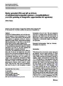
large non-proteinaceous molecule (C129H223N3O54) with a molecular weight of 2680.13 Daltons. The chemical structure of palytoxin is characterized as a polyalcohol consisting of a very long, partially unsaturated aliphatic chain, interspersed with five sugar moieties, beginning with an OH group and ending with an NH2 group[16–20] (fig. 1). It belongs to the group of long continuous carbon atom chain natural toxins, second only to maitotoxin[21, 22]. Because of its very large size and absence of repeating units, determination of the entire structure including stereochemistry is truly a “tour de force”[15]. Palytoxin contains 64 dissymmetric carbon atoms (chiral centers), two amide groups, and six olefinic bonds offering more than one sextillion (1021) possible isomers. Among these, 42-hydroxy-palytoxin and ostreocin-D are the two better characterized palytoxin analogues[21, 22]. Palytoxin has both lipophilic and hydrophilic regions and is referred to as a super-carbonchain compound, since it has the longest chain of continuous carbon atoms in any known natural product. It is heat-stable, not inactivated by boiling, and is stable in neutral aqueous solutions for prolonged periods; however, a rapid decomposition occurs under acid or alkaline conditions, leading to loss of its toxicity[23].
Fig. 1 Chemical structure of marine toxin palytoxin
2 PALYTOXIN SOURCES Palytoxin was first isolated from the soft coral Palythoa toxica in Hawaii, and subsequently from many other organisms such as seaweeds and shellfish. Palytoxin and a series of its analogues, such as homopalytoxin, bishomopalytoxin, neopalytoxin, deoxypalytoxin, and 42-hydroxy-palytoxin, were subsequently identified in the Palythoa species[4]. Recently, palytoxin was also found in a benthic dinoflagellate, Ostreopsis siamensis[24]. Ostreopsis ovata and Ostreopsis siamensis are unicellular algae found in tropical and subtropical coastal waters, and recently along the coast of Europe and in the Mediterranean as well[25–29]. Palytoxin and some other analogues, including ostreocin-D, mascarenotoxin-a, -b, and -c, and ovatoxina, -b, c, -d, and -e, were identified in the benthic
dinoflagellates of the genus Ostreopsis, which have been proposed as producing organisms even though a bacterial origin has been suggested[30, 31]. Both Ostreopsis ovata and Ostreopsis siamensis dinoflagellates produce palytoxin and its analogues that are powerful vasoconstrictors in mammals. Since 2003, Ostreopsis blooms in Italy and Spain have been accompanied by reports of respiratory problems and skin/mucosa irritation in persons in contact with toxic microalgal cells[32]. Occurence of Ostreopsis siamensis and Ostreopsis ovata is linked to extensive death of edible mollusks, and echinoderms[25, 33], and human illnesses[32, 34]. Palytoxin is an extremely toxic product that was first isolated from the zoanthid Palythoa toxica[35]. This toxin also occurred in various other marine organisms living in close association with zoanthid colonies, e.g. sponges (Porifera), soft corals
775
J Huazhong Univ Sci Technol[Med Sci] 35(5):2015
(Alcyonaria), gorgonians (Gorgonaria), mussels, and crustaceans. Predators, e.g. polychaete worms (Hermodice carunculata), starfish (Acanthaster planci) and fish (Chaetodon species) feeding on Palythoa colonies, accumulate high toxin concentrations in their organs, where palytoxin is stored in its active form[36]. 3 PHARMACOLOGY AND TOXICOLOGY During the last four decades, palytoxin has been widely studied for its pharmacological mechanisms and toxicological effects. Much of the scientific evidence comes from in vivo studies conducted on laboratory animals and in in vitro studies. Evidently, palytoxin and its analogues exert multiple adverse effects on humans and animals through several mechanisms of action. Palytoxin and its analogues exert their potent biological activity by altering mechanisms of ion homeostasis in excitable and non-excitable tissues. In an earlier investigation, Haberman proved the presence of the predicted palytoxin-induced ion channel[14]. In a subsequesnt study[11], Frelin and Van Renterghem provided evidence of multiple mechanisms in palytoxininduced toxicity based on electrophysiological and pharmacological findings[12]. The authors suspected that palytoxin has more than one site of action in excitable cells and it may act as an agonist for a family of lowconductance channels that transport Na+/K+, H+, and Ca2+ ions[9]. Other studies further showed that Na+/K+-ATPase is the potential molecular target for the toxicity of palytoxin in sensitive systems[37, 38]. The recognized mechanism of palytoxin activity involves its binding to the extracellular portion of this plasma membrane protein[39]. Thus, it could convert an enzyme carrying ions against their concentration gradients into a nonselective cation channel, allowing passive flow of ions[40]. More findings[6, 7, 38] have indicated that palytoxin would interfere with the otherwise normal strict coupling between inner and outer gates of the pump controlling the ion access to the Na+/K+-ATPase, thus allowing the gates to be simultaneously opened with Na+ influx into the cells and K+ efflux. This causes depolarization and triggers a series of adverse biological effects[6]. The ability of palytoxin to make internal portions of the Na+/K+-ATPase accessible to relatively large molecules has been exploited to further characterize the structurefunction relationship of the pump, leading to a better understanding of its ion translocation pathway[7]. Many natural compounds that have been isolated from marine sources exert cytotoxicity by modulating cytoskeletal properties, in particular those concerning actin filaments[35, 41]. In the study of Louzao et al[35], human neuroblastoma cells were used to study the modifications of ion fluxes associated with palytoxin activities, and the effects on essential cytoskeletal component, the actin system. Their results showed that palytoxin modified membrane permeability as a first step, triggering depolarization and increasing Ca2+ influx. The substantial loss of filamentous actin, and the morphologic alterations elicited by this toxin, are possibly secondary to their action on ion channels. The authors concluded that Ca2+ could be related to actin cytoskeletal organization. However, the potential
mechanism is not elucidated and need further study. In other studies, Del Favero et al[42] found that in epithelial cells from the rabbit duodenum-jejunum, omission of extracellular Ca2+ (nominally Ca2+-free medium) halved the effect of palytoxins on F-actin disassembly. Those data led to the idea that this toxin modified the actin filament system of intestinal cells not only by modulating some signaling pathway activated by external Ca2+, but also by acting on another, still unknown, element. Thus, more studies are urgently needed to elucidate this mechanism. Studies of the cellular cytoskeleton have revealed that the signaling cascade, triggered by palytoxin, leads to actin filament system distortion. For example, palytoxin could activate and converse Na+/K+ pumps to nonselective cation channels, triggering depolarization of Na+ accumulation with subsequent Ca2+ overload. In several in vitro studies, palytoxin has been found to modulate secretion of catecholmines and other neurotransmitters. Tatsumi et al reported that palytoxin (1 nmol/L–1 µmol/L) caused the release of [3H]norepinephrine from clonal rat pheochromocytoma cells in a concentration-dependent manner[43]. The palytoxininduced Ca2+ influx was markedly inhibited by Co2+, whereas the palytoxin-induced Na+ was not affected by tetrodotoxin. At lower concentrations of palytoxin, the [3H]norepinephrine release is due to increasing tetrodotoxin-insensitive Na+ permeability across the cell membrane, while at higher concentrations it was mainly caused by a direct increase in Ca2+ influx into the cells. In a similar study, Yoshizumi et al demonstrated palytoxin-induced secretion of catecholamines from cultured bovine adrenal chromaffin cells[44]. At the same time, Satoh and Nakazato reported that palytoxin, in a dose-dependent manner, increased the release of [3]acetylcholine ([3]ACh), cytosolic free Ca2+ conentration, and uptake of 22Na+, and decreased membrane potential in rat cerebrocortical synaptosomes[45]. Results revealed that at high concentrations (10-6 mol/L), palytoxin increased the release of [3]ACh by directly increasing the influx of Ca2+ into synaptosomes and by releasing Ca2+ from intracellular storage sites via a Na+/Ca2+ exchange mechanism. The release of [3H]ACh induced by concentrations of palytoxin less than 10-10 mol/L is more dependent on the simultaneous presence of both Ca2+ and Na+ than higher concentrations of palytoxin. In an in vitro study, Crinelli et al explored the underlying mechanism in palytoxin-induced inflammation following an exposure through inhalation and/or cutaneous contact[46]. Palytoxin and its analogues have been shown to increase the levels of mRNAs encoding inflammation-related proteins in human macrophages via p38 mitogen activated protein kinase and NF-kB. In essence, these toxins may activate pro-inflammatotory signalling cascades. 4 ANIMAL TOXICITY The toxicity of palytoxin and its analogues has been studied in animals to a much greater extent than in humans. Several experimental studies have described toxicities of palytoxins and its analogues in animals exposed via various routes of administration, such as
776 intravenous (i.v.), intraperitoneal (i.p.), intramuscular (i.m.), subcutaneous (s.c.), intratracheal, and topical applications on eyes and skin. The toxicity of palytoxin in animals has been reviewed and results are summarized in table 1. Palytoxin has been found to be highly toxic after i.v. injection in rabbits, dogs, rhesus monkeys, guinea pigs, and rats, as the LD50 ranged between 25 and 89 ng/kg. Animals died within several minutes due to intense vasoconstriction, increased arterial pressure, and heart failure[2, 47]. The LD50 values of palytoxin by i.p. injection are similar to those after i.m. and s.c. injections. Palytoxin is highly toxic also by intratracheal instillation. The lethal dose of palytoxin for mice by this route exceeds 2 µg/kg in two hours. Palytoxin is much less toxic by the oral route in mice (LD50, 651–767 ng/kg)[5, 48]. The oral toxicity of palytoxin is found to be three times lower than the i.p. toxicity. This is because palytoxin is a very large hydrophilic molecule and therefore absorption is less efficient through the gastrointestinal tract than through the peritoneum[49–51]. Palytoxin is not lethal when topically applied to the skin or eyes, but cause skin irritation and erythema in 50% of mice when applied at a dose of 20 ng/ear[52]. Further details on toxic doses of palytoxin in different species and routes of exposure can be found in a study from Tubaro et al[4]. Toxic symptoms after oral exposure include fever, ataxia, drowsiness, and weakness of limbs, followed by death. Recently, Tubaro et al described some histopathological and clinical chemistry changes induced by palytoxin in experimental animals[4]. Following oral
J Huazhong Univ Sci Technol[Med Sci] 35(5):2015
administration of palytoxin in mice, increased plasma levels of creatine phosphokinase (CPK), lactate dehydrogenase (LDH), and glutamic-oxal(o)acetic transaminase (GOT) were observed[48]. Histological changes include inflammation in the forestomach, liver and pancreas, while cardiac and skeletal muscle cells reveal only ultrastructural alterations under electron microscopy (EM). Following i.p. injection of palytoxin in mice, adhesions are observed in the peritoneum, with ascites and small intestine dilation. Palytoxin cause single-cell necrosis in the small intestine and other cells. EM findings show swelling of mitochondria and separation of organelles in myocytes, loss of microvilli in renal tubules and vacuolation of pancreatic acinar cells[49, 50, 53–55]. Ostreocin-D treatment in mice causes erosion in the stomach and intestines[50]. Tubaro et al reported similar findings with a significant increase in plasma potassium in mice treated with 42-hydroxypalytoxin[56]. In a subacute study, palytoxin treatment in mice (250 ng/kg, 5 times per week; total injections: 5, 10, 15, and 29) caused diarrhea, peritonitis, decreased thymus weights, and increased spleen weights[49]. These changes are reversed within one month after the last dose. In another study[50], Ito and Yasumoto reported that intratracheal instillation of palytoxin in mice caused alveolar hemorrhage, pulmonary edema, gastrointestinal erosion, and glomerular atrophy. Similar lung injuries were observed in ostreocin-D treated mice but showed slower progression and recovery than those treated by palytoxin.
Table 1 Acute toxicity (LD50) of palytoxin Organism Route* Reported LD50 (ng/kg) References Mouse i.p. 50 [77] Mouse i.v. 150 (LD59) [1] Mouse i.v. 740 [78] Mouse oral 767 [48] Mouse oral 651 [56] Mouse oral 510 [5] Rat i.m. 240 [80] Rat i.p. 710 [80] Rat i.v. 89 [80] Rat i.t. 360 [80] Rat Oral > 40 [80] Rat Rectal > 10 [80] Rat s.c. 400 [80] Guinea Pig i.v. 110 [80] Guinea Pig Parenteral 25 [79] Rabbit i.v. 25 [80] Monkey i.p. 78 [80] Dog i.v. 33 [80] Dog Oral 50 [79] Unlisted 62.5 [68] Crab** * : i.p., intraperitoneal; i.v., intravenous; i.t., intratracheal; s.c.; subcutaneous; **: shore crab Carcinus maenas
Palytoxin is a skin tumor promotor[5, 57, 58]. Earlier researches have revealed that palytoxin, like promoter 12-O-tetradecanoylphorbol-13-acetate (TPA), was a skin irritant and a tumor promoter in the multi-stage mouse skin assay. In contrast to TPA, however, palytoxin did not induce ornithine decarboxylase in mouse skin and did not induce HL60 cell adhesion. Moreover, palytoxin did not bind to protein kinase C in vitro and was
therefore classified as a non-TPA-type tumor promoter. Subsequent cell culture studies provided further evidence that palytoxin stimulates signaling pathways that do not require protein kinase C[59, 60]. Palytoxin-stimulated responses that are likely to play a role in carcinogenesis are stimulation of arachidonic acid metabolism and the production of prostaglandins, modulation of the epid-
777
J Huazhong Univ Sci Technol[Med Sci] 35(5):2015
ermal growth factor (EGF) receptor, and modulation of mitogen activated protein (MAP) kinase cascades[57, 58, 60]. 5 HUMAN TOXICITY Palytoxin has been implicated in toxic events in humans following ingestion, inhalation, or cutaneous contact. Rumore and Houst reported a case of palytoxin inhalational toxicity in pediatric siblings following secondary exposure to vapors from cleaning of an aquarium containing zoanthids[60]. Symptoms included fever, tachycardia, leukocytosis and elevated lactic dehydrogenase. Both patients received supportive treatment in the pediatric intensive care unit and were discharged after 48 h. In another report[2], a man in Virginia (USA) presented with presumed inhalational toxin exposure from attempting to remove a colony of medium sized green/brown zoanthids from a rock by pouring boiling water over the infested portion. The zoanthids had been growing in the aquarium for 3 years and had arrived as a contaminant with live rock. During this process, the patient inhaled steam and immediately stopped after noticing a foul odor. The zoanthid containing rock was retained in a separate aquarium. Within 20 min, the patient experienced rhinorrhea and coughing at which time the patient took an antihistamine believing the symptoms to be caused by seasonal allergies. Within 4 h post exposure, the patient experienced dispnea and lightheadedness which progressed to severe fits of coughing and chest pain. More reports of health problems caused by inhalation of palytoxin are summarized by Tubaro et al[61]. Several recent publications have reported similar findings for cutaneous exposure to palytoxin from zoanthids. Hoffmann et al[62] recounted a man in Germany collapsing 16 h after receiving minor cuts to 3 fingers while handling several zoanthid colonies in a home aquarium. Initial symptoms started 2 h after contact which included shivering, myalgias, and general weakness of the extremities, progressing to dizziness and speech disturbance at the time of collapse. At the time of admission to the hospital the patient’s speech was impaired and swelling and erythema were noted at the site of the finger cuts with the numbness and paresthesias of the fingers progressing to involve the whole arm over the next 20 h. Human illness and death arising from consumption of crabs and fish contaminated or suspected to be contaminated with palytoxin have been reported in tropical and subtropical regions[4]. Cases of death and near-death illness resulting from palytoxin have been reported due to consumption of contaminated crabs in Philippines[63], sea urchins in Brazil[64], and fish in Japan, Madagascar, and the USA[65–66]. Palytoxin can cause various clinical effects, including a bitter and metallic taste in the mouth, neurological and gastrointestinal disturbances, abdominal cramps, nausea, vomiting, diarrhea, tremor myalgia, muscle cramps, cardiac dysfunctions, paresthesia, dysgeusia, bradycardia, hypertension, respiratory distress, renal failure, cyanosis, coma, and death[4, 69]. Death occurs due to myocardial injury[41, 70]. The most common complication of palytoxin poisoning is rhabdomyolysis[71]. Palytoxin-induced
rhabdomyolysis and myocardial damage are charactereized by elevated myosin light chain level and serum levels of CK, as well as altered electrocardiogram[66]. 6 RISK ASSESSMENT The majority of palytoxin poisoning incidents in humans are a result of seafood consumption. Due to cooccurrence with other seafood toxins, such as ciguatoxins, saxitoxins, and tetrodotoxin, it has been difficult to assess the true risk of palytoxin poisoning through seafood consumption in humans; however, limited cases have been well documented, with some involving human fatalities[2]. Continued research into the distribution and occurrence of palytoxin and palytoxinlike compounds both in seafood and marine organisms sold in the aquarium trade appears warranted. Although the actual toxicological potential of palytoxins has yet to be evaluated[56], an acute reference dose (ARfD) of 0.2 µg/kg (sum of palytoxin and ostreocin-D) has been established through experimental toxicity data[72]. Based on all animal studies, Riobó et al[26] reported the estimated dose of palytoxin for humans to be between 2.3 and 31.5 µg, and an ARfD was suggested to be 64 µg for a person with an average weight of 60 kg[73]. Recent evidence also suggests that humans are poisoned by palytoxin and its analogues through inhalation and dermal routes[2, 69, 74]. The most common signs after inhalation and cutaneous/systemic exposures are respiratory distress, bronchoconstriction, mild dyspnea, rhinorrhea, cough, fever, and a small incidence of dermatitis and conjunctivitis. 7 TREATMENT Animal studies have shown that vasodilators, such as papaverine and isosorbide dinitrate, can be used as antidotes. However, these antidotes are beneficial only if injected into the heart immediately following palytoxin exposure[75]. Treatment in humans is symptomatic and supportive. After oral administration, treatment such as gastric lavage, forced diuresis therapy, artifical respiration and fluid administration are applied, but in some cases fatalities can not be prevented[4]. After inhalation/dermal exposure, corticosteroids, NSAIDs, histamine antagonists, nebulized β-agonists and/or oxygen therapy can be administered to alleviate the symptoms, with a recovery period of a few hours to days[56]. 8 ECOTOXICOLOGY In the reefs, zoanthid species of the genus Palythoa had been detected. Various other marine organisms live in close association with zoanthid colonies, including sponges (Porifera), soft corals (Alcyonaria), gorgonians (Gorgonaria), mussels, and crustaceans. Predators feeding on Palythoa colonies accumulate high concentration of toxin in their organs, where palytoxin is stored in its active form. The high level of toxin tolerance observed in marine animals may enable the wide distribution of palytoxin in marine biota and its transport and sequestration in food chains[36]. However,
778 one should note that many other animals, including mouse, dog, rabbit, and guinea pig, are sensitive to palytoxin[20, 42]. Palytoxin which has been primarily detected in marine zoanthids (Palythoa sp.), occurs also in a wide range of other animals, e.g. sponges, corals, shellfish, polychaetes and crustaceans, but also in fish, which feed on crustaceans and zoanthids. These animals exhibit a high resistance to the toxin’s action[76]. Bioassays tested for some marine invertebrates and evidence from environmental populations exposed to the toxins also give indications of the high impact that these toxins may have on natural food webs. The recognition of their wide distribution coupled with the poisoning effects that these toxins can have on animals, and especially on humans, have concerned the scientific community[36].
J Huazhong Univ Sci Technol[Med Sci] 35(5):2015
8 9
10
11
12
13
9 CONCLUSIONS Palytoxin and its analogues are the most toxic, complex, and least understood toxins found in nature, especially in seafood. Literature abounds showing that palytoxin may be fatal to humans in the µg range. Adverse effects can occur by inhalation or dermal exposure after contact with aerosolized seawater during Ostreopsis blooms or handling aquaria containing zoanthids. Considering the lack of reliable quantitative data on palytoxins toxicity in humans and the lowestobserved-adverse-effect level (LOAEL) of 200 ng palytoxin/kg for acute oral toxicity in mice as the reference point, an ARfD of 0.2 µg/kg, applied to the sum of palytoxin and ostreocin-D, has been established by the European Food Safety Authority (EFSA) to propose 30 µg/kg as the tolerance limit in shellfish meat[72]. In essence, the detailed information on toxic potential of palytoxin is lacking and there is no antivenin. Conflict of Interest Statement The authors declare that there is no conflict of interest with any financial organization or corporation or individual that can inappropriately influence this work.
14 15
16 17 18 19
20 21
22
23 REFERENCES 1 Moore RE, Scheuer PJ. Palytoxin: a new marine toxin from a coelenterate. Science, 1971,172(3982):495-498 2 Deeds JR, Schwartz MD. Human risk associated with palytoxin exposure. Toxicon, 2010,56(2):150-162 3 Carballeira NM, Emiliano A, Sostre A, et al. Fatty acid composition of bacteria associated with the toxic dinoflagellate Ostreopsis lenticularis and with Caribbean Palythoa species. Lipids, 1998,33(6):627-632 4 Tubaro A, Sosa S, Hungerford J. Toxicology and diversity of marine toxins. In: Gupta RC, ed. Veterinary Toxicology: Basic and Clinical Principles. Amsterdam: Academic Press/Elsevier, 2012, 896-934 5 Aligizaki K, Katikou P, Milandri A, et al. Occurrence of palytoxin-group toxins in seafood and future strategies to complement the present state of the art. Toxicon, 2011,57(3):390-399 6 Munday R. Palytoxin toxicology: Animal studies. Toxicon, 2011,57(3):470-477 7 EFSA. Scientific opinion on marine biotoxins in shellfish – palytoxin group. EFSA J, 2009,1393(1):1-38
24
25
26
27
28
Wu CH. Palytoxin: membrane mechanisms of action. Toxicon, 2009,54(8):1183-1189 Rossini GP, Bigiani A. Palytoxin action on the Na(+),K(+)-ATPase and the disruption of ion equilibria in biological systems. Toxicon, 2011,57(3):429-439 Weidmann S. Effects of palytoxin on the electrical activity of dog and rabbit heart. Experientia, 1977,33(11):14871489 Rossini GP, Bigiani A. Palytoxin action on the Nat,KtATPase and the disruption of ion equilibria in biological systems. Toxicon, 2011,57(3):429-439 Frelin C, Van Renterghem C. Palytoxin. Recent electrophysiological and pharmacological evidence for several mechanisms of action. Gen Pharmacol, 1995,26(1): 33-37 Artigas P, Gadsby DC. Large diameter of palytoxininduced Na/K pump channels and modulation of palytoxin interaction by Na/K pump ligands. J Gen Physiol, 2004,123(4):357-376 Haberman E. Palytoxin acts through Na+/K-ATPase. Toxicon, 1989,27(6):1171-1187 Ares IR, Louzao MC, Vieytes MR, et al. Actin cytoskeleton of rabbit intestinal cells is a target for potent marine phycotoxins. J Exp Biol, 2005,208(22):4345-4354 Shimizu Y. Structural chemistry: Complete structure of palytoxin elucidated. Nature, 1983,302(1):112 Moore RE. Structure of palytoxin. Fortschr Chem Org Naturst, 1985,48(1):81-202 Riobó P, Franco JM. Palytoxins: Biological and chemical determination. Toxicon, 2011,57(3):368-375 Ciminiello P, Dell'Aversano C, Dello Iacovo E, et al. LCMS of palytoxin and its analogues: state of the art and future perspectives. Toxins, 2011,57(3):376-389 Munday R. Palytoxin toxicology: animal studies. Toxicon, 2011,57(3):470-477 Kita M, Uemura D. Marine huge molecules: the longest carbon chains in natural products. Chem Rec, 2010,10(1):48-52 Moore RE, Bartolini G, Barchi J, et al. Absolute stereochemistry of palytoxin. J Am Chem Soc, 1982,104 (13):3776-3779 Ramos V, Vasconcelos V. Palytoxin and analogs: biological and ecological effects. Mar Drugs, 2010,8(7):2021-2037 Fernández DA, Louzao MC, Vilariño N, et al. The kinetic, mechanistic and cytomorphological effects of palytoxin in human intestinal cells (Caco-2) explain its lower-thanparenteral oral toxicity. FEBS J, 2013,280(16):3906-3919 Ciminiello P, Dell'Aversano C, Fattorusso E, et al. The Genoa 2005 outbreak. Determination of putative palytoxin in Mediterranean Ostreopsis ovata by a new liquid chromatography tandem mass spectrometry method. Anal Chem, 2006,78(17):6153-6159 Riobó P, Paz B, Franco JM. Analysis of palytox-in-like in Ostreopsis cultures by liquid chromatography with precolumn derivatization and fluorescence detection. Anal Chim Acta, 2006,566(2):217-223 Monti M, Minocci M, Beran A, Ivena L. First record of Ostreopsis cfr Ovata on macroalgae in the northern Adriatic. Sea. Mar Pol Bull, 2007,54(5):598-601 Aligizaki K, Panagiota K, Nikolaidis G, et al. First episode of shellfish contamination by palytoxin-like compounds from Ostreopsis species (Aegean Sea, Greece). Toxicon, 2008,51(3):418-427
779
J Huazhong Univ Sci Technol[Med Sci] 35(5):2015
29
30
31
32
33
34
35
36
37
38
39
40
41
42
43
44
Rhodes L. World-wide occurrence of the toxic dinoflagellate genus oxytropsis Schmidt. Toxicon, 2011,57(3):400-407 Katikou P. Palytoxin and analogues: etiology and origin, chemistry, metabolism, and chemical analysis. In: Botana LM, ed. Seafood and Freshwater Toxins: Pharmacology, Physiology and Detection. Boca Raton: CRC Press, 2008, 631-663 Ciminiello P, Dell'Aversano C, Dello Iacovo, et al. Complex palytoxin-like profile of Ostreopsis ovata. Identification of four new ovatoxins by high-resolution liquid chromatography/mass spectrometry. Rapid Commun Mass Spectrom, 2010,24(18):2735-2744 Tichadou L, Glaizal M, Armengaud A, et al. Health impact of unicellular algae of the Ostreopsis genus blooms in the Mediterranean Sea: experience of the French Mediterranean coast surveillance network from 2006 to 2009. Clin Toxicol, 2010,48(8):839-844 Sansoni G, Borghini B, Camici G, et al. Fioriture algali di Ostreopsis Ovata (Gonyaulacales: Dinophyceae): Unproblema emergente. Biol Ambientale, 2003,17(1):1723 Gallitelli M, Ungaro N, Addante LM, et al. Respiratory illness as a reaction to tropical algal bloom occurring in a temperate climate. J Am Med Assoc, 2005,293(21):25992600 Louzao MC, Ares IR, Vieytes MR, et al. The cytoskeleton, a structure that is susceptible to the toxic mechanism activated by palytoxins in human excitable cells. FEBS J, 2007,274(8):1991-2004 Gleibs S, Mebs D. Distribution and sequestration of palytoxin in coral reef animals. Toxicon, 1999,37(11): 1521-1527 Harmel N, Apell HJ. Palytoxin-induced effects on partial reactions of the Na,K-ATPase. J Gen Physiol, 2006,128(1):103-118 Rodrigues AM, Infantosi AF, de Almeida AC. Palytoxin and the sodium/potassium pump--phosphorylation and potassium interaction. Phys Biol, 2009,6(3):036010 Vedovato N, Gadsby DC. The two C-terminal tyrosines stabilize occluded Na/K pump conformations containing Na or K ions. J Gen Physiol, 2010,136(1):63-82 Redondo J, Fiedler B, Scheiner-Bobis G. Palytoxininduced Na+ influx into yeast cells expressing the mammalian sodium pump is due to the formation of a channel within the enzyme. Mol Pharmacol, 1996,49(1):49-57 Louzao MC, Ares IR, Cagide E. Marine toxins and the cytoskeleton: a new view of palytoxin toxicity. FEBS J, 2008,275(24):6067-6074 Del Favero G, Beltramo D, Onidi M, et al. Acute oral toxicity of Palytoxin and Okadaic acid in mice. Toxicol Lett, 2012,21(1):S157-S158 Tatsumi M, Takahashi M, Ohizumi Y. Mechanism of palytoxin-induced [3H]norepinephrine release from a rat pheochromocytoma cell line. Mol Pharmacol, 1984,25(3): 379-383 Yoshizumi Y, Nakanishi A, Houchi H, et al. Characterization of palytoxin-induced catecholamine secretion from cultured bovine adrenal chromaffin cells. Effects of Na+- and Ca2+-channel blockers. Biochem Pharmacol, 1991,42(1):17-23
45
46
47
48
49
50
51
52
53
54
55
56
57
58
59
60
61
62
63
Satoh E, Nakazato Y. Mode of action of palytoxin on the release of acetylcholine from rat cerebrocrtical synaptosomes. J Neurochem, 1991,57(4):1276-1280 Crinelli R, Carloni E, Giaconini E, et al. Palytoxin and an Ostreopsis toxin extract increase the levels of mRNAs encoding inflammation-related proteins in human macrophages via p38 MAPK and NF-κB. PloS One, 2012,7(6): e38139 Ito K, Urakawa N, Koike H. Cardiovascular toxicity of palytoxin in anesthetized dogs. Arch Intl de Pharmacod et de Ther, 1982,258(1):146-154 Sosa S, Del Favero G, De Bortoli M, et al. Palytoxin toxicity after acute oral administration in mice. Toxicol Lett, 2009,191(2-3):253-259 Ito E, Ohkusu M, Yasumoto T. Intestinal injuries caused by experimental palytoxicosis in mice. Toxicon, 1996,34(6):643-652 Ito E, Yasumoto T. Toxicological studies on palytoxin and ostreocin-D administered to mice by three different routes. Toxicon, 2009,54(3):244-251 Ramos V, Vasconcelos V. Palytoxin and analogs: biological and ecological effects. Mar Drugs, 2010,8(7):2021-2037 Fujiki H, Suganuma M, Nakayasu M, et al. Palytoxin is a non 12-O-tetradecanoylphorbol-13-acetate type tumor promotor in two-stage skin carcinogenesis. Carcinogenesis, 1986,7(5):707-710 Terao K, Ito E, Yasumoto T. Light and electron microscopic observation of experimental palytoxin poisoning in mice. Bull Soc Pathol Exot, 1992,85(5):494496 Del Favero G, Beltramo D, Sciancalepore M, et al. Toxicity of palytoxin after repeated oral exposure in mice and in vitro effects on cardiomyocytes. Toxicon, 2013,75(1):3-15 Pelin M, Sosa S, Pacor S, et al. The marine toxin palytoxin induces necrotic death in HaCaT cells through a rapid mitochondrial damage. Toxicol Lett, 2014,229(3):440-450 Tubaro A, Del Favero G, Beltramo D, et al. Acute oral toxicity in mice of a new oalytoxin analog: 42-hydroxypalytoxin. Toxicon, 2011,57(5):755-763 Tubaro A, Durando P, Del Favero G, et al. Case definitions for human poisonings postulated to palytoxins exposure. Toxicon, 2011,57(3):478-495 Forino M, Ciminiello P, Fattorusso E, et al. Palytoxins: a still haunting Hawaiian curse. Pytochem Rev, 2010,9(4):491-500 Wattenberg EV, Fujiki H, Rosner MR. Heterologous regulation of the epidermal growth factor receptor by palytoxin, anon-12-O-tetradecanoylphorbol-13-acetatetype tumor promoter. Cancer Res, 1987,47(17):4618-4622 Rumore MM, Houst BM. Palytoxin poisoning via inhalation in pediatric siblings. Int J Case Rep Images, 2014,5(7):501-504 Tubaro A, Durando P, Del Favero G, et al. Case definitions for human poisonings postulated to palytoxins exposure. Toxicon, 2011,57(3):478-495 Hoffmann K, Hermanns-Clausen M, Buhl C, et al. A case of palytoxin poisoning due to contact with zoanthid corals through skin injury. Toxicon, 2008,51(8):1535-1537 Alcala AC, Alcala LC, Garth JS, et al. Human fatality due to ingestion of the crab Demania reynaudii that contained a palytoxin-like toxin. Toxicon, 1988,26(1):105-107
780 64
65
66
67
68
69
70
71
J Huazhong Univ Sci Technol[Med Sci] 35(5):2015
Granéli E, Ferreira CEL, Yasumoto T, et al. Sea urchins poisoning by the benthic dinoflagellate Ostreopsis ovata on the Brazilian coast. In: Book of Abstracts of Xth International Conference on Harmful Algae. St. Petersburg, Florida. 2002 Fukui M, Murata M, Inoue A, et al. Occurrence of palytoxin in the trigger fish Melichtys vidua. Toxicon, 1987,25(10):1121-1124 Okano H, Masuoka H, Kamei S, et al. Rhabdomyolysis and myocardial damage induced by palytoxin, a toxin of blue humphead parrotfish. Int Med, 1998,37(3):330-333 Onuma Y, Satake M, Ukena T, et al. Identification of putative palytoxin as the cause of clupeotoxism. Toxicon, 1999,37(1):55-65 Béress L, Zwick J, Kolkenbrock HJ, et al. A method for the isolation of the caribbean palytoxin (C-PTX) from the coelenterate (zooanthid) Palythoa caribaeorum. Toxicon, 1983,21(2):285-290 Nordt SP, Wu J, Zahller S, et al. Palytoxin poisoning after dermal contact with Zoanthid coral. J Emerg Med, 2009,40(4):397-399 Vasconcelos V, Ramos V. Palytoxin and analogs: Biological and ecological effects. Mar Drugs, 2010,8(7):2021-2037 Yoshimine K, Orita S, Okada S, et al. Two cases of parrotfish poisoning with rhabdomyolysis. Nippon Naika Gakkai Zasshi, 2001,90(7):1339-1341
72
73
74 75
76 77
78
79
80
EFSA (European Food Safety Authority). Scientific opinion on marine biotoxins in shellfish-palytoxin group. EFSA J, 2009,1393:1-38 Rhodes L, Munday R, Briggs L. Ostreopsis siamensis and palytoxin-related compounds in New Zealand: a risk to human health? In: Moestrup Ø, ed. Proceedings of the 12th International Conference on Harmful Algae., Copenhagen: ISSHA and Intergovernmental Oceanographic Commision of UNESCO, 2008:326-329 Majlesi N, Su MK, Chan GM, et al. A case of inhalation exposure to palytoxin. Clin Toxicol, 2008,46(1):673 Wiles J, Vick J, Christensen M. Toxicological evaluation of palytoxin in several animal species. Toxicon, 1974,12(4):427-433 Mebs D. Occurrence and sequestration of toxins in food chains. Toxicon, 1998,36(11):1519-1522 Kaul PN, Daftari P. Marine pharmacology: bioactive molecules from the sea. Annu Rev Pharmacol Toxicol, 1986,26(1):117-142 Mahnir VM, Kozlovskaya EP, Kalinovsky AI. Sea anemone Radianthus macrodactylus--a new source of palytoxin. Toxicon, 1992,30(11):1449-1456 Webber HH, Ruggieri GD. Food-Drugs from the Sea. In: Proceedings of the Conference, 4th, Mayaguez, 1974. Washington, DC: Marine Technology Soc,1974,311 Vick JA, Wiles JS. The mechanism of action and treatment of palytoxin poisoning. Toxicol Appl Pharmacol, 1975,34(2):214-223 (Received Aug. 28, 2014; revised July 6, 2015)