The Journal of Immunology
Transcript Signatures in Experimental Asthma: Identification of STAT6-Dependent and -Independent Pathways1 Nives Zimmermann,* Anil Mishra,* Nina E. King,* Patricia C. Fulkerson,* Matthew P. Doepker,* Nikolaos M. Nikolaidis,* Laura E. Kindinger,* Elizabeth A. Moulton,* Bruce J. Aronow,† and Marc E. Rothenberg2* The analysis of polygenic diseases such as asthma poses a challenging problem. In an effort to provide unbiased insight into disease pathogenesis, we took an empirical approach involving transcript expression profiling of lung tissue from mice with experimental asthma. Asthmatic responses were found to involve sequential induction of 4.7% of the tested genome; notably, there was ectopic expression of a series of genes not previously implicated in allergic or pulmonary responses. Genes were widely distributed throughout all chromosomes, but preferentially included genes involved in immunity, development, and homeostasis. When asthma was induced by two independent experimental regimens, unique gene transcript profiles were found depending upon the mode of disease induction. However, the majority of genes were common to both models representing an asthma signature genome. Analysis of STAT6-deficient mice revealed that an unexpectedly large segment of the asthma genes were STAT6 independent; this correlated with sustained inflammatory events in these mice. Notably, induction of asthma in STAT6-deficient mice resulted in gene induction not seen in wild-type mice. These results raise concern that therapeutic blockade of STAT6 in the asthmatic setting may reprogram the genetic signature, resulting in alternative lung pathology, which we indeed observed in STAT6-deficient mice. These results provide unprecedented insight into the complex steps involved in the pathogenesis of allergic airway responses; as such, these results have significant therapeutic and clinical implications. The Journal of Immunology, 2004, 172: 1815–1824.
D
espite intense ongoing asthma research, there is currently an epidemic of this disease in the western world and the incidence is on the rise (1, 2). Experimentation in the asthma field has largely focused on analysis of the cellular and molecular events induced by allergen exposure in sensitized animals (primarily mice) and humans. These studies have identified elevated production of IgE, mucus hypersecretion, airways obstruction, eosinophilic inflammation, and enhanced bronchial reactivity to spasmogens in the asthmatic response (3–5). Clinical and experimental investigations have demonstrated a strong correlation between the presence of CD4⫹ Th2 lymphocytes (Th2 cells) and disease severity, suggesting an integral role for these cells in the pathophysiology of asthma (6, 7). Th2 cells are thought to induce asthma through the secretion of an array of cytokines that activate inflammatory and residential effector pathways (8 –10). In particular, IL-4 and IL-13 are produced at elevated levels in the asthmatic lung and are thought to be central regulators of many of the hallmark features of disease (11). They share a common receptor subunit, the IL-4R␣ and signaling through STAT6 (12, 13). Divisions of *Allergy and Immunology and †Pediatric Informatics, Department of Pediatrics, Cincinnati Children’s Hospital Medical Center, University of Cincinnati College of Medicine, Cincinnati, OH 45229 Received for publication August 6, 2003. Accepted for publication November 17, 2003. The costs of publication of this article were defrayed in part by the payment of page charges. This article must therefore be hereby marked advertisement in accordance with 18 U.S.C. Section 1734 solely to indicate this fact. 1 This work was supported in part by the American Heart Association Scientist Development (to N.Z.) and postdoctorate fellowship (to N.E.K.) grants, National Institutes of Health Grants R01 AI42242-05 (to M.E.R.), AI45898-04 (to M.E.R.), AI53479-01 (to M.E.R.), the Human Frontier Science Program (to M.E.R), International Life Sciences Institute (to M.E.R.), Burroughs Wellcome Fund (to M.E.R.), and by the University of Cincinnati Center for Environmental Genetics. 2
Address correspondence and reprint requests to Dr. Marc E. Rothenberg, Division of Allergy and Immunology, Cincinnati Children’s Hospital Medical Center, 3333 Burnet Avenue, Cincinnati, OH 45229-3039. E-mail address:
[email protected] Copyright © 2004 by The American Association of Immunologists, Inc.
Mice with targeted deletion of IL-4, IL-13, or STAT6 develop attenuation of certain features of asthma, including inflammatory cell infiltrates and airway hyperresponsiveness (AHR)3 (14, 15). Although these studies have provided the rationale for the development of multiple therapeutic agents that interfere with specific inflammatory pathways (16 –19), the development of the asthma phenotype is likely to be related to the complex interplay of a large number of genes. In this study, we aimed to gain critical insight into the spectrum of genes involved in the pathogenesis of experimental asthma, to define the genetic variability between two “phenotypically similar” asthmatic states, and to use transcript profiling to further uncover the importance of STAT6 pathways in the pathogenesis of disease.
Materials and Methods Experimental asthma induction BALB/c mice (National Cancer Institute, Frederick, MD) and STAT6-deficient mice (20) (BALB/c background, The Jackson Laboratory, Bar Harbor, ME) were housed under specific pathogen-free conditions. Asthma models were induced by two i.p. injections of OVA and aluminum hydroxide, followed by two OVA or saline intranasal challenges 3 days apart, as previously described (21). Mice were sacrificed 3 or 18 h following the first or second allergen challenge. Aspergillus fumigatus Ag-associated asthma was induced by exposing mice intranasally three times a week for 3 wk as described elsewhere (22–24). Mice were sacrificed 18 h following the last challenge. In some experiments, bronchoalveolar lavage fluid (BALF) was collected and infiltrating cells differentiated. However, in experiments in which RNA was collected for microarray analysis, BALF was not performed.
3 Abbreviations used in this paper: AHR, airway hyperresponsiveness; BALF, bronchoalveolar lavage fluid; CXCL, CXC chemokine ligand; CCL, CC chemokine ligand; MRP, myeloid-related protein; ADAM, a disintegrin and metalloprotease; SPRR, small, proline rich.
0022-1767/04/$02.00
1816 Preparation of RNA and microarray hybridization RNA was extracted using the TRIzol reagent as per the manufacturer’s instructions. Following TRIzol purification, RNA was repurified with phenol-chloroform extraction and ethanol precipitation. Microarray hybridization was performed by the Affymetrix GeneChip Core facility at Children’s Hospital Medical Center using the murine U74Av2 GeneChip (Affymetrix, Santa Clara, CA) as previously reported (21). This analysis was performed with one mouse per chip (n ⱖ3 for each allergen challenge condition and n ⱖ2 for each saline challenge condition). A further description of the methodology according to the Minimum Information About a Microarray Experiment guidelines (available at www.mged.org/Workgroups/MIAME/ miame.html) is provided in supplementary information.
Microarray data analysis From data image files, gene transcript levels were determined using algorithms in the Microarray Analysis Suite version 5 software (Affymetrix). Global scaling was performed to compare genes from chip to chip, as previously reported (21). Differences between saline- and OVA-treated mice were also determined using the GeneSpring software (Silicon Genetics, Redwood City, CA). Data for each allergen challenge time point was normalized to the average of the saline-treated mice. Gene lists were created as previously reported (21). Student’s t test with the multiple testing correction of Benjamini and Hochberg false discovery rate was used. Functional classifications were based on the Gene Ontology classification (25) obtained through the NetAffx server (available at www.netaffx.com) and public information in GenBank. Supplementary Tables I4 through 6 contain the gene lists presented in this article and analyzed as described here. Raw data used for supplementary Tables III (OVA) and 4 (Aspergillus) are the same as the one used for supplementary Tables II and III in our previous publication (21). However, analysis for the current article was performed in the updated Microarray Suite, version 5. Several genes are represented by more than one probe set on the chip; thus, the actual number of genes on a certain list may be lower due to duplication.
Northern blot analysis The cDNA probes, generated by PCR or from commercially available vectors (American Type Culture Collection, Manassas, VA) were sequence confirmed, radiolabeled with 32P, and hybridized as previously reported (24, 26, 27).
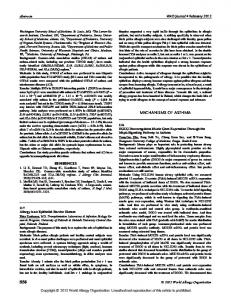
Results Kinetic analysis of OVA-induced experimental asthma We were interested in reproducibly and accurately identifying genes that were differentially expressed in a well-established model of asthma. Thus, mice were sensitized and challenged using the OVA model (Fig. 1A). We first analyzed global gene expression profiles at an early time point (3 h) after the first allergen encounter. Indeed, only 36 genes were induced by early encounter with allergen (Fig. 1B, Table I, and supplementary Table I). Genes encoding for molecules involved in early recognition of foreign pathogens were induced, including CD14 and a C-type lectin (28). Genes involved in initiating innate responses were induced, including CD83 (a dendritic cell activation marker), members of the IL-1 family (IL-1␣, IL-1, and IL-1R antagonist), numerous chemoattractants including chemokines active on neutrophils (CXC chemokine ligand (CXCL) 1, CXCL2), and myeloid-related proteins (MRP) 8 and MRP 14 (29) (Fig. 1, C and D, and data not shown). Consistent with myeloid cell recruitment, the gene for G-CSF receptor was elevated in the early response. Interestingly, genes not known to be expressed in the lung were among the early allergen-induced genes. For example, there was ectopic expression of several members of the schlafen family of growth-regulating genes, previously shown to be expressed only in the thymus (30). We next focused our attention on the genes that were induced 18 h after the first allergen challenge. At this later time point, there was a progressive induction of 57 genes (supplementary Table II), many of which were not evident acutely after allergen challenge. 4
The on-line version of this article contains supplemental material.
TRANSCRIPT SIGNATURES IN ASTHMA Indeed, 11 of the early activation genes remained elevated, whereas 46 additional genes increased (Fig. 1). Those include the gene for ADAM-8 (a disintegrin and metalloprotease) (31) and acidic chitinase (32), genes not previously reported in asthma but related to proteins associated with tissue remodeling. A role for specific members of the ADAM family (ADAM-10 and ADAM17) has been demonstrated in the immune system where they are involved in processing of the cell surface precursor form of TNF-␣; indeed, a member of this family of molecules (ADAM-33) has recently been identified as a human asthma gene (33). Trefoil factor 2 and Gob5, also among the genes induced 18 h after the initial challenge, have a role in mucus production (34, 35). This indicates that the genetic program for chronic processes, such as mucus production and airway remodeling, is already underway 18 h after the first allergen challenge. Consistent with this, unlike the acute time point, there was not a unique “genetic signature” compared with genes induced 18 h after two allergen challenges. In fact, most genes that were induced 18 h after the first allergen challenge were further increased following the second challenge. Additionally, similar to early genes, there was apparent ectopic expression of genes not previously shown to be expressed in the lung such as intelectin (an intestinal lectin) and trefoil factor 2 (an intestinal gene) (36 –38). We next focused our attention on characterizing the genes induced following the second allergen challenge. A different set of chemokines (e.g., eotaxin (CC chemokine ligand (CCL11), macrophage chemoattractant protein-2 (CCL8)) was induced following second allergen challenge (supplementary Table III). Consistent with the target cells of the induced chemokines, the genes encoding for macrophage-associated markers were induced (CCR5, CCR2, and F4/80). In addition, specific genes involved in cytoskeleton rearrangement, such as scinderin, were induced. Genes regulating cell proliferation such as cyclin B1 and B2, Ki-67, topoisomerase II, and spermidine synthase were elevated. A series of relevant transcription factors were induced such as the proinflammatory NF of activated T cells (NFAT)c. Additionally, the late asthma gene set encoded for a variety of serine proteases (including complement proteins (C1q, Properdin) and coagulation factors VII and X). Indeed, fibrin deposition and complement activation in the asthmatic lung have been reported (39 – 42). In addition, the late asthma gene set contained numerous protease inhibitors implicated in tissue repair such as the cathepsin genes. Additionally, a series of small proline-rich (SPRR) proteins involved in squamous cell differentiation and remodeling (43, 44) were induced. Table I provides an extensive summary of dynamically regulated genes and verification of representative early and late genes by Northern blot analysis is shown in Fig. 1D. Over the course of our studies, we have used Northern blot analysis to examine expression of a series of genes identified by microarray analysis. In particular, we have examined 30 of the putatively OVA-induced genes, and all were shown to be increased by Northern blot analysis. Those genes included SPRR2, Kreisler, arginase I and II, granzyme B, ADAM-8, heme oxygenase, acidic chitinase, trefoil factor 2, RELM-, matrix metalloprotease 12, TDAG8, 12/15-lipoxygenase, cationic amino acid transporter 2, chemokines CCL2, CCL5, CCL6, CCL7, CCL8, CCL9, CCL11, CCL12, CXCL1, CXCL5, CXCL9, CXCL10, CXCL12, and chemokine receptors CCR1, CCR2, and CCR5. Importantly, at least half of these genes were unexpected (i.e., they were not previously known to be expressed in the lung or induced by allergen challenge). Collectively, these results indicate a very low rate of false positives in the allergen-induced gene lists.
The Journal of Immunology
1817
FIGURE 1. Overlap of induced genes at specific phases of experimental asthma. A, A schematic representation of the allergen challenge protocol is depicted. Mice received two i.p. injections of OVA and alum. Subsequently, mice were challenged with OVA or saline intranasally and analyzed 3 or 18 h after the first or second allergen challenge. B, Microarray analysis was performed and gene lists were created for each time point. The overlap of those lists is presented in a Venn diagram format. C, Quantitative analysis (average difference values) of the induction of a representative early and late gene, CXCL2 (top) and SPRR2 (bottom), respectively, is shown. Saline-challenged mice are depicted with open bars and allergen-challenged mice with filled bars. Time points are as follows: 3H, one challenge, 3 h; 18H, one challenge, 18 h; 2C, two challenges, 18 h. The values represent mean ⫾ SD. D, Northern blot analysis of CXCL2 and SPRR2 cDNA expression is shown. The ethidium bromide (EtBr)-stained gel and position of 18S are shown. Each lane represents a separate mouse. E, The distribution of genes induced after two OVA challenges on chromosomes is shown. The number was normalized to the total number of genes for each particular chromosome (based on MGSCv3 release 3, NT release build 29) and is presented as percentage of total genes. Specific clusters are highlighted with a star. The p value for chromosome 6 is indicated. IgH, Ig H chain; Ig-, Ig chain; H2, histocompatibility-2.
To systematically analyze genes induced by allergen challenge, we assigned them into functional categories (Table II). Additionally, we identified and classified 62 genes that were decreased by at least 2-fold compared with saline-challenged mice. Immunerelated genes were predominantly in the increased group (39%) compared with the decreased group (2%). In contrast, genes involved in development and homeostasis composed the majority of
decreased genes (67%), while only comprising 24% of the increased genes. Since many of the genes induced were related to the immune response, we hypothesized that distinct chromosomal loci (rich for immune-related gene families) were induced by allergen challenge. Thus, we analyzed the chromosomal location of genes induced following two allergen challenges (Fig. 1E). Initially, we
1818 Table I. Kinetics and stratification of genes up-regulated in OVAinduced asthma modela
TRANSCRIPT SIGNATURES IN ASTHMA represented. When the total number of genes induced by allergen challenge on a particular chromosome was normalized for the number of genes present on that chromosome, we found that the majority of chromosomes contained a comparable amount of genes induced by allergen challenge. Fisher’s test for overrepresentation of increased genes on individual chromosomes revealed that only chromosome 6 had a significant increase ( p ⫽ 0.03). Comparison of OVA and A. fumigatus-induced experimental asthma We were next interested in comparing global transcript profiles in two independent models of asthma. We analyzed experimental asthma induced by A. fumigatus Ags because this model involves a unique mucosal sensitization route (intranasal) compared with the OVA model (45) and because A. fumigatus is a ubiquitous and common aeroallergen. Importantly, both asthma models have similar phenotypic and histological features including Th2-associated eosinophilic inflammation, mucus production, collagen deposition, and AHR (21–23, 41– 47). Eighteen hours after nine doses of intranasal A. fumigatus allergen challenge, lung RNA was subjected to the same microarray and data processing analyses as those performed after OVA challenges. Compared with mice challenged with intranasal saline, A. fumigatus-challenged mice had 387 genes induced (Fig. 2A and supplementary Table IV). This was compared with the 436 genes that were induced following OVA challenges, irrespective of the time and dose. The majority (56% of OVA and 63% of Aspergillus) of the induced transcripts overlapped between the two experimental asthma models. However, 194 (44% of the 436 genes increased following OVA challenges) and 145 (37% of genes increased by Aspergillus) genes were unique for the OVA and A. fumigatus models, respectively. The genes for several cell-specific markers including granzymes A and B (normally present in cytotoxic cells and NK cells) (48 –50) and mouse mast cell proteases 2 and were only seen in the OVA model (Fig. 2A). Northern blot analysis confirmed specific granzyme B mRNA accumulation in the OVA model (Fig. 2, B and C). In contrast, in A. fumigatus-induced asthma, the genes that were specifically up-regulated included the genes for coagulation factor III, complement component I, and the extracellular adhesion molecule laminin thought to be involved in remodeling processes. Northern blot analysis confirmed Aspergillus-specific expression of CIDE-B (cell death-inducing DFF45-like effector B), a member of a novel family of cell death activators that has been demonstrated to be expressed in hemopoietic organs (51). Thus, despite roughly similar asthma phenotypes, two independent asthma models were characterized by a large number of uniquely dysregulated genes.
Table II. Functional classification of dysregulated genesa a Gene expression was labeled as ⫹ if a gene received a present call in at least two samples, was increased ⬎2-fold and p ⬍ 0.05. When gene expression was changed at a higher level compared to the lowest time point, it was labeled as ⫹⫹ or ⫹⫹⫹ (each ⫹ denotes increased expression by ⬃2-fold). A ⫹/⫺ was given to genes that met only one of the two criteria: either ⬎2-fold or P ⬍ 0.05; and a ⫺ indicates no change.
searched for clusters (more than five probe sets per 2 cM) and five were identified. For instance, on chromosome 6, transcripts for the Ig L chain were overrepresented among the increased genes. Similarly, on chromosomes 12 and 17 there were several transcripts from the Ig H chain locus and the major histocompatibility locus (H2), respectively. Finally, both the CC and CXC chemokine clusters (on chromosomes 11 and 5, respectively) were heavily
Classification
Increased (%)
Decreased (%)
Development and homeostasis Immune related Transcription factor Signal transduction Structural Kinases and phosphatases Proteases and inhibitors Extracellular matrix and adhesion Complement and hemostasis Cell cycle
24 39 4.7 7.1 3.5 4.7 3.2 2.6 3.2 7.6
67 1.9 1.9 1.9 5.8 11.5 1.9 0 1.9 5.8
a Genes were classified based on the Gene Ontology classification and public information available in GenBank. Unknown genes (expressed sequence tags in Unigene build 74) were excluded from this classification. The analysis was performed with genes induced after two OVA challenges. There were 74 (17.9%) unknown genes in the increased group and 10 (16.1%) in the decreased group.
The Journal of Immunology
1819
FIGURE 2. Expression of allergenspecific genes. A, Overlap of genes induced during OVA and Aspergillus-induced experimental asthma and representative sets of genes in each category is shown below the corresponding segment of the Venn diagram. Induction of granzyme B and cell death-inducing DFF45-like effector B (CIDE-B) in allergen-challenged mice as measured by microarray analysis (B) and Northern blot analysis (C) is shown. B, The average difference for the hybridization signal following saline (u) and allergen (f) challenge is depicted. Error bars represent the SD. The values represent mean ⫾ SD. Raw data used for OVA and Aspergillus are the same as the ones used in our previous publication (21). However, bioinformatics processing and validation are different. C, The ethidium bromide (EtBr)-stained gel and position of 18S are shown. Each lane represents a separate mouse.
In addition, our results identified a set of 242 genes that were commonly involved in disease pathogenesis, rather than unique to a particular allergen or mode of disease induction. For instance, fibrosis is a pathological feature of both human asthma and animal models of asthma. A previous publication (52) revealed a set of four genes (tenascin C, osteopontin, heme oxygenase, and tropoelastin) considered to play a significant role in experimental lung fibrosis. Of these four genes, tenascin C, osteopontin, and heme oxygenase were among the asthma signature genes and tropoelastin was only increased by Aspergillus. Additionally, collagen type III and VI were among the asthma signature genes, consistent with profibrotic events. Role of STAT6 in experimental asthma A central question in the asthma field is to elucidate the role of the IL-4R␣/STAT6 pathway since these molecules seem to be essential for many of the hallmark features of asthma. We initially hypothesized that the vast majority of asthma signature genes would be STAT6 dependent. To test this hypothesis, we subjected
STAT6-deficient mice to the OVA protocol (with two allergen or saline challenges). Lung RNA was analyzed by microarray analysis and compared with wild-type mice subjected to the same protocol. Consistent with our hypothesis, only 60 genes increased in OVA compared with saline-challenged STAT6-deficient mice (Fig. 3A and supplementary Table V). This is in sharp contrast to 414 genes induced by allergen challenge in wild-type mice. Overlap of the two groups of genes is depicted in Fig. 3A. This analysis established 373 STAT6-dependent genes (i.e., they were not increased in STAT6-deficient OVA-challenged compared with saline-challenged mice). In contrast, there were 41 STAT6-independent genes (i.e., they were increased in STAT6-deficient OVAchallenged compared with saline-challenged mice). Examples of genes identified in the STAT6-dependent group include eotaxin-1 and arginase I, consistent with previous studies (21, 24), and SPRR2A (Fig. 3, B and C). Additionally, granzyme B and mouse mast cell protease 8, previously mentioned as OVA-specific genes, were also STAT6 dependent. Thus, it appears that genes from
1820
TRANSCRIPT SIGNATURES IN ASTHMA
FIGURE 3. Dependence of OVA-induced genes on STAT6. A, Overlap of genes increased in STAT6KO and wild-type mice following OVA-induced experimental asthma is depicted in a Venn diagram. A set of representative genes in the STAT6-dependent and -independent category is shown below. B, Microarray data and C, Northern blot data for SPRR2 and IFN-␥-inducible protein 10 is shown. B, The average difference for the hybridization signal following saline (䡺) and allergen (f) challenge is depicted. The values represent mean ⫾ SD. Each lane represents a separate mouse. D, Composition of the BALF following induction of experimental asthma with OVA is shown. A representative experiment is shown; values represent mean ⫾ SEM.
multiple source cells, including epithelial, NK, CTL, and mast cells, are dependent on STAT6-mediated transcription. Among the STAT6-independent genes, we observed several chemokines, mostly of the CXCL family, macrophage scavenger receptor and a C-type lectin. The STAT6-independent chemokines included CXCL10 (IFN-inducible protein 10, Fig. 3, B and C), a CXCR3
ligand that exhibits an inhibitory effect on CCR3-mediated eosinophil functions (53). Induction of CXCR3 ligands in allergic asthma has been demonstrated to be IFN-␥ dependent (54), which is consistent with our finding that STAT6 is not required. Collectively, our data indicate that the majority of OVA-induced genes are dependent on STAT6, consistent with the observed decrease in
The Journal of Immunology eosinophilic infiltration in the BALF (Fig. 3D), peribronchial inflammation, mucus production, and AHR (14, 55) in STAT6-deficient mice. However, a smaller portion of genes is STAT6 independent, suggesting alternative cytokine signaling events, such as IFN-␥, in experimental asthma. Notably, the sustained allergeninduced BALF neutrophilia (Fig. 3D) is likely explained by these STAT6-independent genes, at least in part. Compared with the OVA-induced asthma model, in the Aspergillus-induced asthma model, a larger portion of the induced genes was STAT6 independent. Remarkably, 285 genes were increased in STAT6-deficient mice (Fig. 4A and supplementary Table VI) compared with 387 in the wild-type mice. The overlap of the two lists is shown in Fig. 4A. This analysis identified 245
1821 STAT6-dependent genes including arginase I and eotaxin-1. Additionally, genes mentioned earlier, such as acidic chitinase and SPRR2, were among the STAT6-dependent genes (Fig. 4 and data not shown). Notably, 142 STAT6-independent genes were identified, including arginase II and C1q. Finally, in contrast to the OVA model, there was a large portion of genes (143) that was uniquely induced only in the STAT6-deficient mice, suggesting that STAT6 may also mediate repression of genes directly or indirectly (56). Examples of these genes included vanin-1, orosomucoid, CD6, and CCR7 (Fig. 4 and data not shown). This is consistent with the previous demonstration of increased CCR7 expression in STAT6-deficient B cells (56). Thus, this analysis identified a larger portion of
FIGURE 4. Dependence of Aspergillus-induced genes on STAT6. A, Overlap of genes increased in STAT6KO and wild-type mice following Aspergillus-induced experimental asthma is depicted in a Venn diagram. A set of representative genes in the STAT6-dependent and -independent category as well as new genetic program is shown below. MMP3, Matrix metalloprotease 3; Itin4, inter-␣-trypsin inhibitor, H chain 4; Irf7, IFN regulatory factor 7; RGS19, regulator of G protein signaling 19; MDR1, multidrug resistance 1. B, Microarray data and C, Northern blot data for acidic chitinase, vanin-1, and CXCL5 is shown. B, The average difference for the hybridization signal following saline (u) and allergen (f) challenge is depicted. The values represent mean ⫾ SD. C, The ethidium bromide (EtBr)-stained gel is shown. Each lane represents a separate mouse. D, Composition of the BALF following induction of experimental asthma with Aspergillus is shown. A representative experiment is shown; values represent mean ⫾ SEM.
1822 STAT6-independent genes than in the OVA model, and, importantly, a new genetic program, involving 1.2% of the tested genome, induced by Aspergillus in STAT6-deficient mice. Interestingly, a large portion of STAT6-independent genes included numerous chemokines (Fig. 4 and data not shown). For instance, while eotaxin-1 was STAT6 dependent in both models, monocyte chemoattractant protein 3 (CCL7) was STAT6 dependent in the OVA model, but STAT6 independent in the Aspergillus model. This is consistent with our observation that BALF infiltration of inflammatory cells is different between the two models in STAT6deficient mice. For instance, while STAT6-deficient mice have a significant decrease in eosinophil infiltration (95% decrease) in the OVA-induced asthma model, there is only a 57% decrease in eosinophil infiltration in the Aspergillus-induced asthma model (Figs. 3D and 4D). Overall, Aspergillus-challenged STAT6deficient mice display sustained inflammation, consistent with the maintenance of marked gene induction (Fig. 4D). In contrast, AHR and mucus production are STAT6 dependent in both models (Refs. 14 and 55 and data not shown), consistent with a portion of the genes dependent on STAT6.
Discussion DNA microarray profile analysis of mice undergoing experimental asthma has revealed unprecedented insight into the complex pathways involved in disease pathogenesis. The determination that asthmatic responses involve the dynamic expression of ⬃4.7% of the tested genome indicates that a vast number of gene products, many of which were not known to be expressed in the lung, contribute to disease pathogenesis at least to some degree. Although there are particular “hot spots” for asthma gene transcription (primarily chemokine, H2, and Ig loci), the allergen-induced genes span the entire genome. Functional classification identified a predominance of immunity genes, but also a surprisingly large number of genes involved in development and homeostasis and significant numbers of genes involved in diverse processes including transcription, cell cycle regulation, and signal transduction. Initially, we characterized the early time points following allergen challenge and demonstrated that genes involved in early recognition of foreign pathogens and initiating immune responses were predominant. This is consistent with innate immune responses active early in the process. Conversely, at later time points, there was a preponderance of genes involved in adaptive immunity as well as pathophysiological processes associated with chronic asthma, such as fibrosis. The finding that similar asthma states in mice are composed of a large number of nonoverlapping dysregulated genes indicates that the genetic programs are dependent upon the mode of disease induction, at least in part. Our experimental induction protocols utilized largely different experimental regimens. Indeed, diverse induction modes (with variable involvement of natural adjuvants such as endotoxin or diesel exhaust particles) are likely to be responsible for human asthma. In our study, we used two independent protocols. Although Aspergillus is a mixture of multiple proteins, including ones with protease activity, the OVA preparation is more uniform. Furthermore, differential contamination with LPS or other pattern recognition molecules may account for some of the gene induction activity. Indeed, recently low levels of LPS present in OVA have been shown to effect induction of allergic airway inflammation (57). Thus, contaminating LPS in the OVA or Aspergillus preparations could have effects on the observed gene inductions. However, our preparations of OVA and Aspergillus have no detectable LPS as measured by the Limulus assay (limit of detection ⫽ 2 pg/ml); additionally, we have observed that treatment of the Ag preparations with an endotoxin removal column (polymixin B) had no impact on the ability of these Ag prepara-
TRANSCRIPT SIGNATURES IN ASTHMA tions to induce experimental asthma (data not shown). Additionally, the route of sensitization (mucosal for Aspergillus and systemic for OVA) is different between the two protocols. Finally, the timing of sensitization and challenge also differs between the two models. Notably, despite these differences, the phenotype observed in the lungs of mice is similar (Refs. 21–23, 46 and 47 and Figs. 3 and 4). Taken together, our analysis has identified genes common to two independent protocols consistent with similar phenotypes observed as well as genes unique to a specific protocol, suggesting independent mechanisms can lead to allergic airway inflammation, at least in part. The dissection of global transcript profiles provides a valuable opportunity to identify new pathways. For example, the identification of the involvement of the SPRR proteins and members of the chitinase family represents novel pathways that are likely involved in processes relevant to airway remodeling and inflammation. Proteins of the SPRR family appear to be involved in differentiation of epithelial cells. Although the majority of studies have focused on the role of SPRR proteins in the epithelium of skin and upper gastrointestinal tract, we find induction of SPRR2 in the bronchial epithelium (by in situ hybridization, data not shown). Similarly, we demonstrate that several members of the chitinase family are significantly induced during experimental asthma. Since chitin is a common element in parasites and fungi, but does not occur in mammalian tissues, chitinases may serve an antimicrobial role or participate in the removal of chitin-containing Ags. The related chitinases, gp39 and gp38K, have been demonstrated to play a role in tissue remodeling (58, 59); thus, while chitinases in asthma may be induced as part of the innate mechanism to clear chitin, these enzyme are likely to be contributing in disease pathogenesis. It has not escaped our attention that the asthma signature genes include products of alternatively activated macrophages (i.e., arginase I, 15-lipoxygenase, and YM-1). Although alternatively activated macrophages have been primarily classified by the expression of these IL-4/IL-13-induced genes in vitro, our results draw attention to the participation of alternatively activated macrophages in asthmatic responses in vivo. Additionally, we observed induction of MRP 8 and MRP 14, myeloid chemoattractants of the calcium S100 family, that have not been previously implicated in allergic or asthmatic responses (29). We have determined that a large portion of the asthma signature genome is STAT6 independent. This is an unexpected result in view of the central requirement of IL-4/IL-13/STAT6 signaling in allergic airway inflammation (14, 55). However, these results are consistent with recent studies demonstrating that certain aspects of asthma, specifically in the chronic phase of the disease, are relatively STAT6 independent (34, 60, 61). Interestingly, a subset of genes is STAT6 dependent in the OVA model and STAT6 independent in the Aspergillus model. Although the mechanism of this finding warrants future investigations, it is interesting to speculate on several possibilities. First, it remains possible that specific genes are produced by different cell types in the two models; each cell type may have distinct STAT6 dependence due to the absence or presence of STAT6 or IL-4R␣. Second, the Aspergillus model represents a more chronic model compared with the OVA model, and there may thus be distinct regulation pathways (at different relative time points), as we have observed with TFF2 (34). Third, certain genes may be regulated by cytokines and transcription factors that are Th2 associated (STAT6), but not Th2 restricted (e.g., NF-B); thus, there may be alternative signaling pathways induced by Aspergillus in the absence of STAT6. Although we have not directly addressed the role of IL-4 and IL-13, our results suggest that asthma can bypass the classic IL-4/IL-13/STAT6 pathway. It is notable that in the absence of STAT6, a new asthma program is
The Journal of Immunology elicited, suggesting that certain elements of airway inflammation and disease may be sustained or exacerbated by blockade of STAT6. Identification of STAT6-independent genes and pathways will facilitate taking new approaches in the scientific analysis of the asthma problem and rational design of future therapeutics. Previous studies have determined that Th2 responses can occur independent of STAT6 in the absence of the Bcl6 or CTLA4 molecules (62, 63). Thus, the observed STAT6-independent transcription may arise from suppression of Bcl6-associated transcription and/or by strong T cell costimulation. These pathways will need to be considered for rational design of future asthma therapies. Although we are at the early stages of transcript profile analysis of allergic airway inflammation, these findings have important clinical and therapeutic ramifications.
Acknowledgments We thank Dr. Fred Finkelman for helpful discussions, Andrea Lippelman for editorial assistance, Dr. Mario Medvedovic for guidance with statistical analysis, and Lily Rosa-Rosa for helpful discussions regarding Affymetrix microarray analysis.
References 1. ISAAC. 1998. Worldwide variation in prevalence of symptoms of asthma, allergic rhinoconjunctivitis, and atopic eczema: ISAAC. The International Study of Asthma and Allergies in Childhood (ISAAC) Steering Committee. Lancet 351:1225. 2. Umetsu, D. T., J. J. McIntire, O. Akbari, C. Macaubas, and R. H. DeKruyff. 2002. Asthma: an epidemic of dysregulated immunity. Nat. Immunol. 3:715. 3. Broide, D. H. 2001. Molecular and cellular mechanisms of allergic disease. J. Allergy Clin. Immunol. 108:S65. 4. Busse, W. W., and R. F. Lemanske, Jr. 2001. Asthma. N. Engl. J. Med. 344:350. 5. Lee, N. A., E. W. Gelfand, and J. J. Lee. 2001. Pulmonary T cells and eosinophils: coconspirators or independent triggers of allergic respiratory pathology?. J. Allergy Clin. Immunol. 107:945. 6. Robinson, D. S., Q. Hamid, S. Ying, A. Tsicopoulos, J. Barkans, A. M. Bentley, C. Corrigan, S. R. Durham, and A. B. Kay. 1992. Predominant TH2-like bronchoalveolar T-lymphocyte population in atopic asthma. N. Engl. J. Med. 326:298. 7. Hogan, S. P., A. Koskinen, K. I. Matthaei, I. G. Young, and P. S. Foster. 1998. Interleukin-5-producing CD4⫹ T cells play a pivotal role in aeroallergen-induced eosinophilia, bronchial hyperreactivity, and lung damage in mice. Am. J. Respir. Crit. Care Med. 157:210. 8. Drazen, J. M., J. P. Arm, and K. F. Austen. 1996. Sorting out the cytokines of asthma. J. Exp. Med. 183:1. 9. Ray, A., and L. Cohn. 1999. Th2 cells and GATA-3 in asthma: new insights into the regulation of airway inflammation. J. Clin. Invest. 104:985. 10. Corry, D. B. 1999. IL-13 in allergy: home at last. Curr. Opin. Immunol. 11:610. 11. Wills-Karp, M. 2001. IL-12/IL-13 axis in allergic asthma. J. Allergy Clin. Immunol. 107:9. 12. Murata, T., P. D. Noguchi, and R. K. Puri. 1996. IL-13 induces phosphorylation and activation of JAK2 Janus kinase in human colon carcinoma cell lines: similarities between IL-4 and IL-13 signaling. J. Immunol. 156:2972. 13. Takeda, K., T. Tanaka, W. Shi, M. Matsumoto, M. Minami, S. Kashiwamura, K. Nakanishi, N. Yoshida, T. Kishimoto, and S. Akira. 1996. Essential role of Stat6 in IL-4 signalling. Nature 380:627. 14. Akimoto, T., F. Numata, M. Tamura, Y. Takata, N. Higashida, T. Takashi, K. Takeda, and S. Akira. 1998. Abrogation of bronchial eosinophilic inflammation and airway hyperreactivity in signal transducers and activators of transcription (STAT)6-deficient mice. J. Exp. Med. 187:1537. 15. Webb, D. C., A. N. McKenzie, A. M. Koskinen, M. Yang, J. Mattes, and P. S. Foster. 2000. Integrated signals between IL-13, IL-4, and IL-5 regulate airways hyperreactivity. J. Immunol. 165:108. 16. Milgrom, H., R. B. Fick, Jr., J. Q. Su, J. D. Reimann, R. K. Bush, M. L. Watrous, and W. J. Metzger. 1999. Treatment of allergic asthma with monoclonal anti-IgE antibody: rhuMAb-E25 Study Group. N. Engl. J. Med. 341:1966. 17. Leckie, M. J., A. ten Brinke, J. Khan, Z. Diamant, B. J. O’Connor, C. M. Walls, A. K. Mathur, H. C. Cowley, K. F. Chung, R. Djukanovic, et al. 2000. Effects of an interleukin-5 blocking monoclonal antibody on eosinophils, airway hyperresponsiveness, and the late asthmatic response. Lancet 356:2144. 18. Boushey, H. A., Jr. Experiences with monoclonal antibody therapy for allergic asthma. J. Allergy Clin. Immunol. 108:S77, 2001. 19. Barnes, P. J. 2001. Cytokine-directed therapies for asthma. J. Allergy Clin. Immunol. 108:S72. 20. Shimoda, K., J. van Deursen, M. Y. Sangster, S. R. Sarawar, R. T. Carson, R. A. Tripp, C. Chu, F. W. Quelle, T. Nosaka, D. A. Vignali, et al. 1996. Lack of IL-4-induced Th2 response and IgE class switching in mice with disrupted Stat6 gene. Nature 380:630. 21. Zimmermann, N., N. E. King, J. Laporte, M. Yang, A. Mishra, S. M. Pope, E. E. Muntel, D. P. Witte, A. A. Pegg, P. S. Foster, et al. 2003. Dissection of experimental asthma with DNA microarray analysis identifies arginase in asthma pathogenesis. J. Clin. Invest. 111:1863.
1823 22. Mehlhop, P. D., M. van de Rijn, A. B. Goldberg, J. P. Brewer, V. P. Kurup, T. R. Martin, and H. C. Oettgen. 1997. Allergen-induced bronchial hyperreactivity and eosinophilic inflammation occur in the absence of IgE in a mouse model of asthma. Proc. Natl. Acad. Sci. USA 94:1344. 23. Kurup, V. P., B. W. Seymour, H. Choi, and R. L. Coffman. 1994. Particulate Aspergillus fumigatus antigens elicit a TH2 response in BALB/c mice. J. Allergy Clin. Immunol. 93:1013. 24. Zimmermann, N., S. P. Hogan, A. Mishra, E. B. Brandt, T. R. Bodette, S. M. Pope, F. D. Finkelman, and M. E. Rothenberg. 2000. Murine eotaxin-2: a constitutive eosinophil chemokine induced by allergen challenge and IL-4 overexpression. J. Immunol. 165:5839. 25. Ashburner, M., C. A. Ball, J. A. Blake, D. Botstein, H. Butler, J. M. Cherry, A. P. Davis, K. Dolinski, S. S. Dwight, J. T. Eppig, et al. 2000. Gene ontology: tool for the unification of biology. The Gene Ontology Consortium. Nat. Genet. 25:25. 26. Pope, S. M., E. B. Brandt, A. Mishra, S. P. Hogan, N. Zimmermann, K. I. Matthaei, P. S. Foster, and M. E. Rothenberg. 2001. IL-13 induces eosinophil recruitment into the lung by an IL-5- and eotaxin-dependent mechanism. J. Allergy Clin. Immunol. 108:594. 27. Yang, M., S. P. Hogan, P. J. Henry, K. I. Matthaei, A. N. McKenzie, I. G. Young, M. E. Rothenberg, and P. S. Foster. 2001. Interleukin-13 mediates airways hyperreactivity through the IL-4 receptor-␣ chain and STAT-6 independently of IL-5 and eotaxin. Am. J. Respir. Cell Mol. Biol. 25:522. 28. Balch, S. G., A. J. McKnight, M. F. Seldin, and S. Gordon. 1998. Cloning of a novel C-type lectin expressed by murine macrophages. J. Biol. Chem. 273:18656. 29. Hessian, P. A., J. Edgeworth, and N. Hogg. 1993. MRP-8 and MRP-14, two abundant Ca2⫹-binding proteins of neutrophils and monocytes. J. Leukocyte Biol. 53:197. 30. Schwarz, D. A., C. D. Katayama, and S. M. Hedrick. 1998. Schlafen, a new family of growth regulatory genes that affect thymocyte development. Immunity 9:657. 31. Yoshiyama, K., Y. Higuchi, M. Kataoka, K. Matsuura, and S. Yamamoto. 1997. CD156 (human ADAM8): expression, primary amino acid sequence, and gene location. Genomics 41:56. 32. Boot, R. G., E. F. Blommaart, E. Swart, K. Ghauharali-van der Vlugt, N. Bijl, C. Moe, A. Place, and J. M. Aerts. 2001. Identification of a novel acidic mammalian chitinase distinct from chitotriosidase. J. Biol. Chem. 276:6770. 33. Van Eerdewegh, P., R. D. Little, J. Dupuis, R. G. Del Mastro, K. Falls, J. Simon, D. Torrey, S. Pandit, J. McKenny, K. Braunschweiger, et al. 2002. Association of the ADAM33 gene with asthma and bronchial hyperresponsiveness. Nature 418:426. 34. Nikolaidis, N. M., N. Zimmermann, N. E. King, A. Mishra, S. M. Pope, F. D. Finkelman, and M. E. Rothenberg. 2003. 2003. Trefoil factor-2 is an allergen-induced gene regulated by TH2 cytokines and STAT6 in the lung. Am. J. Respir. Cell Mol. Biol. 29:458. 35. Nakanishi, A., S. Morita, H. Iwashita, Y. Sagiya, Y. Ashida, H. Shirafuji, Y. Fujisawa, O. Nishimura, and M. Fujino. 2001. Role of gob-5 in mucus overproduction and airway hyperresponsiveness in asthma. Proc. Natl. Acad. Sci. USA 98:5175. 36. Thim, L. 1997. Trefoil peptides: from structure to function. Cell Mol. Life Sci. 53:888. 37. Podolsky, D. K. 2000. Mechanisms of regulatory peptide action in the gastrointestinal tract: trefoil peptides. J. Gastroenterol. 35:69. 38. Komiya, T., Y. Tanigawa, and S. Hirohashi. 1998. Cloning of the novel gene intelectin, which is expressed in intestinal paneth cells in mice. Biochem. Biophys. Res. Commun. 251:759. 39. Behrens, B. L., R. A. Clark, D. M. Presley, J. P. Graves, D. C. Feldsien, and G. L. Larsen. 1987. Comparison of the evolving histopathology of early and late cutaneous and asthmatic responses in rabbits after a single antigen challenge. Lab. Invest. 56:101. 40. Oh, C. K., B. Ariue, R. F. Alban, B. Shaw, and S. H. Cho. 2002. PAI-1 promotes extracellular matrix deposition in the airways of a murine asthma model. Biochem. Biophys. Res. Commun. 294:1155. 41. Karp, C. L., A. Grupe, E. Schadt, S. L. Ewart, M. Keane-Moore, P. J. Cuomo, J. Kohl, L. Wahl, D. Kuperman, S. Germer, et al. 2000. Identification of complement factor 5 as a susceptibility locus for experimental allergic asthma. Nat. Immunol. 1:221. 42. Humbles, A. A., B. Lu, C. A. Nilsson, C. Lilly, E. Israel, Y. Fujiwara, N. P. Gerard, and C. Gerard. 2000. A role for the C3a anaphylatoxin receptor in the effector phase of asthma. Nature 406:998. 43. Tesfaigzi, J., and D. M. Carlson. 1999. Expression, regulation, and function of the SPR family of proteins: a review. Cell Biochem. Biophys. 30:243. 44. Koizumi, H., T. Kartasova, H. Tanaka, A. Ohkawara, and T. Kuroki. 1996. Differentiation-associated localization of small proline-rich protein in normal and diseased human skin. Br. J. Dermatol. 134:686. 45. Huang, W. W., E. A. Garcia-Zepeda, A. Sauty, H. C. Oettgen, M. E. Rothenberg, and A. D. Luster. 1998. Molecular and biological characterization of the murine leukotriene B4 receptor expressed on eosinophils. J. Exp. Med. 188:1063. 46. Mishra, A., T. E. Weaver, D. C. Beck, and M. E. Rothenberg. 2001. Interleukin5-mediated allergic airway inflammation inhibits the human surfactant protein C promoter in transgenic mice. J. Biol. Chem. 276:8453. 47. Rothenberg, M. E., J. A. MacLean, E. Pearlman, A. D. Luster, and P. Leder. 1997. Targeted disruption of the chemokine eotaxin partially reduces antigen induced tissue eosinophilia. J. Exp. Med. 185:785. 48. Jenne, D. E., and J. Tschopp. 1988. Granzymes, a family of serine proteases released from granules of cytolytic T lymphocytes upon T cell receptor stimulation. Immunol. Rev. 103:53.
1824 49. Berke, G. 1994. The binding and lysis of target cells by cytotoxic lymphocytes: molecular and cellular aspects. Annu. Rev. Immunol. 12:735. 50. Kam, C. M., D. Hudig, and J. C. Powers. 2000. Granzymes (lymphocyte serine proteases): characterization with natural and synthetic substrates and inhibitors. Biochim. Biophys. Acta 1477:307. 51. Inohara, N., T. Koseki, S. Chen, X. Wu, and G. Nunez. 1998. CIDE, a novel family of cell death activators with homology to the 45 kDa subunit of the DNA fragmentation factor. EMBO J. 17:2526. 52. Kaminski, N., J. D. Allard, J. F. Pittet, F. Zuo, M. J. Griffiths, D. Morris, X. Huang, D. Sheppard, and R. A. Heller. 2000. Global analysis of gene expression in pulmonary fibrosis reveals distinct programs regulating lung inflammation and fibrosis. Proc. Natl. Acad. Sci. USA 97:1778. 53. Loetscher, P., A. Pellegrino, J. H. Gong, I. Mattioli, M. Loetscher, G. Bardi, M. Baggiolini, and I. Clark-Lewis. 2001. The ligands of CXC chemokine receptor 3, I-TAC, Mig, and IP10, are natural antagonists for CCR3. J. Biol. Chem. 276:2986. 54. Medoff, B. D., A. Sauty, A. M. Tager, J. A. Maclean, R. N. Smith, A. Mathew, J. H. Dufour, and A. D. Luster. 2002. IFN-␥-inducible protein 10 (CXCL10) contributes to airway hyperreactivity and airway inflammation in a mouse model of asthma. J. Immunol. 168:5278. 55. Mathew, A., J. A. MacLean, E. DeHaan, A. M. Tager, F. H. Green, and A. D. Luster. 2001. Signal transducer and activator of transcription 6 controls chemokine production and T helper cell type 2 cell trafficking in allergic pulmonary inflammation. J. Exp. Med. 193:1087. 56. Schroder, A. J., P. Pavlidis, A. Arimura, D. Capece, and P. B. Rothman. 2002. Cutting edge: STAT6 serves as a positive and negative regulator of gene expression in IL-4-stimulated B lymphocytes. J. Immunol. 168:996.
TRANSCRIPT SIGNATURES IN ASTHMA 57. Eisenbarth, S. C., D. A. Piggott, J. W. Huleatt, I. Visintin, C. A. Herrick, and K. Bottomly. 2002. Lipopolysaccharide-enhanced, Toll-like receptor 4-dependent T helper cell type 2 responses to inhaled antigen. J. Exp. Med. 196:1645. 58. Malinda, K. M., L. Ponce, H. K. Kleinman, L. M. Shackelton, and A. J. Millis. 1999. Gp38k, a protein synthesized by vascular smooth muscle cells, stimulates directional migration of human umbilical vein endothelial cells. Exp. Cell Res. 250:168. 59. Rejman, J. J., and W. L. Hurley. 1988. Isolation and characterization of a novel 39 kilodalton whey protein from bovine mammary secretions collected during the nonlactating period. Biochem. Biophys. Res. Commun. 150:329. 60. Foster, P. S., D. C. Webb, M. Yang, C. Herbert, and R. K. Kumar. 2003. Dissociation of T helper type 2 cytokine-dependent airway lesions from signal transducer and activator of transcription 6 signalling in experimental chronic asthma. Clin. Exp. Allergy 33:688. 61. Blease, K., J. M. Schuh, C. Jakubzick, N. W. Lukacs, S. L. Kunkel, B. H. Joshi, R. K. Puri, M. H. Kaplan, and C. M. Hogaboam. 2002. Stat6-deficient mice develop airway hyperresponsiveness and peribronchial fibrosis during chronic fungal asthma. Am. J. Pathol. 160:481. 62. Dent, A. L., J. Hu-Li, W. E. Paul, and L. M. Staudt. 1998. T helper type 2 inflammatory disease in the absence of interleukin 4 and transcription factor STAT6. Proc. Natl. Acad. Sci. USA 95:13823. 63. Bour-Jordan, H., J. L. Grogan, Q. Tang, J. A. Auger, R. M. Locksley, and J. A. Bluestone. 2003. CTLA-4 regulates the requirement for cytokine-induced signals in TH2 lineage commitment. Nat. Immunol. 4:182.