JOURNAL OF BACTERIOLOGY, May 2000, p. 2778–2786 0021-9193/00/$04.00⫹0 Copyright © 2000, American Society for Microbiology. All Rights Reserved.
Vol. 182, No. 10
Transcriptional Control of Expression of Genes for Photosynthetic Reaction Center and Light-Harvesting Proteins in the Purple Bacterium Rhodovulum sulfidophilum SHINJI MASUDA,* KENJI V. P. NAGASHIMA, KEIZO SHIMADA,
AND
KATSUMI MATSUURA
Department of Biology, Tokyo Metropolitan University, Minamiohsawa, Hachioji, Tokyo 192-0397, Japan Received 8 December 1999/Accepted 29 February 2000
The purple photosynthetic bacterium Rhodovulum sulfidophilum synthesizes photosynthetic apparatus even under highly aerated conditions in the dark. To understand the oxygen-independent expression of photosynthetic genes, the expression of the puf operon coding for the light-harvesting 1 and reaction center proteins was analyzed. Northern blot hybridization analysis showed that puf mRNA synthesis was not significantly repressed by oxygen in this bacterium. High-resolution 5ⴕ mapping of the puf mRNA transcriptional initiation sites and DNA sequence analysis of the puf upstream regulatory region indicated that there are three possible promoters for the puf operon expression, two of which have a high degree of sequence similarity with those of Rhodobacter capsulatus, which shows a high level of oxygen repression of photosystem synthesis. Deletion analysis showed that the third promoter is oxygen independent, but the activity of this promoter was not enough to explain the aerobic level of mRNA. The posttranscriptional puf mRNA degradation is not significantly influenced by oxygen in R. sulfidophilum. From these results, we conclude that puf operon expression in R. sulfidophilum is weakly repressed by oxygen, perhaps as a result of the following: (i) there are three promoters for puf operon transcription, at least one of which is oxygen independent; (ii) readthrough transcripts which may not be affected by oxygen may be significant in maintaining the puf mRNA levels; and (iii) the puf mRNA is fairly stable even under aerobic conditions. The purple photosynthetic bacterium Rhodovulum sulfidophilum is a marine bacterium that can grow by either photosynthesis or respiration on a wide range of organic compounds (20). Like those of other purple bacteria, the photosynthetic apparatus of R. sulfidophilum is composed of three membrane-spanning pigment protein complexes known as the reaction center (RC) and the light-harvesting 1 and 2 complexes (LH1 and LH2, respectively). The light energy captured by LH1 and LH2 is transferred to the RC, where the primary photochemical reaction takes place. The RC complexes of most purple bacteria are known to consist of at least L, M, and H subunits. The light-harvesting complexes are composed of two membrane-spanning polypeptides, ␣ and  subunits, which bind bacteriochlorophylls (BChls) and carotenoids (13, 15, 48). It is known that these proteins are encoded by three operons: the puf operon, which encodes the LH1 ␣ and  polypeptides and the RC-L and -M polypeptides; the puc operon, which encodes the LH2 ␣ and  polypeptides; and the puh operon, which encodes RC-H polypeptide (9, 26, 44, 46, 47). Some purple bacteria have a pufC gene in the puf operon that encodes an RC-bound cytochrome subunit with four ctype hemes. Recently, we sequenced the whole puf operon of R. sulfidophilum and showed that this bacterium has a pufC gene in the puf operon encoding a unique cytochrome subunit that contains only three possible heme-binding sites (30). In some purple bacteria such as Rhodobacter species, the synthesis of the photosynthetic apparatus is regulated by oxygen concentration and light intensity. Many studies using Rhodobacter capsulatus and Rhodobacter sphaeroides have demonstrated that photosystem synthesis is controlled at a
number of different levels in the cells (2–4, 11, 15, 24, 34, 45). Much of the environmental regulation appears to be exerted at the level of photosynthesis gene transcription (2, 3, 34, 45). The importance of the posttranscriptional mRNA degradative processes and posttranslational effects in determining final levels of membrane-bound photosynthetic apparatus has also been reported (15, 24). In contrast to Rhodobacter species, R. sulfidophilum synthesizes the photosynthetic apparatus even under highly aerated conditions in the dark (14). A study of the puc operon of R. sulfidophilum showed that puc mRNA synthesis is weakly repressed by oxygen but markedly suppressed by highintensity light (19). This is distinctly different from the results shown for the Rhodobacter species, which show a high degree of repression by oxygen and a weak repression by light (2, 3). The RegA-RegB two-component regulatory system was identified as a transcriptional factor that anaerobically activates the expression of the puf, puc, and puh operons in R. capsulatus (31, 37). Genes homologous to regA and regB were also found in R. sphaeroides and named prrA and prrB, respectively (17, 36). Recently, we have found and characterized the RegA-RegB regulatory system in R. sulfidophilum and showed that it controls the photosynthetic gene expression in this bacterium (29), as it does in R. capsulatus (37). We also demonstrated that the species-dependent difference in the repression of photosynthetic gene expression under aerobic conditions is not the result of altered redox sensing by the sensor kinase protein, RegB (29). These observations suggested the presence of another redox-responding protein that affects the RegB activity. To date, high-resolution mRNA mapping for the identification of transcription initiation sites in combination with detailed promoter deletion studies for the puf operon has been undertaken only with Rhodobacter species, although the location of the 5⬘ ends of puf mRNA has been reported for several purple bacteria (1, 5, 6, 12, 22, 27, 32, 33, 41). Genetic analyses
* Corresponding author. Mailing address: Department of Biology, Tokyo Metropolitan University, Minamiohsawa, Hachioji, Tokyo 1920397, Japan. Phone: 81-426-77-2582. Fax: 81-426-77-2559. E-mail:
[email protected]. 2778
EXPRESSION OF R. SULFIDOPHILUM puf OPERON
VOL. 182, 2000
2779
TABLE 1. Bacteria and plasmids used in this study Strain or plasmid
E. coli DH5␣
Characteristic(s)
Source or reference
F⫺ recA1 endA1 hsdR17(rK⫺ mK⫹) supE44 gyr96 relA1 ⌬(lacZYA-argF)U169 80dlacZ⌬M15 deoR thi Tpr Smr hsdR pro recA RP4-2-Tc::Mu-Km::Tn7 in chromosome
Bethesda Research Laboratories 38
R. sulfidophilum W4 RESA1
Wild type regA⌬PinAI-PinAI::Kmr
20 29
R. capsulatus ATCC 11166
Wild type
35
Apr, multiple cloning sites in lacZ⬘ pUC118 with 10-kb EcoRI insert encoding R. sulfidophilum puf operon 2,580-bp EcoRI-BamHI fragment containing pufQ, pufB, pufA, and part of pufL from pUFS101 cloned into EcoRI-BamHI-cut pUC119 Transcriptional fusion vector containing unique PstI, NotI, NsiI, AvrII, StuI, BspMII, and XbaI restriction sites between the 2.0-kb ⍀ Smr/Spr and the 5.1-kb lacZYA⬘, IncQ/IncP4 2.4-kb HincII-XbaI fragment from pUFS001 cloned into StuI-XbaI-cut pCF1010 2.1-kb NruI-XbaI fragment from pUFS001 cloned into StuI-XbaI-cut pCF1010 1.9-kb SmaI-XbaI fragment from pUFS001 cloned into StuI-XbaI-cut pCF1010 1.5-kb PstI-XbaI fragment from pUFS001 cloned into PstI-XbaI-cut pCF1010 1.4-kb BalI-XbaI fragment from pUFS001 cloned into StuI-XbaI-cut pCF1010 1.0-kb HincII-BalI fragment from pUFS001 cloned into StuI-cut pCF1010 656-bp PCR product from pUFS001 template cloned into StuI-XbaI-cut pCF1010 374-bp PCR product from pUFS001 template cloned into StuI-XbaI-cut pCF1010
42 30 This study
S17-1
Plasmids pUC119 pUFS101 pUFS001 pCF1010 pPS001 pPS002 pPS003 pPS004 pPS005 pPS100 pPS200 pPS300
of the puf operon promoter from R. capsulatus have indicated that transcription initiation sites and a cis-acting regulatory site involved in oxygen and light control of promoter activity are located far upstream from the 5⬘ end of the stable puf mRNA (1, 5). Our previous study showed that R. sulfidophilum has a pufQ gene as the first gene of the operon, as in R. capsulatus, and that the DNA sequences upstream of the operons of R. sulfidophilum and R. capsulatus showed high similarity (30). Thus, comparative studies of the R. sulfidophilum puf operon promoter would be useful for understanding the molecular basis of the regulation of puf operon expression with respect to the oxygen-independent synthesis of the photosynthetic apparatus in this bacterium. In this study, we identified and characterized the R. sulfidophilum puf operon promoters. The results suggest that the R. sulfidophilum puf operon has three possible transcription initiation sites, one of which is oxygen and regA independent. The promoter region was compared with that of the R. capsulatus puf promoter. MATERIALS AND METHODS Bacterial strains and cultures. The bacterial strains and plasmids used in this study are listed in Table 1. R. capsulatus was anaerobically grown at 30°C in 30-ml screw-cap bottles filled with RCV medium (39). Wild-type R. sulfidophilum W4 and the regA-disrupted strain RESA1 were grown under the same conditions as R. capsulatus in RCV medium supplemented with 0.35 M sodium chloride. Aerobic growth of R. capsulatus and R. sulfidophilum was achieved by shaking a 10-ml culture in a 100-ml conical flask at 200 rpm. Illumination was provided by 60-W tungsten lamps. Escherichia coli strains DH5␣ and S17-1 were grown at 37°C in Luria-Bertani medium. Antibiotics were added at the final concentrations given: to E. coli cultures, ampicillin (100 g/ml) or tetracycline (20 g/ml), and to R. sulfidophilum cultures, kanamycin (50 g/ml) or streptomycin (20 g/ml), where necessary. Conjugation techniques. The pCF1010-derived plasmids were mobilized into R. sulfidophilum cells by conjugation with the mobilizing strain E. coli S17-1 (38). Plasmid construction. The plasmid pCF1010 (28) was used to make transcriptional fusions of the R. sulfidophilum puf operon promoter region to a promoterless lacZ gene including a ribosome-binding site. First, a 2,580-bp EcoRIBamHI fragment containing pufQ, pufB, pufA, and part of pufL as well as 1,073
28 This This This This This This This This
study study study study study study study study
bp upstream of pufQ was cut out from the plasmid pUFS101 (30) and cloned into EcoRI/BamHI-cut pUC119, resulting in pUFS001. This plasmid has a unique XbaI site just downstream of the BamHI site (in the polycloning site). DNA fragments digested by HincII-XbaI, NruI-XbaI, SmaI-XbaI, and BalI-XbaI from pUFS001 (see Fig. 3) were cloned into StuI/XbaI-cut pCF1010 to construct the puf-lacZ fusions named pPS001, pPS002, pPS003, and pPS005, respectively. A PstI-XbaI fragment from pUFS001 was cloned into PstI/XbaI-cut pCF1010 to construct pPS004. A HincII-BalI fragment from pUFS001 was cloned into StuIcut pCF1010 and named pPS100. For the construction of pPS200 and pPS300, two DNA fragments were amplified by PCR using pUFS001 as a template DNA. The fragments were amplified by the combination of forward primer M13-F (5⬘-CGACGTTGTAAAACGACGGCCAGT-3⬘) and reverse primer Z1200RXbaI (5⬘-TTTCTAGACCGAGCGGCAGGATATGAG-3⬘) and forward primer M13-F and reverse primer Z940RXbaI (5⬘-TTTCTAGAGCAAA GGCGCAGGGCAGCC-3⬘). Both Z940RXbaI and Z1200RXbaI primers have additional polynucleotides at the 5⬘ ends to give the XbaI sites in the fragments (underlined). The DNA fragments amplified by M13-F and Z1200RXbaI primers and M13-F and Z940RXbaI primers were digested with HincII and XbaI and then ligated into the StuI/XbaI-cut pCF1010, resulting in pPS200 and pPS300, respectively. The sequence accuracy of the amplified fragments was confirmed by sequencing using a 310A Genetic Analyzer (Applied Biosystems). -Galactosidase assays. Cell extracts were prepared from 10-ml cultures showing an optical density of 0.5 to 0.6 at 660 nm, and the -galactosidase activity of the extracts was determined as described by Young et al. (43). Protein content determination was performed with a Bradford assay kit (Bio-Rad Laboratories). Final results were obtained as the amount of o-nitrophenyl--galactoside (ONPG) hydrolyzed per minute per milligram of total protein. Results are the averages based on at least three independent assays. Northern hybridization analysis. Total RNA of R. sulfidophilum and R. capsulatus cells grown till the logarithmic growth phase, showing an optical density of 0.5 to 0.6 at 660 nm, was extracted using an RNeasy kit (Qiagen). The total amounts of RNA obtained from the same amounts of the cells estimated from the optical density varied by less than 15% among the various conditions. Electrophoresis of the total RNA was performed in 1.0% agarose gels containing formaldehyde (40 mM MOPS [morpholinepropanesulfonic acid], 10 mM sodium acetate, 1 mM EDTA, and 2.2 M formaldehyde, pH 7.0). After electrophoresis, the RNA was transferred to positively charged nylon membranes (Boehringer Mannheim) or a Hybond-N⫹ positively charged nylon membrane (Amersham). Four probes were used (probes A, B, C, and D, shown in Fig. 1a). Probe A is a PCR product amplified with forward primer ZENDF (5⬘-AGAGGGAGCTCG CATGT-3⬘) and reverse primer L350R (5⬘-CCGGGTTTGTAGTGGAA-3⬘) from cell lysates of R. sulfidophilum or R. capsulatus as described previously (30). Probe B is a 1.2-kb DNA fragment excised from pUFS101 by ApaI endonuclease. Probe C is a PCR product amplified with forward primer Z0F (5⬘-CGGAGTT
2780
MASUDA ET AL.
J. BACTERIOL.
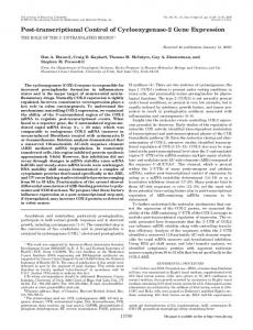
FIG. 1. Physical map and expression of the puf operon of R. sulfidophilum. (a) Gene arrangement of the R. sulfidophilum puf operon. The genes are indicated by open boxes: Q, pufQ; B, pufB; A, pufA; L, pufL; M, pufM; C, pufC. The positions of putative hairpin structures are indicated by open circles. DNA probes used in Northern hybridization experiments are indicated by thick lines. (b) Northern hybridization analysis of mRNA transcripts with four different specific probes for the R. sulfidophilum puf operon. Total RNA was extracted from R. sulfidophilum grown under anaerobic light conditions. The digoxigenin-dUTP-labeled probe A, probe B, probe C, and probe D were used. The lengths of the standard RNAs are indicated on the left. (c) Effects of oxygen and light on puf operon transcription in R. sulfidophilum and R. capsulatus. Three micrograms of total RNA isolated from cells grown under four different conditions (LL-ANA, anaerobic low-intensity light [3 W/m2]; HL-ANA, anaerobic high-intensity light [100 W/m2]; DR-AER, aerobic dark; and HL-AER, aerobic high-intensity light [100 W/m2]) was loaded on each lane. Northern hybridization was performed with the 32P-labeled probe A of R. sulfidophilum or R. capsulatus. Ethidium bromide staining of 16S rRNA (lower panel, rRNA) was used to confirm equivalent loading of total RNA.
CATGGTCTATT-3⬘) and reverse primer B140R (5⬘-CCACGGGCGCCACTG CCA-3⬘) using pUFS001 as a template DNA. Probe D is a PCR product amplified with forward primer Z590F (5⬘-CCATGTCGCCGAGATGCG-3⬘) and reverse primer Z0R (5⬘-AATAGACCATGAACTCCG-3⬘) using pUFS001 as a template DNA. These DNA fragments were labeled with digoxigenin-dUTP as instructed by the manufacturer (Boehringer Mannheim). Probe A of R. sulfidophilum and R. capsulatus was also labeled with [␣-32P]dCTP using a Random Primer DNA labeling kit (Takara). RNA Molecular Weight Marker I (Boehringer Mannheim) was used as a molecular weight standard in some cases. Hybridization was carried out with DIG Easy Hyb Granules (Boehringer Mannheim) at 50°C for over 12 h. After hybridization, the membrane was washed twice with 2⫻ SSC (20⫻ SSC is 3 M sodium chloride and 0.3 M sodium citrate, pH 7.0) and 0.1% sodium dodecyl sulfate (SDS) for 5 min at room temperature and twice with 0.1⫻ SSC and 0.1% SDS for 15 min at 65°C. For quantification of the radiolabeled bands, a BAS 2000 photoimaging system (Fuji film) was used. Half-life measurement of puf mRNA by Northern blot analysis. Cells of R. sulfidophilum were grown till the logarithmic growth phase, showing an optical density of 0.5 to 0.6 at 660 nm. Then, rifampin was added to the cultures (500 g/ml), and cells were collected at various time points. Total RNA was isolated with an RNeasy kit. Total RNA (3 g per lane) was electrophoresed on 1.0% formaldehyde agarose gels and transferred to a Hybond-N⫹ positively charged nylon membrane. Probe C was used as a puf-specific DNA probe (see “Northern hybridization analysis” and Fig. 1a). The probe was labeled with [␣-32P]dCTP using a Random Primer DNA labeling kit. DNA-RNA hybridization was carried out with DIG Easy Hyb at 50°C as instructed by the manufacturer. After hybridization, the membrane was washed twice with 2⫻ SSC and 0.1% SDS for 5 min at room temperature and twice with 0.1⫻ SSC and 0.1% SDS for 15 min at 65°C. For half-life measurement, the radiolabeled bands were quantified using a BAS 2000 photoimaging system. Primer extension experiment. Total RNA of R. sulfidophilum grown till the logarithmic growth phase, showing an optical density of 0.5 to 0.6 at 660 nm, was extracted with an RNeasy kit from aerobically and photosynthetically grown cells. Primers used in this experiment were Z1100R (5⬘-GTCGAGCAGTG CCTGGGCGTCCTC-3⬘), Z0R (5⬘-AATAGACCATGAACTCCG-3⬘), and AENDR (5⬘-TCATGGGCGTGATGATCC-3⬘) (nucleotide numbers of the 5⬘ ends of the primers in the sequence with GenBank accession no. AB020784 are 975, 1218, and 1353, respectively). These primers were labeled at the 5⬘ end with [␥-32P]ATP using a DNA MEGALABEL labeling kit (Takara). To determine the 5⬘ end of the puf operon mRNA, a mixture containing 20 g of RNA, 50 mM Tris hydrochloride (pH 8.3), 50 mM KCl, 10 mM MgCl2, and 0.5 pmol of primer in a final volume of 50 l was incubated at 80°C for 2 min and then at 60°C for 45 min for DNA-RNA hybridization. Then, four deoxynucleoside triphosphates
(final concentration, 0.5 mM each) and 20 U of reverse transcriptase (Rousassociated virus-2) were added to the mixture, and the primer extension reaction proceeded at 42°C for 1 h. The DNA synthesized was extracted with phenolchloroform (1:1), precipitated with ethanol, and suspended in Tris-EDTA buffer. DNA was electrophoresed on a 6% polyacrylamide gel containing 8 M urea with a sequencing ladder, using the same DNA primers. Sequencing gels were dried and exposed to film with an intensifying screen at ⫺80°C. Measurement of BChl content. Membranes were obtained by sonicating cells grown until mid-logarithmic phase, showing an optical density of 0.5 to 0.6 at 660 nm, followed by differential ultracentrifugation as described previously (30). BChl contents in the membranes were determined using acetone-methanol (7:2) extract as described previously (10). Membrane protein content was determined with a Lowry assay kit (Bio-Rad Laboratories). Then, the relative values of BChl content per membrane protein in each sample were calculated. Materials. Restriction endonucleases and reverse transcriptase were purchased from Takara. Synthetic oligonucleotide primers were purchased from Life Technologies, Inc. [␥-32P]ATP and [␣-32P]dCTP were purchased from Amersham.
RESULTS Northern blot analysis. Figure 1a shows the structure of the R. sulfidophilum puf operon, which consists of six puf genes, pufQ, -B, -A, -L, -M, and -C (30). The puf operon transcripts were analyzed by Northern blot hybridization with the four puf-specific probes (probes A to D). As shown in Fig. 1b, probe A, corresponding to pufQBA and part of pufL (Fig. 1a), was hybridized with a 0.5-kb mRNA and also weakly with an approximately 3.5-kb mRNA. Probe B, corresponding to pufC (Fig. 1a), was hybridized with the 3.5-kb mRNA but not with the 0.5-kb mRNA. These two bands were not detected with probe D, which consisted mainly of the 3⬘ end of bchZ and a small 5⬘ segment of the pufQ gene, but they were detected with probe C, which consisted of pufB and part of the pufQ gene (Fig. 1b). The transcription of the puf operon appeared to be terminated after pufC because the signal nucleotide sequence for the typical Rho-independent transcriptional termination
EXPRESSION OF R. SULFIDOPHILUM puf OPERON
VOL. 182, 2000
2781
TABLE 2. Effects of oxygen and light on puf mRNA levels, pufQ- and pufL-lacZ expression, and photopigment synthesisa Organism
Growth conditionb
R. sulfidophilum
R. capsulatus
mRNA contentc
pufQ-lacZe
pufL-lacZf
BChlg
100 114 85 21
100 109 33 2
100 135 35 7
100 128 84 51
100 115 33 21
NTh NT NT NT
NT NT NT NT
100 130 ⬍1 ⬍1
0.5 kb
3.5 (2.7) kbd
HL-ANA LL-ANA DK-AER HL-AER
100 127 86 39
HL-ANA LL-ANA DK-AER HL-AER
100 136 30 20
a
Indicated as percentages of the values under anaerobic high-intensity-light conditions (set as 100). HL-ANA, anaerobic high-intensity light (100 W/m2); LL-ANA, anaerobic low-intensity light (3 W/m2); DK-AER, aerobic dark; HL-AER, aerobic high-intensity light (100 W/m2). c The relative amounts of mRNA were measured by Northern hybridization and quantified by the photoimaging system (Fig. 1c). d 3.5 kb, R. sulfidophilum puf mRNA; 2.7 kb, R. capsulatus puf mRNA (Fig. 1c). e The relative activities of the pufQ-lacZ transcriptional fusion were calculated based on the -galactosidase activity of pPS100 in R. sulfidophilum in Fig. 4. Results are based on the averages of at least three independent assays, and the uncertainty limits in single measurements are within 20%. f The relative activities of the pufL-lacZ transcriptional fusion were calculated based on the -galactosidase activity of pPS001 in R. sulfidophilum in Fig. 4. Results are based on the averages of at least three independent assays, and the uncertainty limits in single measurements are within 15%. g Relative BChl contents in membrane preparations were determined as described in Materials and Methods. Results are based on at least three independent assays, with single measurements varying less than 5% from that value. h NT, not tested. b
was found downstream of the puf operon (30). These observations indicated that the large transcript (3.5 kb) and the small transcript (0.5 kb) in Fig. 1b encode pufBALMC and pufBA, respectively. The small transcript (0.5 kb) was more abundant than the large transcript (3.5 kb). This difference may be a factor that is thought to adjust the ratio of LH1 peptides to RC proteins, as previously suggested for R. capsulatus (7, 25, 47). To test the effects of oxygen and light on the expression of the R. sulfidophilum puf operon, the levels of the two puf mRNA transcripts were analyzed by Northern hybridization under four different growth conditions (Fig. 1c). Total RNAs were isolated from cells grown under anaerobic low-intensitylight (3 W/m2), anaerobic high-intensity-light (100 W/m2), aerobic dark, and aerobic high-intensity-light (100 W/m2) conditions (LL-ANA, HL-ANA, DR-AER, and HL-AER, respectively, in Fig. 1c). The analyses were also performed with R. capsulatus (Fig. 1c). Because R. capsulatus has a pufX instead of a pufC gene in the puf operon (44), the large transcript of the puf operon of R. capsulatus is shorter (2.7 kb) than that of R. sulfidophilum (3.5 kb) (Fig. 1c) (46, 47). Quantitative values digitized by a photoimaging system are shown in Table 2. The puf-specific mRNAs were also monitored by dot blot hybridization, and the results are identical to those of the Northern hybridization analysis (standard deviations are ⬍20%). Under anaerobic conditions, the decrease in light intensity from 100 to 3 W/m2 resulted in increased levels of the puf mRNA (about a 30 and 15% increase for the small and large transcripts, respectively) in both R. sulfidophilum and R. capsulatus (Table 2). However, the relative levels of puf mRNA under aerobic growth conditions were different between these two species. In R. capsulatus, the relative levels of the small and large transcripts were about 30 and 20% for aerobic dark and aerobic high-intensity-light conditions, respectively, and only a trace amount of photopigment was observed in such conditions (Table 2). The results were in agreement with previous studies (7, 26, 46, 47) in the sense that not only photopigment synthesis but also the cellular levels of puf mRNA were strongly affected by oxygen. On the other hand, the relative level of puf mRNA was about 85% for aerobic dark conditions in R. sulfidophilum (Table 2), which was consistent with the relative BChl content
in cells grown under the same conditions (84%) (Table 2). Thus, in contrast to R. capsulatus, a high level of puf mRNA was still maintained in R. sulfidophilum even under aerobic conditions in the dark, although a significant decrease was observed when high light was used under aerobic conditions (Table 2). The BChl content in R. sulfidophilum cells grown under aerobic high-intensity-light conditions was 51% of that under anaerobic high-intensity-light conditions, whereas in R. capsulatus the relative BChl content was ⬍1%, although both species contain significant amounts of puf mRNA (⬃20 to 39%) (Table 2). This observation implies that regulation of BChl synthesis differs in these two species. Primer extension analysis. To determine the possible transcription initiation site of the R. sulfidophilum puf operon, we performed RNA 5⬘-end mapping by primer extension analysis. The 32P-labeled primers (Z1100R, Z0R, and AENDR: see Materials and Methods) were hybridized to the total RNA extracted from cells grown under anaerobic light and aerobic dark conditions (Fig. 2, ⫺ and ⫹, respectively). Using a Z0R primer, two 5⬘ ends of puf mRNA were observed. One had a less intense signal corresponding to 11 bp downstream from the pufQ start codon (pufP3), and the other showed a more intense signal corresponding to 132 bp upstream from the pufQ start codon (pufP2) (Fig. 2B). Results obtained with the AENDR primer also showed the presence of the two 5⬘ ends of the puf mRNA at the corresponding positions (data not shown). A third 5⬘ end of the puf mRNA was detected by the Z1100R primer (pufP1) (Fig. 2A). This site was resolved to a position much farther upstream (332 bp) from the pufQ start codon. These 5⬘ ends of puf mRNA are indicated in Fig. 3 as P1, P2, and P3. All three transcripts were detected in both anaerobically and aerobically grown cells, but the relative band intensity between the conditions was different depending on the initiation sites. Deletion analysis of the puf operon promoter. In order to analyze the R. sulfidophilum puf operon promoter, a nested set of deletions was constructed in the region of DNA upstream from the pufL gene. The deleted DNA fragments were cloned into the promoter testing vector, pCF1010 (28), to construct puf-lacZ transcriptional fusions (Fig. 3). These constructs were
2782
MASUDA ET AL.
FIG. 2. Primer extension mapping of the 5⬘ end of puf operon transcripts. Total cellular RNA isolated from R. sulfidophilum grown under aerobic dark conditions (⫹) and anaerobic light conditions (⫺) was hybridized with 5⬘-endlabeled Z1100R primer (A) or Z0R primer (B) (see Materials and Methods). Lanes G, A, T, and C show sequence ladders using the same synthetic primers. The numbers of 5⬘ and 3⬘ ends shown in the nucleotide sequences are the nucleotide numbering in the GenBank sequence with accession no. AB020784.
introduced into R. sulfidophilum cells and assayed for -galactosidase activity levels under anaerobic low-intensity-light (3 W/m2), anaerobic high-intensity-light (100 W/m2), aerobic dark, and aerobic high-intensity-light (100 W/m2) conditions (LL-ANA, HL-ANA, DR-AER, and HL-AER, respectively, in Fig. 3). The largest fragment, which extended from 584 bp upstream of pufP1 to pufL (pPS001), showed the highest lacZ expression under anaerobic low-intensity-light conditions. -Galactosidase activity of the plasmid was reduced by aeration and high-light illumination, suggesting that the transcriptional activity assayed with this lacZ fusion was repressed by oxygen and high-intensity light. High-light repression of lacZ activity was more apparent under aerobic conditions than under anaerobic conditions for this construct. Deletion of 303 and 540 bp from the 5⬘ end of the largest fragment (pPS002 and pPS003, respectively) resulted in different levels of lacZ activity. A comparison of the activities with the largest fragment (pPS001) indicates that the values were reduced under anaerobic low-light, anaerobic high-light, and aerobic dark conditions (⬃30 to 50%) but increased under aerobic highlight conditions (⬃280 to 480%). The transcriptional activity was detected in the construct pPS004, which contained one of the three 5⬘ ends (pufP3). Removal of all the sites of the mRNA 5⬘ ends, yielding the construct pPS005, caused a very low level of lacZ expression (approximately 50-fold reduction relative to the most active fragment, pPS001). Deletion of 1.4 kb from the 3⬘ end of the largest fragment, yielding the construct designated pPS100,
J. BACTERIOL.
resulted in reduced lacZ activity (⬃25 to 60%) although the fragment contained the same 5⬘ end and all three sites of the mRNA 5⬘ ends. The fragment of pPS200 containing only one of the three 5⬘ ends (pufP1) showed significant lacZ activity, but the removal of an additional 282 nucleotides with pufP1, yielding the plasmid pPS300, caused no lacZ activity. The in vivo expression activities observed with these vectors support the presence of at least two possible promoters (pufP1 and pufP3) based on the results of the primer extension analysis. The effect of light intensities on lacZ activity in some constructs was the opposite under aerobic conditions from what it was under anaerobic conditions. The transcriptional activities of pufQ-lacZ (pPS100) and pufL-lacZ (pPS001) under four different growth conditions are summarized in Table 2 as values relative to those for anaerobic high-light conditions. The relative BChl contents in cells grown under four different growth conditions were in general agreement with the puf mRNA contents, although most of the BChl in the membrane could be present in the puc-encoded LH2 complex. However, the puf operon expression measured by the pufQ- or pufL-lacZ transcriptional fusions was not directly correlated with the BChl levels (Table 2). This may be due to the lack of upstream sequences on these plasmid-borne fusions. A readthrough transcription from the upstream region may be present in the R. sulfidophilum puf operon (see Discussion). Decay rate of puf mRNA in R. sulfidophilum. In order to examine the posttranscriptional control of puf operon expression in R. sulfidophilum, the half-lives of both small (0.5-kb) and large (3.5-kb) puf transcripts were determined under anaerobic and aerobic growth conditions by Northern hybridization with the 32P-labeled probe C (Fig. 1a) corresponding to pufB and part of the pufQ gene. Results are shown in Fig. 4. The half-lives of 3.5-kb puf mRNA were about 10 and 13 min in aerobic and anaerobic conditions, respectively, whereas the half-lives of 0.5-kb mRNA isolated from aerobically and anaerobically grown cultures did not show a significant difference, with values of about 40 min. We also performed corresponding measurements with R. capsulatus (data not shown). The results obtained were similar to those of the previous study, which showed that the half-lives of the larger puf transcript (2.7 kb) were about 3 and 8 min in aerobically and anaerobically grown cultures, respectively, and that those of the smaller RNA (0.5 kb) were about 30 min under both aerobic and anaerobic conditions (23). The larger transcript was degraded about twice as fast under aerobic conditions in R. capsulatus but only about 1.3 times faster under aerobic than under anaerobic conditions in R. sulfidophilum. The stability of puf mRNA was only weakly influenced by oxygen tension in R. sulfidophilum. Influence of regA mutation on the control of transcription of R. sulfidophilum puf operon. We previously constructed a regA deletion mutant (RESA1) from R. sulfidophilum and showed that the RegA-RegB regulatory cascade has an important role for the expression of photosynthesis genes in this bacterium, as in the Rhodobacter species (29). To test the effects of the regulatory mutant on the promoter activity of puf operon in R. sulfidophilum, we assayed the -galactosidase activity of various puf-lacZ fusions in RESA1. As shown in Fig. 5, the effects of -galactosidase activity caused by regA mutation fell into three types. In one type, the transcriptional pufL-lacZ fusions containing all three promoters (pPS001, pPS002, and pPS003 [Fig. 3]) exhibited nearly baseline levels of lacZ activity in RESA1 (Fig. 5A for pPS001; data for pPS002 and pPS003 are not shown). In contrast, a construct containing only the pufP3 promoter (pPS004 [Fig. 3]) retained the -galactosidase activity in the regA mutant as much as in the wild-type cells (Fig. 5B). The third type is a pufQ-lacZ transcriptional fusion from
EXPRESSION OF R. SULFIDOPHILUM puf OPERON
VOL. 182, 2000
2783
FIG. 3. Deletion analysis of R. sulfidophilum puf operon. 5⬘ and 3⬘ deletions were constructed by using DNA-restricted and PCR-generated fragments (see Materials and Methods). These deletions were fused to the lacZ gene to construct various puf-lacZ transcriptional fusions. -Galactosidase activities associated with the transcriptional fusions on plasmids present in R. sulfidophilum cells were determined under different growth conditions (LL-ANA, anaerobic low-intensity light [3 W/m2]; HL-ANA, anaerobic high-intensity light [100 W/m2]; DK-AER, aerobic dark; HL-AER, aerobic high-intensity light [100 W/m2]). Values in parentheses are the standard deviations of at least three independent assays. No activity was detected for the sequence without insertion of the lacZ fusion (vector only). The positions of three 5⬘ ends of puf mRNA determined in experiments in Fig. 2 are indicated by arrows (P1, pufP1; P2, pufP2; P3, pufP3).
which the 3⬘ end was deleted from a DNA fragment of pPS001 (pPS100 [Fig. 3]) and which showed a low level of -galactosidase activity even in the mutant while still containing the same 5⬘ end of pPS001 (16 and 28% of those in wild-type cells under anaerobic and aerobic conditions, respectively [Fig. 5C]). DISCUSSION Three possible puf operon promoters in R. sulfidophilum. In this study, the R. sulfidophilum puf operon and its promoters were analyzed to clarify the mechanism of the oxygen-independent expression of photosynthetic genes. Primer extension analysis showed that there are three 5⬘ ends of puf mRNA (pufP1, pufP2, and pufP3 in Fig. 2). By employing deletion analyses, two of the three 5⬘ ends of puf mRNA were mapped as the possible transcription initiation sites at positions 332 bp upstream and 11 bp downstream of the pufQ start codon (pufP1 and pufP3, respectively [Fig. 2 and 3]), because the deletion of pufP1 or pufP3 from the inserted fragment of lacZ fusions resulted in significant reduction of the lacZ activity (Fig. 3). The other 5⬘ end found in the primer extension analysis (pufP2; 132 bp upstream from pufQ) was not directly supported as a transcription initiation site by the deletion analysis because of the lack of appropriate restriction sites. In R. capsulatus, two puf transcription initiation sites have been reported and mapped to positions 316 and 129 bp upstream of the pufQ start codon. The nucleotide sequence alignment of puf operon promoter regions of R. capsulatus and R. sulfidophilum revealed that the two puf transcription initiation sites of R. capsulatus are located near the corresponding sites of pufP1 and pufP2 of R. sulfidophilum (separated by 9 and 4 bp, re-
spectively) (data not shown) (1, 5). The regions are highly conserved between these two species, suggesting that the pufP2 of R. sulfidophilum is also a transcription initiation site, as in R. capsulatus. Oxygen regulation on the puf operon expression in R. sulfidophilum. Northern blot hybridization experiments showed that the mRNA levels of the R. sulfidophilum puf operon were mostly maintained even under aerobic dark conditions (Fig. 1c), which was consistent with the relative BChl content in the cells (Table 2). This observation is in contrast to that in the Rhodobacter species (Fig. 1c) (7, 26, 46, 47), indicating that the aerobic synthesis of the photosynthetic apparatus in R. sulfidophilum seems to be mostly due to the puf operon expression, which is weakly sensitive to oxygen and regulated at the transcriptional level. Deletion analysis showed that one of the possible promoters, pufP3, was expressed independently of oxygen (Fig. 3, pPS004). The activity of the promoter can contribute to the aerobic expression of the puf operon. However, the activity of the major promoter for the puf operon transcription located far upstream from the pufB gene, pufP1, showed fivefold-lower levels under aerobic dark than under anaerobic high-intensitylight conditions (Fig. 3, pPS200). Probably, as a result, the transcriptional activities of both pufQ-lacZ (pPS100) and pufLlacZ (pPS001) fusions were repressed by oxygen by a factor of three when the activities were compared between the aerobic dark and anaerobic high-intensity-light conditions (Table 2 and Fig. 3). On the other hand, only a 15% decrease in the amount of mRNA was observed under aerobic dark conditions (Table 2). A similar difference was also observed under aerobic highintensity-light conditions. The transcriptional pufQ-lacZ and pufL-lacZ activities showed very low levels under such condi-
2784
MASUDA ET AL.
J. BACTERIOL.
FIG. 4. puf mRNA decay kinetics in R. sulfidophilum. Three micrograms of total RNA isolated from rifampin-treated R. sulfidophilum cells grown under aerobic dark and anaerobic light conditions was loaded on each lane. Hybridization was performed with the 32P-labeled probe C (Fig. 1a). The optical density of the bands was plotted against the time of RNA isolation (squares, aerobic conditions; circles, anaerobic conditions). The half-lives calculated from these blots for the 3.5-kb mRNA were about 10 min under aerobic conditions (dashed line) and about 13 min under anaerobic conditions (solid line). For the 0.5-kb mRNA, the half-lives were about 40 min under both aerobic conditions (dashed line) and anaerobic conditions (solid line).
tions (2 and 7%, respectively, compared to values for anaerobic high-intensity-light conditions) (Table 2). However, the amounts of the two puf mRNA transcripts were 39 and 21% for the small and large transcript, respectively, when those two growth conditions were compared (Table 2). Thus, the pufspecific transcriptional activity does not directly reflect the level of puf mRNA under aerobic growth conditions (Table 2). The transcriptional overlap (readthrough transcription) must be a factor causing the absence of direct correlation between the level of chromosomally derived puf mRNA and transcriptional activity of DNA segments inserted into the lacZ fusion plasmid. Since the stop codon of bchZ overlaps the start codon of pufQ in R. sulfidophilum (30) and the puf promoters (at least pufP1) are located in the bchZ gene (Fig. 3), the readthrough should exist between the transcription for bchZ and that for the puf operon, as shown for R. capsulatus (40, 43). Previous studies using R. capsulatus have shown that decay of mRNA is an important factor in controlling puf operon expression (1, 18, 21, 23, 24). In this study, we measured the decay time of puf operon transcripts under aerobic and anaerobic conditions. For R. sulfidophilum, the large puf mRNA was less stable by a factor of 0.77 (Fig. 4) under aerobic conditions than under anaerobic conditions. This number, however, was 0.38 (data not shown) in R. capsulatus (23). The difference in relative stability of this molecule between the two bacterial species seems to contribute to the high content of the puf mRNA under aerobic conditions in R. sulfidophilum. However, this contribution is not sufficient to explain the higher content of puf mRNA than that expected from the puf promoter activity (Table 2). Light regulation of puf operon expression in R. sulfidophilum. Light regulation of puf operon expression is apparent under aerobic conditions in R. sulfidophilum (Fig. 1c and Table 2). pufQ- and pufL-lacZ activities were highly repressed by
FIG. 5. Measurement of puf operon transcriptional activity in wild-type (W.T.) and regA-disrupted R. sulfidophilum strains. Bars represent -galactosidase activities associated with the puf-lacZ transcriptional fusions, pPS001 (A), pPS004 (B), and pPS100 (C) (Fig. 3), in cells grown under anaerobic highintensity light (100 W/m2) (⫺O2) or aerobic dark conditions (⫹O2). Data for the wild type are taken from the results shown in Fig. 3. Units of -galactosidase activity represent micromoles of ONPG hydrolyzed per minute per milligram of total protein.
high-intensity light under aerobic conditions (⬃6 to 20% of those under aerobic dark conditions). Similarly, high-light illumination also resulted in lower levels of chromosomally derived puf mRNA under such conditions (⬃24 to 45% of those under aerobic dark conditions). Since the major fraction of puf mRNA seems to be derived from the readthrough transcription under aerobic conditions as discussed above, the readthrough activity may also be repressed to some extent by high-intensity light under aerobic conditions. The effect of light on the puf operon-specific promoters was
EXPRESSION OF R. SULFIDOPHILUM puf OPERON
VOL. 182, 2000
analyzed by deletion analysis, and the results were a little complicated. The strains containing pPS004 covering only pufP3 showed the high-light repression under aerobic conditions, whereas pPS200, covering only pufP1, and pPS003, from which the 5⬘ end of 540 bp was deleted from the largest fragment (pPS001), did not exhibit the light repression (Fig. 3). Plasmid pPS002, from which 303 bp was deleted from the 5⬘ end of pPS001, showed a unique phenotype in that high-intensity light activated the promoter activity under aerobic conditions (Fig. 3). These observations suggest that there are several light-dependent cis regulatory sites on the puf operon promoters. It was also suggested that the HincII-NruI region (584 to 281 bp upstream from the first transcription initiation site, respectively) is an important regulatory site responsible not only for the elevation of the puf operon transcription responding to anaerobiosis but also for the light regulation of the puf operon expression (Fig. 3). Unidentified regulatory factors may bind to this region and interact with RNA polymerase mediated by other protein factors. RegA-RegB regulatory system for puf operon expression in R. sulfidophilum. As mentioned previously, two promoters in the R. capsulatus puf operon have been reported. The R. capsulatus upstream promoter corresponding to pufP1 of R. sulfidophilum is highly expressed and regulated (1, 5). It was recently reported that the response regulator RegA binds to this promoter region (8, 16). It is clear from puf-lacZ fusion analysis that RegA is involved in activating puf operon transcription in R. sulfidophilum through association with the upstream region of the PstI site located between pufP1 and pufP3 (Fig. 5A and B). The promoter activity of pufP1 may be regulated by the RegA-RegB regulatory system in R. sulfidophilum, as in R. capsulatus (8, 16). Comparison of activities obtained with pPS100 with those of pPS001 indicates that deletion of the BalI-BamHI region located between pufQ and pufL (Fig. 3) resulted in reduced lacZ activity (⬃25 to 60%) although the fragment contained the same 5⬘ end and all possible promoters. In addition, deletion of this region recovered some puf operon transcriptional activity in a regA mutant (Fig. 5A and C), suggesting that the region has a possible cis site which is involved in the transcriptional control of puf operon expression by the RegA-RegB regulatory system. Further genetic characterization of the R. sulfidophilum puf operon should be useful in clarifying the details of the control mechanisms of puf operon expression in purple bacteria. ACKNOWLEDGMENTS We thank Alastair G. McEwan and Anthony L. Shaw (University of Queensland) for generous provision of materials. This work was supported in part by grants from the Ministry of Education, Science, and Culture of Japan and a special grant (1999) from Tokyo Metropolitan University. REFERENCES 1. Adams, C. W., M. E. Forrest, S. N. Cohen, and J. T. Beatty. 1989. Structural and functional analysis of transcriptional control of the Rhodobacter capsulatus puf operon. J. Bacteriol. 171:473–482. 2. Bauer, C. E. 1995. Regulation of photosynthesis gene expression, p. 1221– 1234. In R. E. Blankenship, M. T. Madigan, and C. E. Bauer (ed.), Anoxygenic photosynthetic bacteria. Kluwer Academic Publishers, Dordrecht, The Netherlands. 3. Bauer, C. E., and T. H. Bird. 1996. Regulatory circuits controlling photosynthesis gene expression. Cell 85:5–8. 4. Bauer, C. E., J. Buggy, and C. Mosley. 1993. Control of photosystem genes in Rhodobacter capsulatus. Trends Genet. 9:56–60. 5. Bauer, C. E., D. A. Young, and B. L. Marrs. 1988. Analysis of the Rhodobacter capsulatus puf operon. J. Biol. Chem. 263:4820–4827. 6. Belanger, G., and G. Gingras. 1988. Structure and expression of the puf operon messenger RNA in Rhodospirillum rubrum. J. Biol. Chem. 263:7639– 7645.
2785
7. Belasco, J. G., T. J. Beatty, C. W. Adams, A. Von Gabain, and S. N. Cohen. 1985. Differential expression of photosynthesis genes in R. capsulatus results from segmental differences in stability within the polycistronic rxcA transcript. Cell 40:171–181. 8. Bowman, W. C., S. Du, C. E. Bauer, and R. G. Kranz. 1999. In vitro activation and repression of photosynthesis gene expression in Rhodobacter capsulatus. Mol. Microbiol. 33:429–437. 9. Clark, W. G., E. Davidson, and B. L. Marrs. 1984. Variation of levels of mRNA coding for antenna and reaction center polypeptides in Rhodopseudomonas capsulata in response to changes in oxygen concentration. J. Bacteriol. 157:945–948. 10. Clayton, R. K. 1966. Spectroscopic analysis of bacteriochlorophylls in vitro and in vivo. Photochem. Photobiol. 5:669–677. 11. Cohen-Bazire, G., W. R. Sistrom, and R. Y. Stanier. 1957. Kinetic studies of pigment synthesis by non-sulfur purple photosynthetic bacteria. J. Cell Comp. Physiol. 49:25–68. 12. DeHoff, B. S., J. K. Lee, T. J. Donohue, R. I. Gumport, and S. Kaplan. 1988. In vivo analysis of puf operon expression in Rhodobacter sphaeroides following deletion of a putative inter-cistronic terminator. J. Bacteriol. 170:4681– 4692. 13. Deisenhofer, J., O. Epp, K. Miki, R. Huber, and H. Michel. 1985. Structure of the protein subunit in the photosynthetic reaction centre of Rhodopseudomonas viridis at 3A resolution. Nature 318:618–624. 14. Doi, M., Y. Shioi, N. Gad’on, J. R. Golecki, and G. Drews. 1991. Spectroscopical studies on the light-harvesting pigment-protein complex II from dark-aerobic and light-anaerobic grown cells of Rhodobacter sulfidophilus. Biochim. Biophys. Acta 1058:235–241. 15. Drews, G., and R. J. Golecki. 1995. Structure, molecular organization, and biosynthesis of membranes of purple bacteria, P. 231–257. In R. E. Blankenship, M. T. Madigan, and C. E. Bauer (ed.), Anoxygenic photosynthetic bacteria. Kluwer Academic Publishers, Dordrecht, The Netherlands. 16. Du, S., T. H. Bird, and C. E. Bauer. 1998. DNA-binding characteristics of RegA*: a constitutively active anaerobic activator of photosynthesis gene expression in Rhodobacter capsulatus. J. Biol. Chem. 273:18509–18513. 17. Eraso, J. M., and S. Kaplan. 1994. prrA, a putative response regulator involved in oxygen regulation of photosynthesis gene expression in Rhodobacter sphaeroides. J. Bacteriol. 176:32–43. 18. Fritsch, J., R. Rothfuchs, R. Rauhut, and G. Klug. 1995. Identification of an mRNA element promoting rate-limiting cleavage of the polycistronic puf mRNA in Rhodobacter capsulatus by an enzyme similar to RNase E. Mol. Microbiol. 15:1017–1029. 19. Hagemann, G. E., E. K. Katsiou, H. Forkl, A. C. J. Steindorf, and M. H. Tadros. 1997. Gene cloning and regulation of gene expression of the puc operon from Rhodovulum sulfidophilum. Biochim. Biophys. Acta 1351:341– 358. 20. Hansen, T. A., and H. Veldkamp. 1973. Rhodopseudomonas sulfidophila, nov. spec., a new species of the purple nonsulfur bacteria. Arch. Mikrobiol. 92:45–58. 21. Heck, C., R. Rothfuchs, A. Jager, R. Rauhut, and G. Klug. 1996. Effect of the pufQ-pufB intercistronic region on puf mRNA stability in Rhodobacter capsulatus. Mol. Microbiol. 20:1165–1178. 22. Hunter, C. N., P. McGlynn, M. K. Ashby, J. G. Burgess, and J. D. Olsen. 1991. DNA sequencing and complementation/deletion analysis of the bchCA-puf operon region of Rhodobacter sphaeroides: in vivo mapping of the oxygen-regulated puf promoter. Mol. Microbiol. 5:2649–2661. 23. Klug, G. 1991. Endnucleolytic degradation of puf mRNA in Rhodobacter capsulatus is influenced by oxygen. Proc. Natl. Acad. Sci. USA 88:1765–1769. 24. Klug, G. 1995. Post-transcriptional control of photosynthesis gene expression, p. 1235–1244. In R. E. Blankenship, M. T. Madigan, and C. E. Bauer (ed.), Anoxygenic photosynthetic bacteria. Kluwer Academic Publishers, Dordrecht, The Netherlands. 25. Klug, G., C. W. Adams, J. Belasco, B. Doerge, and S. N. Cohen. 1987. Biological consequences of segmental alterations in mRNA stability: effects of deletion of the inter-cistronic hairpin loop region of Rhodobacter capsulatus puf operon. EMBO J. 6:3515–3520. 26. Klug, G., N. Kaufmann, and G. Drews. 1985. Gene expression of pigmentbinding proteins of the bacterial photosynthetic apparatus: transcription and assembly in the membrane of Rhodopseudomonas capsulata. Proc. Natl. Acad. Sci. USA 82:6485–6489. 27. Lee, J. K., B. S. DeHoff, T. J. Donohue, R. I. Gumport, and S. Kaplan. 1989. Transcriptional analysis of puf operon expression in Rhodobacter sphaeroides 2.4.1 and an intercistronic transcription terminator mutant. J. Biol. Chem. 264:19354–19365. 28. Lee, J. K., and S. Kaplan. 1995. Transcriptional regulation of puc operon expression in Rhodobacter sphaeroides. J. Biol. Chem. 270:20453–20458. 29. Masuda, S., Y. Matsumoto, K. V. P. Nagashima, K. Shimada, K. Inoue, C. E. Bauer, and K. Matsuura. 1999. Structural and functional analyses of photosynthetic regulatory genes regA and regB from Rhodovulum sulfidophilum, Roseobacter denitrificans, and Rhodobacter capsulatus. J. Bacteriol. 181:4205– 4215. 30. Masuda, S., M. Yoshida, K. V. P. Nagashima, K. Shimada, and K. Matsuura. 1999. A new cytochrome subunit bound to the photosynthetic reac-
2786
31.
32.
33. 34. 35. 36. 37. 38. 39. 40.
MASUDA ET AL.
tion center in the purple bacterium, Rhodovulum sulfidophilum. J. Biol. Chem. 274:10795–10801. Mosley, C. S., J. Y. Suzuki, and C. E. Bauer. 1994. Identification and molecular genetic characterization of a sensor kinase responsible for coordinately regulating light harvesting and reaction center gene expression in anaerobiosis. J. Bacteriol. 176:7566–7573. Nagashima, K. V. P., K. Matsuura, S. Ohyama, and K. Shimada. 1994. Primary structure and transcription of genes encoding B870 and photosynthetic reaction center apoproteins from Rubrivivax gelatinosus. J. Biol. Chem. 269:2477–2484. Nishimura, K., H. Shimada, H. Ohta, T. Masuda, Y. Shioi, and K. Takamiya. 1996. Expression of the puf operon in an aerobic photosynthetic bacterium, Roseobacter denitrificans. Plant Cell Physiol. 37:153–159. Pemberton, J. M., I. M. Horne, and A. G. McEwan. 1998. Regulation of photosynthetic gene expression in purple bacteria. Microbiology 144:267– 278. Pfenning, N., and H. G. Truper. 1971. Type and neotype strains of the species of phototrophic bacteria maintained in pure culture. Int. J. Syst. Bacteriol. 21:19–24. Phillips-Jones, M. K., and C. N. Hunter. 1994. Cloning and nucleotide sequence of regA, a putative response regulator gene of Rhodobacter sphaeroides. FEBS Lett. 116:269–276. Sganga, M. W., and C. E. Bauer. 1992. Regulatory factors controlling photosynthetic reaction center and light-harvesting gene expression in Rhodobacter capsulatus. Cell 68:945–954. Simon, R., U. Priefer, and A. Puhler. 1983. A broad host range mobilization system for in vivo genetic engineering: transposon mutagenesis in Gram negative bacteria. Bio/Technology 1:37–45. Weaver, P. F., J. D. Wall, and H. Gest. 1975. Characterization of Rhodopseudomonas capsulata. Arch. Microbiol. 105:207–216. Wellington, C. L., and J. T. Beatty. 1991. Overlapping mRNA transcripts of
J. BACTERIOL.
41.
42. 43.
44.
45. 46.
47. 48.
photosynthesis gene operons in Rhodobacter capsulatus. J. Bacteriol. 173: 1432–1443. Wiessner, C., I. Dunger, and H. Michel. 1990. Structure and transcription of the genes encoding the B1015 light-harvesting complex  and ␣ subunits and the photosynthetic reaction center L, M, and cytochrome c subunits from Rhodopseudomonas viridis. J. Bacteriol. 172:2877–2887. Yanisch-Perron, C., J. Vieira, and J. Messing. 1985. Improved M13 phage cloning vectors and host strains: nucleotide sequences of the M13mp18 and pUC19 vectors. Gene 33:103–119. Young, D. A., C. E. Bauer, J. C. Williams, and B. L. Marrs. 1989. Genetic evidence for superoperonal organization of genes for photosynthetic pigments and pigment binding proteins in Rhodobacter capsulatus. Mol. Gen. Genet. 218:1–12. Youvan, D. C., E. J. Bylina, M. Alberti, H. Begusch, and J. E. Hearst. 1984. Nucleotide and deduced polypeptide sequence of the photosynthetic reaction center, B870 antenna and flanking polypeptides from R. capsulata. Cell 37:949–957. Zeilstra-Ryalls, J., J. M. Eraso, M. Gomelsky, A. Yeliseev, J. O’Gara, and S. Kaplan. 1998. Control of photosystem formation in Rhodobacter sphaeroides. J. Bacteriol. 180:2801–2809. Zhu, Y. S., and J. E. Hearst. 1986. Regulation of expression of genes for light-harvesting antenna proteins LH-I and LH-II; reaction center for polypeptides RC-L, RC-M, and RC-H; and enzymes of bacteriochlorophyll and carotenoid biosynthesis in Rhodobacter capsulatus by light and oxygen. Proc. Natl. Acad. Sci. USA 83:7613–7617. Zhu, Y. S., P. L. Kiley, T. J. Donohue, and S. Kaplan. 1986. Origin of the mRNA stoichiometry of the puf operon in Rhodobacter sphaeroides. J. Biol. Chem. 261:10366–10374. Zuber, H., and R. J. Cogdell. 1995. Structure and organization of purple bacterial antenna complexes, p. 315–348. In R. E. Blankenship, M. T. Madigan, and C. E. Bauer (ed.), Anoxygenic photosynthetic bacteria. Kluwer Academic Publishers, Dordrecht, The Netherlands.