The luciferase gene from the firefly, Photinus pyralis, was used as a reporter of ... complementary DNA clone of the firefly luciferase gene under the control of a ...
ing (Ti) plasmid of Agrobacterium tumefaciens (6), has permitted investigations into the regulation of organ-specific gene expression in plants, the development of pathogen
Transient and Stable Expression of the Firefly Luciferase Gene in Plant Cells and Transgenic Plants DAVID W. OW, KEITH V. WOOD, MARLENE DELUCA, JEFFREY R. DE WET,* DONALD R. HELINSKI, STEPHEN H. HOWELLt The luciferase gene from the firefly, Photinus pyralis, was used as a reporter of gene expression by light production in transfected plant cells and transgenic plants. A complementary DNA clone of the firefly luciferase gene under the control of a plant virus promoter (cauliflower mosaic virus 35S RNA promoter) was introduced into plant protoplast cells (Daucus carota) by electroporation and into plants (Nicotiana tabacum) by use of the Arobacterum tumefaciens tumor-inducing plasmid. Extracts from electroporated celLs (;4 bours after the introduction of DNA) and from transgenic plants produce light when mixed with the substrates luciferin and adenosine triphosphate. Light produced by the action of luciferase was also detected in undisrupted leaves or cells in culture from transgenic plants incubated in luciferin and in whole transgenic plants "watered" with luciferin. Although light was detected in most organs in intact, transgenic plants (leaves, stems, and roots), the pattern of luminescence appeared to reflect both the organ-specific distribution of luciferase and the pathway for uptake of luciferin through the vasculature of the plant.
or herbicide resistance, and the control of gene expression linked to plant physiological processes including responses to light
(7).
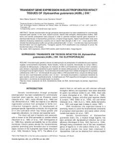
To test the activity of the firefly luciferase gene in plant cells in a transient expression assay, we used electroporation (5) to introduce a luciferase cDNA construct into protoplasts of cultured carrot cells (8). The basic plasmid pDO432 (Fig. 1) contains an "intronless" luciferase gene created by de Wet et al. (3) from a partial cDNA clone linked at an Xba I site (129 bp downstream from the start of translation) to the 5' end of the firefly gene. The fusion was created because a full-length cDNA covering all of the translated region was not obtained in the initial cDNA cloning effort (2). The luciferase gene was spliced upstream, via a Bam HI site, to the cauliflower mosaic virus (CaMV) 35S RNA promoter, making the expected start of transcription 81 bp upstream from the predicted start of translation of the luciferase gene (Fig. 1). The luciferase gene was spliced on its downstream side, via a Bam HI site, to a segment at the 3' end of the nopaline synthase (nos) gene (9). The plasmid pDO432 was introduced into carrot protoplasts by electroporation,
because unlike the bacterial lux gene system which encodes an enzyme that cata- (4), only one protein is needed for light lyzes the light-producing, adenosine production if the substrates ATP, 02, and triphosphate (ATP)-dependent oxidation luciferin are available (1). of luciferin (1), is a powerful reporter gene The introduction of DNA into plants for for assessing gene expression in eukaryotic testing the expression of gene constructs has organisms. We report that light emitted been made possible by the development of D. W. Ow, J. R. de Wet, D. R. Helinski, S. H. Howell, from firefly luciferase was detected in plants new methods for transforming plants and Department of Biology, University of California, San La Jolla, CA 92093. and plant cells in culture transformed with a plant cells. Several procedures, including Diego, K. V. Wood and M. DeLuca, Department of Chemistry, luciferase complementary DNA (cDNA) electroporation, have been developed to in- Univcrsity of California, San Diego, La Jolla, CA 92093. construct driven by a plant virus promoter. troduce naked DNA into plant cells for The firefly luciferase gene has been ex- testing the transient expression of gene con*Present address: Stanford University, Stanford, Califorpressed in bacteria (2) but is ideal for use as structs (5). The production of transgenic nia 94305. a reporter gene in eukaryotic organisms (3), plants, transformed with the tumor-induc- tTo whom all correspondence should be addressed.
T HE FIREFLY LUCIFERASE GENE,
Fig. 1. The structure of the CaMV 35S RNA promoter-luciferase-nos 3'end construct in pUC19 (pDO432). The 35S RNA promoter (35S p) was obtained by creating a Bam HI site with oligonucleotide-directed mutagenesis of pCaMV10 (from CaMV isolate CM1841) (14) at the start of 35S RNA transcription (15) (position 7431 as underlined, GGACAC-4 GGATCC). The 35S RNA promoter (1586 bases) extends from this newly created Bam HI site upstream from the Hind III site at position 5849 on the map of CaMV-CM1841 (14). The luciferase gene was obtained as a Hind III-Bam HI fragment (1886 bases) from pJD201 (3). The upstream Hind III site was filled in and converted to a Bam HI site by the addition of Bam HI linkers. The luciferase gene in pJD201 is an intronless gene created by a fusion between a partial cDNA clone (Luc23) and the 5' end of the corresponding firefly luciferase gene, joined in the coding region at an Xba I site (3). The presumed start of translation is 81 bases downstream from the newly created Bam HI site, the point offusion of the luciferase gene and the 35S RNA promoter. A 1028-bp fragment containing polyadenylation sites from the 3' end of the A. tumefaciens nos gene (9) was inserted downstream from the luciferase gene. The 3' end of the luciferase cDNA, in particular, the 139 bp Ssp I-Bam HI fragment, carries a consensus polyadenylation signal (AATAAA) 28 bases upstream from its 3' end. Plasmid constructs were tested for activity in a transient expression assay in which 10 ,ug of plasmid DNA and 500 ,ug of carrier calf thymus DNA were introduced by clectroporation (5) into protoplasts prepared from carrot cells growing in suspension (8). Extracts were prepared 24 hours after electroporation by repeated cycles of freezing and thawing of0.5 x 107 to 1 x 10⁷ carrot cells in
856
Luciferase
35S Promoter
-1585
IfL pDO4332
r35
pD041 pDO4446
ZZ
+1 +81 _T
in
nos 3'
+1868 +1731 TA Poly A
+2510
R elatIve
Poly A
+
activity
-
I|
.
0%
Inversion
r
66%
pDO4 455 Hind III
100%
IIII Bam
Hi
Ssp Kpn Bam HI
8%
Kpn
luciferase extraction buffer, 100 mM potassium phosphate buffer (pH 7.5), and 1 mM dithiothreitol, followed by centrifugation for 5 minutes in a microcentrifuge (Eppendorf) at 4°C. For luciferase assays, 1/10 volume of the supematant fluid was diluted into 400 ,u1 of assay buffer [14 mM glycylglycine buffer (pH 7.8), 14 mM MgCI2, and 6 mM ATP], to which 100 ,ul of 1 mM luciferin was added by injection. The peak intensity of the resulting light flash was measured in a luminometer (LKB, model 1250), and the data from a single representative experiment are expressed in light units relative to that produced by pDO432. SCIENCE, VOL. 234
Downloaded from www.sciencemag.org on April 4, 2011
WI
I4 NOVEMBER I986
2
1
3
5
4
7
6
Fig. 2. Blot of genomic DNA from transgenic tobacco plants. DNA was extracted from the leaves of primary transformants as indicated, cleaved with Hind III, fractionated on a 1% agarose gel, transferred to a nitrocellulose filter, and hybridized with the nick-translated 1.6-kb Hind III-Bam HI fragment from pDO432 containing the CaMV 35S promoter (16). Hind III cleavage of integrated pDO432 DNA releases a linear 7-kb fragment. Gel migration position of the 7-kb fragment is indicated by the arrow on the left. (Lanes 1 and 2) Copy number standards. Hind III-cut pDO432 DNA in amounts (110 and 22 pg) equivalent to ten and two copies per tetraploid tobacco genome; (lanes 3 to 7) 10 jig of Hind III-cut DNA from an untransformed tobacco plant (lane 3) and transformed plants B10 (lane 4), B15 (lane 5), B22 (lane 6), and B28 (lane 7).
manner by dropping a small leaf cutting (-5 tein species of a size expected for firefly mm2) in a reaction tube containing sub- luciferase (12). Protein blots of leaf extracts strates and reading the light output in a from plant B 15 showed the presence of a 62-kD protein band that reacts with antiseluminometer. rum and migrates with firefly luciferase. transformants (transgenic Several primary The selected transgenic plants (primary plants B10, B15, B22, and B28) with demonstrated luciferase activity were chosen for transformants), grown in sterile culture, further study. DNA blots indicated the pres- were examined for the distribution of lucifence of pDO432 in the plant genome, erase activity in various plant organs. We which appeared as a 7-kb band in Hind III- found that extracts from all organs-leaves, cleaved DNA fragments from transformed roots and stems-expressed activity to difplants but not from untransformed plants. fering extents (Fig. 3). Considerable variaThe number of copies of pDO432 was quite tion in activity was observed from leaf to similar in the selected transformants and was leaf, with young, uppermost leaves generally equivalent in reconstructions to one to sev- showing higher specific activities than older, eral copies per tobacco genome (Fig. 2). lower leaves. In general, roots and stems The transgenic plants also produced a pro- (upper stems were tested) had more lucifer2500 * Plant Bl0 * Plant BI5 O Plant B22 O Plant B28
@0 2000
._
0. 0
EX 1500 o, .E
X 1000 ._
0
coM 500
._i
0
1
2
3
4
5
8 7 6 Leaf number
9
10
11
StemsRoots
Fig. 3. Distribution of luciferase activity in the organs of transgenic plants. Plants were grown in glass jars under sterile conditions in a modified Murashige and Skoog agar medium (MS medium) (17) and extracts were prepared when plants reached a height of 12 to 15 cm. Extracts from various plant organs were prepared by grinding a small amount of tissue, frozen in liquid N2 in a 1.5-mi microcentrifuge tube, with the extraction buffer described in Fig. 1. The soluble extract obtained after centrifuging for 5 minutes at 4°C in a microcentrifuge was assayed for luciferase activity and protein content (18). Leaf number is the position of the leaf on the plant, starting from the younger leaves at the top of the plant progressing to the older leaves on the bottom. REPORTS 857
Downloaded from www.sciencemag.org on April 4, 2011
and extracts were assayed for activity, usually 24 hours later. Extracts produced a light flash detected in a luminometer in the presence of the substrates luciferin and ATP. In the presence of excess substrates, light production, measured as peak height, was proportional to the amount of plant extract containing luciferase activity. No light above machine background [