[Downloaded free from http://www.ijem.in on Wednesday, September 28, 2016, IP: 188.24.116.207]
Original Article
Trimester specific reference intervals for thyroid function tests in normal Indian pregnant women Tarun Sekhri1, Juhi Agarwal Juhi1,2, Reena Wilfred1, Ratnesh S. Kanwar1, Jyoti Sethi1, Kuntal Bhadra1, Sirimavo Nair2, Satveer Singh1 Division of Endocrinology and Thyroid Research, Institute of Nuclear Medicine and Allied Sciences, Timarpur, New Delhi, 2Department of Foods and Nutrition, Division of Endocrine Physiology, Faculty of Family and Community Sciences, The Maharaja Sayajirao University of Baroda, Vadodara, Gujarat, India 1
A B S T R A C T Context: Accurate assessment of thyroid function during pregnancy is critical, for initiation of thyroid hormone therapy, as well as for adjustment of thyroid hormone dose in hypothyroid cases. Aims: We evaluated pregnant women who had no past history of thyroid disorders and studied their thyroid function in each trimester. Settings and Design: 86 normal pregnant women in the first trimester of pregnancy were selected for setting reference intervals. All were healthy, euthyroid and negative for thyroid peroxidase antibody (TPOAb). These women were serially followed throughout pregnancy. 124 normal nonpregnant subjects were selected for comparison. Material and methods: Thyrotropin (TSH), free thyroxine (FT4), free triiodothyronine (FT3) and anti-TPO were measured using Roche Elecsys 1010 analyzer. Urinary iodine content was determined by simple microplate method. The 2.5th and 97.5th percentiles were calculated as the reference intervals for thyroid hormone levels during each trimester. Statistical Analysis: SPSS (version 14.0, SPSS Inc., Chicago, IL, USA) was used for data processing and analysis. Results: The reference intervals for the first, second and third trimesters for the following parameters: TSH 0.09-6.65, 0.51-6.66, 0.91-4.86 µIU/mL, FT4 9.81-18.53, 8.52-19.43, 7.39-18.28 pM/L and FT3 3.1-6.35, 2.39-5.12, 2.57-5.68 pM/L respectively. Thyroid hormone concentrations significantly differed during pregnancy at different stages of gestation. The pregnant women in the study had median urinary iodine concentration of 150-200 µg/l during each trimester. Conclusions: The trimester-specific reference intervals for thyroid tests during pregnancy have been established for pregnant Indian women serially followed during pregnancy using 2.5th and 97.5th percentiles. Key words: Anti‑thyroid peroxidase, pregnant women, reference intervals, thyroid function tests, trimester
Introduction Maternal and perinatal morbidities are well‑documented complications of pregnancy in women with thyroid dysfunction, both clinical and subclinical. About 2–5% of pregnant women suffer from thyroid disorders, and Corresponding Author: Dr. Tarun Sekhri, Division of Endocrinology and Thyroid Research, Institute of Nuclear Medicine and Allied Sciences, Brig. S.K. Mazumdar Marg, Timarpur, New Delhi ‑ 110 054, India. E‑mail:
[email protected] Access this article online Quick Response Code: Website: www.ijem.in DOI: 10.4103/2230-8210.172239
timely intervention can be done if detected early.[1] It has been proven that maternal thyroid disorders influence the outcome of mother and fetus, during and also after pregnancy. Maternal hypothyroidism is the most frequent thyroid disorder in pregnancy and is associated with fetal loss, placental abruptions, preeclampsia, preterm delivery, and reduced intellectual function in the offspring.[2‑6] In pregnancy, overt hypothyroidism is seen in 0.2% cases[4] and subclinical hypothyroidism in 2.3% cases.[7] Overt hyperthyroidism complicates pregnancy by fetal loss, fetal growth restriction, preeclampsia, and preterm delivery. This is an open access article distributed under the terms of the Creative Commons Attribution‑NonCommercial‑ShareAlike 3.0 License, which allows others to remix, tweak, and build upon the work non‑commercially, as long as the author is credited and the new creations are licensed under the identical terms. For reprints contact:
[email protected] Cite this article as: Sekhri T, Juhi JA, Wilfred R, Kanwar RS, Sethi J, Bhadra K, et al. Trimester specific reference intervals for thyroid function tests in normal Indian pregnant women. Indian J Endocr Metab 2016;20:101-7.
© 2016 Indian Journal of Endocrinology and Metabolism | Published by Wolters Kluwer - Medknow
101
[Downloaded free from http://www.ijem.in on Wednesday, September 28, 2016, IP: 188.24.116.207]
Sekhri, et al.: Trimester‑specific thyroid profile of Indian pregnant women
Mild or subclinical hyperthyroidism is seen in 1.7% of pregnancies and is not associated with adverse outcomes.[8] Increasing attention has, therefore, focused on the diagnosis and treatment of maternal thyroid dysfunction during pregnancy.
women.[16] Because of the current limited availability of reference intervals for thyroid function tests in pregnant Indian women, we decided to evaluate this in our study for more accurate and appropriate interpretation of thyroid hormone levels in pregnant women.
Physiological alterations in the homeostatic control of thyroid hormones cause changes in thyroid function tests in pregnant women. These alterations in thyroid function tests are caused by an increase in thyroxine‑binding globulin (TBG), thyroid stimulation by human chorionic gonadotropin and increased renal iodide clearance.[9,10] The rise in TBG can increase total thyroxine and total triiodothyronine levels to approximately 1.5 times the nonpregnant level by 16 weeks of gestation. Levels of free thyroxine (FT4) and free triiodothyronine (FT3) peak at 10–12 weeks of gestation and decline thereafter during second and third trimester to normal level. Because of physiological changes values of thyroid hormones during pregnancy differ from nonpregnant values. Values in pregnancy also vary between stages of gestation. Many factors such as ethnicity, age, manufacturer’s methodology, iodine status, and calculation method may affect the establishment of reference intervals for thyroid function tests.[11,12] This necessitates the establishment of gestational age‑specific and method‑specific reference intervals for thyroid tests during pregnancy.
Subjects
Several studies from different regions of the world[13‑15] are available, but only one study till now has presented the trimester‑specific thyroid function values in Indian
and
Methods
Settings
The longitudinal study was conducted by the Division of Endocrine Research at the Institute of Nuclear Medicine and Allied Sciences (INMAS), Delhi between June 2007 and June 2010. The subjects were enrolled from various hospitals in Delhi: Gokalpuri Urban Health Centre under Maulana Azad Medical College, St. Stephen’s Hospital, and Hindu Rao Hospital. The Institutional Ethics Committee at INMAS approved the study protocol, and the subjects were recruited after prior informed written consent. Subjects
A total of 553 healthy women (403 normal singleton pregnant women and 150 nonpregnant women) in the age group of 18–45 years consuming iodized salt were recruited. Pregnant women in the first trimester (0–13 weeks) of gestation were enrolled. Women with personal and/or family history of thyroid disorders, palpable goiter, and on medication altering thyroid function were excluded from the study. Obstetric history was elicited to know duration of gestation, gravida, parity, and number of abortions by the gynecologist on duty in the clinic we visited for recruitment of subjects. Physical examination included anthropometry, goiter grading, general, and systemic examination. Further women with the positive
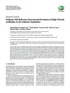
Total number of subjects evaluated 553
Pregnant Subjects: 403
Subjects Excluded: 317 [On the basis of thyroid dysfunction, goiter, autoimmunity & irregular follow-up]
Subjects included for the study: 86
Non Pregnant Subjects: 150
Subjects included for the study: 124
Subjects excluded: 26 [On the basis of thyroid dysfunction, goiter, and autoimmunity]
Trimester specific tests for Thyroid Functions were done and evaluation of results Flow Chart: Flow chart of the subjects enrolled for the study
102
Indian Journal of Endocrinology and Metabolism / Jan-Feb 2016 / Vol 20 | Issue 1
[Downloaded free from http://www.ijem.in on Wednesday, September 28, 2016, IP: 188.24.116.207]
Sekhri, et al.: Trimester‑specific thyroid profile of Indian pregnant women
serum thyroid peroxidase (TPO) antibody (TPO Ab >34 IU/ ml) were excluded on the basis of laboratory results. All the women were serially followed throughout pregnancy. The women who missed any of the visits during pregnancy were also excluded from the analysis. The Flow chart of subject enrollment in study is depicted below. The values of serum FT3, FT4, and thyroid stimulating hormone (TSH) from this pregnant women group (n = 86) and nonpregnant women (n = 124) who were considered as “normal subjects” were used to calculate thyroid function test reference intervals.
of iodine in urine was calculated as median because of skewed data distribution. Mean with standard deviation, median, and 2.5th and 97.5th percentiles (i.e., a central 95% interval) were calculated for serum FT3 and FT4 at each trimester in comparison with nonpregnancy. TSH did not follow a normal distribution and has to be normalized using log‑transformation and summarized as geometric mean (95% confidence interval). For data comparisons between pregnancy and nonpregnancy, and among three trimesters One‑Way ANOVA with further Bonferroni post‑hoc analysis was applied. A two‑tailed P