Accepted Article
DR. SZU-TAH
Article type
CHEN (Orcid ID : 0000-0003-2262-1775)
: Original Article
Original Article Title: Tryptophan as a Surrogate Prognostic Marker for Diabetic Nephropathy Authors: Chien-An Chou1¶#a, Chia-Ni Lin2,3¶, Daniel Tsun-Yee Chiu3,4,5*, I-Wen Chen1, Szu-Tah Chen1*#a 
Chien-An Chou and Chia-Ni Lin have contributed equally to this study.
Affiliations: 1. Division of Endocrinology and Metabolism, Department of Internal Medicine, Chang Gung Memorial Hospital, Chang Gung University, Taoyuan, Taiwan 2. Department of Laboratory Medicine, Chang Gung Memorial Hospital Linkou Branch, Chang Gung University, Taoyuan, Taiwan 3. Department of Medical Biotechnology and Laboratory Science, College of Medicine, Chang Gung University, Taoyuan City, Taiwan 4. Healthy Aging Research Center, Chang Gung University, Taoyuan City, Taiwan 5. Department of Pediatric Hematology, Chang Gung Memorial Hospital, Lin-Kou, Taiwan
#a
Current Address: Division of Endocrinology and Metabolism, Chang-Gung Memorial
Hospital, No.5, Fuxing St., Guishan Dist., Taoyuan City 333, Taiwan (R.O.C.)
This article has been accepted for publication and undergone full peer review but has not been through the copyediting, typesetting, pagination and proofreading process, which may lead to differences between this version and the Version of Record. Please cite this article as doi: 10.1111/jdi.12707 This article is protected by copyright. All rights reserved.
Accepted Article
*
Corresponding Author:
Szu-Tah Chen Division of Endocrinology and Metabolism, Chang-Gung Memorial Hospital, No.5, Fuxing St., Guishan Dist., Taoyuan City 333, Taiwan (R.O.C.) Tel: +886-3-3281200#8821 Fax: +886-3-3288257 E-mail address:
[email protected]
Short running title: Tryptophan and Diabetic Nephropathy
Abstract: Aims/Introduction: Diabetic nephropathy is one of the leading causes of end-stage renal disease. Unfortunately, reliable surrogate markers for predicting the prognostic outcome of diabetic nephropathy were as yet absent. In order to find new markers in predicting the progression of diabetic nephropathy, we conducted a prospective study by investigating the correlation between serum metabolites and the annual change of estimated glomerular filtration rate (eGFR).
Materials and Methods: From September 2013 to September 2015, 52 diabetic patients at various stages of chronic kidney disease (CKD) were enrolled. While serum levels of 175 metabolites were measured by AbsoluteIDQ™ p180 kit, only those with significant difference in advancing CKD stages were selected. After then, serial renal function change of these patients was followed up for
This article is protected by copyright. All rights reserved.
Accepted Article
12 months, outcome of renal function with each selected metabolite was compared according to the occurrence of a rapid decline (sustained annual decrement rate ≥5%) of eGFR.
Results: 26 metabolites were found to be significantly associated with the severity of CKD. Tryptophan (Trp) showed significant association with the event of rapid decline in eGFR (P=0.036). Serum concentration of Trp below 44.20 μM showed the most valuable predictive value with 55.6% sensitivity and 87% specificity.
Conclusions: Lower level of Trp, especially below 44.20μM was related to rapid decline in eGFR. Accordingly, Trp may be regarded as a potential prognostic marker for diabetic nephropathy.
Key words: Diabetic nephropathy, Prognostic Marker, Tryptophan,
Introduction: Diabetes mellitus (DM) has become a worldwide health issue with an ever-increasing incidence in the recent years owing to the highly developed socio-economic status and sedentary life styles1. Complications of DM, such as diabetic nephropathy (DN), could increase all-cause morbidities and mortalities even under intensive blood sugar control, and indeed, DN is one of the leading causes of end-stage renal disease (ESRD) globally2. As a long-term complication of diabetes, DN has been reported to occur in 5% to 40% of patients with type 1 or type 2 DM3,4. Once nephropathy progresses, approximately 20% to 40% of patients inevitably develop ESRD.
This article is protected by copyright. All rights reserved.
Accepted Article
DN might have plethoric clinical manifestations, such as albuminuria, elevated blood pressure, and eventually diminished renal function. It is a great challenge to predict the risk of disease progression among patients with DN. Currently, the estimated glomerular filtration rate (eGFR) is the most frequently used method to measure and categorize renal function, which is calculated based on the serum creatinine level, age, race, and sex. Accordingly, deterioration of renal function can be divided into five stages based on the change of eGFR. Nevertheless, limitation use of eGFR exists clinically; for example, renal hyperfiltration may mislead to extraordinary high eGFR in the course of DN progression5; moreover, the accuracy of eGFR in advanced kidney dysfunction has been questioned6; and most of all, the exact rate of renal function deterioration is seldom predictable by eGFR.
In addition to the alteration of eGFR, DN frequently leads to changes in many metabolites since that kidneys are not only characterized by high efficiency of excretory and absorptive functions, but also responsible for rapid protein synthesis, and amino acid oxidation7. The metabolism of biomolecules thus could be fluctuated even in the early stages of DN. Thanks to the rapid development of proteomic techniques, the metabolized peptides can be identified in tissues8 and various biological entities recently9. To our interest, metabolic approach shows significant relationship between DM and specific metabolites, which indicated changes in sugar, amino acid, and lipid metabolism10. Since metabolomics is the latest development in the omic technology providing the unbiased identification and quantification of small molecules in biological fluids, this approach can complement the proteomic assay to predict the outcome of DN with high sensitivity and specificity.
This article is protected by copyright. All rights reserved.
Accepted Article
In this study, we analyzed the plasma metabolites in patients at different stages of CKD to explore any association between the concentration of serum metabolites and degeneration of renal function. Our aim was to investigate the changes in metabolites at various stages of CKD, and most importantly, to use these metabolites as potentially predictive surrogate markers for DN.
Materials and Methods: Patients With informed consent approved by the Institutional Review Board of Chang Gung Memorial Hospital (IRB No. 104-6411C), we recruited participants at various stages of chronic kidney disease for monitoring renal function change of type 2 diabetes from September 2013 to September 2015. We included clinically stable patients over 18 years of age with regular follow up in this hospital. Patients with acute inflammatory diseases, ever admitted to a hospital in recent 2 months and/or during the follow up periods; and lost regular follow up were all excluded. The sample size required for predicting renal function change by metabolomics in this study was not estimated because the rarity of previous reports. Primary endpoint was the occurrence of rapid decline in renal function in 12 months with treatment adjusted according to the local guidelines. Finally, a total of 52 patients fulfilling the criteria and able to complete an at least one year follow-up were included (Table 1). For evaluation, renal function was estimated by the isotope dilution mass spectrometry traceable
This article is protected by copyright. All rights reserved.
Accepted Article
Modification of Diet in Renal Disease (MDRD) 4-variable equation11 [MDRD eGFR (mL/min/1.73 m2) =175 × Cr-1.154 × Age-0.203(×0.742 if female patient was being observed)]. The CKD staging was defined according to the National Kidney Foundation Kidney Disease Outcomes Quality Initiative (NFK/DOQI) guidelines12, that is, CKD stages 1, 2, 3, 4, and 5, were considered if eGFR values were > 90 mL/min/1.73 m2, 60-89 mL/min/1.73 m2, 30-59 mL/min/1.73 m2, 15-29 mL/min/1.73 m2, and < 15 mL/min/1.73 m2, respectively. Albuminuria was measured by spot urine albumin and creatinine ratio (UACR). All patients except two were subsequently followed up, and their laboratory data were obtained from our hospital for at least 12 months. All blood samples for metabolomic study were collected in the early morning after overnight fasting. Patients were separated to five groups according to their initial CKD stages. At the end of follow-up, the change of eGFR was expressed as percentage change after compared to the initial status. A rapid decline of eGFR was defined as a more than 5% annual decrement according to the previous reports13,14.
Metabolomic approach A targeted quantitative metabolomic approach was employed in combination with liquid chromatography–tandem mass spectrometry (LC-MS/MS) assay and direct flow injection assay (AbsoluteIDQTM180 kit from Biocrates Life Science, Austria) for the metabolomic analyses of the samples. This kit was designed to be used with triple-quad mass spectrometry and allows identification and quantification of more than 180 metabolites. The assay was performed by using Waters Acquity Xevo TQ-S (Waters Corp., MA, USA) instrument This article is protected by copyright. All rights reserved.
Accepted Article
according to manufacturer’s instructions. Briefly, the samples were thawed, vortex-mixed, and centrifuged at 13,000 g. In total, 10 µL of sample supernatant was loaded on a filter paper on top of the kit and dried under nitrogen flow. Further, 20 µL of 5% phenyl isothiocyanate was added for derivatization. After 20-min incubation, the filter spots were dried under nitrogen flow for 45 min. By using 300 µL of methanol containing 5 mM ammonium acetate as extraction solvent, the extracts were obtained in 96-well plate by centrifugation. Subsequently, the extracts were delivered to LC-MS/MS for analysis. In total, 175 known small-molecule metabolites were simultaneously quantified based on multiple reaction monitoring. The metabolomics dataset contained 40 acylcarnitine, 21 amino acids, 15 biogenic amines, 14 sphingomyelins, and 85 glycerophospholipids. Data were analyzed using a web-based server MetaboAnalyst (www.metaboanalyst.ca) to find variables that were correlated across the samples.
Statistical Analysis Continuous data were presented as mean ± SD. The association between initial CKD staging and metabolites was examined using the Spearman Rank correlation. All metabolites were corrected for initial CKD stage and plasma concentration by the Kruskal-Wallis one-way ANOVA. Serum concentration of each metabolite related initial stage of CKD was compared with the occurrence of rapid decline in eGFR by univariate binary logistic regression. Multivariate binary logistic regression was performed to clarify the interactions between each metabolite, and to adjust other possible confounding factors. The receiver operating characteristic (ROC) curve and Youden index were performed to find out the most predictive concentration of the metabolite. All data were analyzed by using the Statistical Package for Social Sciences (SPSS version 19; SPSS Inc., Chicago, IL, USA).
This article is protected by copyright. All rights reserved.
Accepted Article
Results: In total, 52 patients (26 men and 26 women) participated in this study (Table 1). All except two patients were followed-up for more than 12 months. Patients included in this study had a mean age of 56.4 ± 14.4 years old with mean duration of DM of 11.0 ± 8.5 years. Baseline eGFR was 73 ± 37 ml·min-1·1.73m-2 and UACR was 709.1±1603.4 mg/g. Average glycated hemoglobin (HbA1c) level was 7.7±1.6%. Approximately 73% (n=38) of the patients had hypertension and 31 of these patients have received angiotensin converting enzyme inhibitor (ACEI) or angiotensin II receptor blocker (ARB). Of the 52 patients, 11 experienced coronary artery disease, and 8 had cerebral vascular accident previously. Patients were separated into five groups according to initial CKD stages (18 in group 1, 15 in group 2, 12 in group 3, 3 in group 4 and 4 in group 5). In patients with advanced CKD stage, significantly older (P=0.001) and longer DM duration (P=0.036) were noticed. Elevated UACR (P=0.004) and decreased hemoglobin (Hb, P=0.002) showed significant association with CKD stage indicated the severity of DN. No obvious differences were found in body mass index (BMI), prevalence of hypertension, and the usage of ACEI or ARB among the five groups. After a 12-month follow-up, the average eGFR became 70±40 ml·min-1·1.73m-2, without significant difference as compared to the initial eGFR (P=0.055, Wilcoxon signed ranks test). Eventually, 52% (n=27) of the patients had rapid decline in eGFR (11/18 in group 1; 5/15 in group 2; 7/12 in group 3; 1/3 in group 4; and 3/4 in group 5). Out of the 175 metabolites that were analyzed, significant differences were observed in 26 metabolites based on the Kruskal-Wallis one-way ANOVA (Table 2). Among these metabolites, C14, C14:1-OH, C14:2-OH, C16-OH, C16:1, C16:1-OH, C16:2, C16:2-OH, C3-OH, C4, C4:1, C5-DC/C6:OH, C5:1, C7-DC, arginine (Arg), aspartate (Asp), citrulline (Cit), creatinine, kynurenine (Kyn), t4-OH-Pro, and symmetric dimethylarginine (SDMA) This article is protected by copyright. All rights reserved.
Accepted Article
levels increased with the progression of kidney dysfunction; whereas serine (Ser), tryptophan (Trp), tyrosine (Tyr), valine (Val), and PC aa C38:6 levels decreased with the progression of kidney dysfunction (Figs 1 and 2). Creatinine was excluded in further analysis because it is a known marker for renal function. After comparing the aforementioned metabolites with occurrence of rapid eGFR decline, Trp was the only metabolite showing significant difference (P = 0.036, Table 3) by multivariate analysis. Once again, Trp remained significantly associated with rapid eGFR decline after adjusted to confounding factors including gender, age, duration of diabetes, HbA1c, Hb, UACR, and usage of ACEI or ARB (Table 4). To detect the optimal cut point of Trp on predicting diabetic nephropathy progression, ROC curve analysis and Youden index were applied with a cutoff value of 42.20 μM revealing a sensitivity of 55.6% and a specificity of 87%, respectively (Fig 3).
Discussion: DN is one of the leading causes of ESRD globally2. More than what it seems to be, people with DN are at significant risk for ESRD, and the risk for cardiovascular morbidity and mortality has concomitantly increased. Although eGFR sensitively reflects the renal function at the time of the test, it seldom predicts the exact rate of DN deteriorations when the serum creatinine levels have risen already. As expected, a more effective method that can precisely predict and prevent the development and progression of DN at earlier stages might become a cornerstone of diabetic treatment. In this study, we found 26 metabolites significantly associated with eGFR change; among which, levels of 21 metabolites (C14, C14:1-OH, C14:2-OH, C16-OH, C16:1, C16:1-OH, C16:2, C16:2-OH, C3-OH, C4, C4:1, C5-DC/C6:OH, C5:1, C7-DC, Arg, Asp, Cit, Creatinine, Kyn, t4-OH-Pro, and SDMA) increased, and levels of 5 metabolites (Ser, Trp, Tyr, This article is protected by copyright. All rights reserved.
Accepted Article
Val, and PC aa C38:6) decreased in patients with DN. C14, C14:1-OH, C14:2-OH, C16-OH, C16:1, C16:1-OH, C16:2, C16:2-OH, C3-OH, C4, C4:1, C5-DC/C6:OH, C5:1, C7-DC are acylcarnitines; Arg, Asp, Cit, Ser, Trp, Tyr, and Val are amino acids; creatinine, Kyn, t4-OH-Pro, and SDMA are biogenic amines; whereas PC aa C38:6 are glycerophospholipids. Acylcarnitines are filtered through the kidney, and nearly 75% are excreted into urine; as a consequence, serum acylcarnitines increased in patient with CKD15 when the renal excretory function was progressively lost, and mainly short-chain acylcarnitine levels were significantly changed in advanced DN16. Pathophysiologically, DN is a result of complex interaction between metabolic, inflammatory, and hemodynamic change. Concomitantly, reactive oxygen species produced during series of the above reactions precipitate the development of DN17,18. It has been reported that several energy pathway-related metabolites including fatty acids, citrate cycle intermediates, glycerophospholipids, 3‐indoxyl sulfate, and Trp act as potential biomarkers for DN19. Other studies have additionally shown decreased concentration of the serum branched amino acids and Trp in experimental studies and in patients with advanced CKD7,20,21. In this study, the changes of amino acids, especially Trp (an essential non-polar aromatic amino acid that contains a side chain) showed a significant association with rapid decline in eGFR. Our study is the first study revealed a direct evidence of the prognostic value of Trp in DN. Whether a decreased Trp level simply reflects a deteriorating renal function or whether Trp itself contributes as an exacerbating and/or protecting factor remains controversial. For instance, a recent finding of significantly increased chlorination and oxidation of Trp residues in the noncollagenous hexamer (a key connection module of collagen IV networks) of diabetic patients suggests that Trp might be involved in renal oxidation stress attributing to unstable global or local hexamer structures and increased proteolytic degradation22. This article is protected by copyright. All rights reserved.
Accepted Article
Furthermore, indoxyl sulfate, a metabolite of Trp has been shown to play an important role in renal function regression23. After digestion, dietary Trp could be changed to indole by the microbiota in the colon, and further metabolized to indoxyl sulfate in the liver. Accumulation of indoxyl sulfate occurred in patients with CKD towing to decreased function of secretion24. Elevated concentration of indoxyl sulfate could lead to increased oxidative stress25, increased cellular senescence by activation of NF-kappa B26, and increased tubulointerstitial fibrosis by decreasing the expression of klotho, an anti-aging gene27. We hypothesize that disequilibrium in oxidative and anti-oxidative circumstances due to altered enzyme activities, Trp residual chlorination, and oxidation-induced structural instability of collagen hexamer, and the deposition of uremic toxins such as indoxyl sulfate are possible etiologic factors to precipitate renal function deterioration. However, several limitations exist in this study; first of all, serum concentration of the oxidative metabolites and indoxyl sulfate have not been evaluated in this study; thus, a direct evidence to support the association between Trp concentration, renal oxidative stress, and deterioration of renal function is lacking. Secondly, while studies indicated that function of kynurenine pathway reveals a potential value to early predict chronic transplant dysfunction in patient received renal allograft28, it did not show significant difference (P=0.589) in rapid decline in eGFR in our study. One possible mechanism is that the four Kyn metabolizing enzymes (kynurenine formamidase,
kynureninase,
3-hydroxyanthranilate
dioxygenase,
and
kynurenine
aminotransferase) might not be equally affected by renal function impairment29-31. Thirdly, as an essential amino acid, daily intake of Trp was not fully quantified in this examination. Variation might be observed if extremely high or low protein intake occurs. Fourthly, relative small sample size with majority in early stage of CKD was noticed in our study. All patients received only 12 months of following up. Prolonged following up period with expanded patient number would made our result more confident. Finally, although we acknowledged
This article is protected by copyright. All rights reserved.
Accepted Article
that increased Hb turnover may contribute to lower HbA1c in advanced CKD and mislead to clinical judgement; however, alternative method, such as glycoalbumin, was not available in this hospital. In conclusion, among the 175 metabolites screened in this study, 26 metabolites were affected by renal function change in type 2 diabetes. Lower level of Trp (below 44.2μM) may be regarded as a potential surrogate prognostic marker for diabetic nephropathy.
Acknowledgement: The authors wish to thank Prof. Ming-Shi Shiao (Chang-Gung University) for his kind support and valuable suggestions. The authors also thank the Ministry of Science and Technology of Taiwan (MOST103-2320-B-182-026-MY2), the Ministry of Education of Taiwan (EMRPD1E1651, EMRPD1F0261), and Chang-Gung Memorial Hospital (CMRPD1C0771, CMRPD1C0772, CMRPD1C0773, CMRPD3D0161, CMRPD3D0162) for the continuous financial support of this research.
Disclosure: The authors declare that they have no conflicts of interest.
References: 1.
Hu FB. Globalization of diabetes: the role of diet, lifestyle, and genes. Diabetes Care. 2011;34(6):1249-1257.
2.
Kurokawa K, Nangaku M, Saito A, Inagi R, Miyata T. Current issues and future
This article is protected by copyright. All rights reserved.
Accepted Article
perspectives of chronic renal failure. J Am Soc Nephrol. 2002;13 Suppl 1:S3-6. 3.
Ismail N, Becker B, Strzelczyk P, Ritz E. Renal disease and hypertension in non-insulin-dependent diabetes mellitus. Kidney Int. 1999;55(1):1-28.
4.
Parving HH, Hommel E, Mathiesen E, et al. Prevalence of microalbuminuria, arterial hypertension, retinopathy and neuropathy in patients with insulin dependent diabetes. Br Med J (Clin Res Ed). 1988;296(6616):156-160.
5.
Wolf G. Diabetic Nephropathy in Type 2 Diabetes Prevention and Patient Management. Journal of the American Society of Nephrology. 2003;14(5):1396-1405.
6.
Prigent A. Monitoring renal function and limitations of renal function tests. Semin Nucl Med. 2008;38(1):32-46.
7.
Garibotto G, Sofia A, Saffioti S, Bonanni A, Mannucci I, Verzola D. Amino acid and protein metabolism in the human kidney and in patients with chronic kidney disease. Clin Nutr. 2010;29(4):424-433.
8.
Deininger SO, Ebert MP, Futterer A, Gerhard M, Rocken C. MALDI imaging combined with hierarchical clustering as a new tool for the interpretation of complex human cancers. J Proteome Res. 2008;7(12):5230-5236.
9.
Rodriguez-Suarez E, Siwy J, Zurbig P, Mischak H. Urine as a source for clinical proteome analysis: From discovery to clinical application. Biochimica Et Biophysica Acta-Proteins and Proteomics. 2014;1844(5):884-898.
This article is protected by copyright. All rights reserved.
Accepted Article
10.
Floegel A, Stefan N, Yu Z, et al. Identification of serum metabolites associated with risk of type 2 diabetes using a targeted metabolomic approach. Diabetes. 2013;62(2):639-648.
11.
Levey AS, Coresh J, Greene T, et al. Expressing the Modification of Diet in Renal Disease Study equation for estimating glomerular filtration rate with standardized serum creatinine values. Clin Chem. 2007;53(4):766-772.
12.
Hogg RJ, Furth S, Lemley KV, et al. National Kidney Foundation's Kidney Disease Outcomes Quality Initiative clinical practice guidelines for chronic kidney disease in children and adolescents: Evaluation, classification, and stratification. Pediatrics. 2003;111(6):1416-1421.
13.
Clark WF, Macnab JJ, Sontrop JM, et al. Dipstick Proteinuria as a Screening Strategy to Identify Rapid Renal Decline. Journal of the American Society of Nephrology. 2011;22(9):1729-1736.
14.
Zoppini G, Targher G, Chonchol M, et al. Predictors of estimated GFR decline in patients with type 2 diabetes and preserved kidney function. Clin J Am Soc Nephrol. 2012;7(3):401-408.
15.
Fouque D, Holt S, Guebre-Kgziabher F, et al. Relationship between serum carnitine, acylcarnitines, and renal function in patients with chronic renal disease. Journal of Renal Nutrition. 2006;16(2):125-131.
This article is protected by copyright. All rights reserved.
Accepted Article
16.
Baynes JW, Thorpe SR. Role of oxidative stress in diabetic complications: a new perspective on an old paradigm. Diabetes. 1999;48(1):1-9.
17.
Brownlee M. Biochemistry and molecular cell biology of diabetic complications. Nature. 2001;414(6865):813-820.
18.
Sheetz MJ, King GL. Molecular understanding of hyperglycemia's adverse effects for diabetic complications. Jama-Journal of the American Medical Association. 2002;288(20):2579-2588.
19.
Wettersten HI, Weiss RH. Applications of metabolomics for kidney disease research: from biomarkers to therapeutic targets. Organogenesis. 2013;9(1):11-18.
20.
Rhee EP, Souza A, Farrell L, et al. Metabolite profiling identifies markers of uremia. J Am Soc Nephrol. 2010;21(6):1041-1051.
21.
Cano NJ, Fouque D, Leverve XM. Application of branched-chain amino acids in human pathological states: renal failure. J Nutr. 2006;136(1 Suppl):299S-307S.
22.
Brown KL, Darris C, Rose KL, et al. Hypohalous acids contribute to renal extracellular
matrix
damage
in
experimental
diabetes.
Diabetes.
2015;64(6):2242-2253. 23.
Ramezani A, Raj DS. The gut microbiome, kidney disease, and targeted interventions. J Am Soc Nephrol. 2014;25(4):657-670.
24.
Watanabe H, Miyamoto Y, Otagiri M, Maruyama T. Update on the pharmacokinetics
This article is protected by copyright. All rights reserved.
Accepted Article
and
redox
properties
of
protein-bound
uremic
toxins.
J
Pharm
Sci.
2011;100(9):3682-3695. 25.
Fujii H, Nakai K, Fukagawa M. Role of oxidative stress and indoxyl sulfate in progression of cardiovascular disease in chronic kidney disease. Ther Apher Dial. 2011;15(2):125-128.
26.
Motojima M, Hosokawa A, Yamato H, Muraki T, Yoshioka T. Uremic toxins of organic anions up-regulate PAI-1 expression by induction of NF-kappaB and free radical in proximal tubular cells. Kidney Int. 2003;63(5):1671-1680.
27.
Shimizu H, Bolati D, Adijiang A, et al. Indoxyl sulfate downregulates renal expression of Klotho through production of ROS and activation of nuclear factor-kB. Am J Nephrol. 2011;33(4):319-324.
28.
Vavrincova-Yaghi D, Seelen MA, Kema IP, et al. Early Posttransplant Tryptophan Metabolism Predicts Long-term Outcome of Human Kidney Transplantation. Transplantation. 2015;99(8):e97-104.
29.
Schefold JC, Zeden JP, Fotopoulou C, et al. Increased indoleamine 2,3-dioxygenase (IDO) activity and elevated serum levels of tryptophan catabolites in patients with chronic kidney disease: a possible link between chronic inflammation and uraemic symptoms. Nephrol Dial Transplant. 2009;24(6):1901-1908.
30.
Nakamura TS, H.; Ichihara, A. Insulin and glucagon as a new regulator system for
This article is protected by copyright. All rights reserved.
Accepted Article
tryptophan oxygenase activity demonstrated in primary cultured rat hepatocytes. Journal of Biological Chemistry. 1980;255(16):7533-7535. 31.
Saito K, Fujigaki S, Heyes MP, et al. Mechanism of increases in L-kynurenine and quinolinic
acid
in
renal
insufficiency.
Am
J
Physiol
Renal
Physiol.
2000;279(3):F565-572.
Figure Legends: Figure 1. Chronic kidney disease associated Acylcarnitines By Kruskal-Wallis one-way ANOVA, acylcarnitines detected by AbsoluteIDQ p180 kit showed significant differences in patients with DN at progressing CKD stages. The range of serum acylcarnitines were presented with box-and-whisker plot. Abbreviation: DN = diabetic nephropathy; CKD = chronic kidney disease
Figure 2. Correlations of serum aminoacids, biogenic amines, and glycerophospholipids with various stages of chronic kidney disease. Aminoacids, biogenic amines, and glycerophospholipids detected by AbsoluteIDQ p180 kit showing significant differences in patients with DN at progressing CKD stages. The range of serum aminoacids, biogenic amines, and glycerophospholipids were presented with box-and-whisker plot. Abbreviation: DN = diabetic nephropathy; CKD = chronic kidney disease; Arg = arginine; Asp = aspartate; Cit = citrulline; Ser = serine; Trp = tryptophan; Tyr = tyrosine; Val = valine; SDMA = symmetric dimethylarginine
This article is protected by copyright. All rights reserved.
Accepted Article
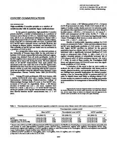
Figure 3. ROC curve analysis was performed by SPSS to determine the best discrimination point of serum tryptophan and rapid progression of diabetic nephropathy. The best discrimination point of serum tryptophan concentration determined by Youden index located at 44.20 μM with a sensitivity of 0.556 and a specificity of 0.870. Area under ROC curve was 0.682 with 95% confidence interval within 0.532 to 0.832; P value = 0.028; standard error = 0.077. Abbreviation: ROC = receiver operating characteristic; eGFR = estimated glomerular filtration rate
This article is protected by copyright. All rights reserved.
Accepted Article
Table 1. Demographic characteristics of 52 diabetic patients at various stages of chronic kidney disease. CKD 1 (n=18)
CKD 2
CKD 3
CKD 4
CKD 5
P
(n=15)
(n=12)
(n=3)
(n=4)
value
Gender( male),n(%)
5(27.78)
11(73.33)
8(66.67)
2(66.67)
0(0)
0.014*
Age(years)
46.3±10.7
56.5±11.8
66.0±15.9
71.0±10.1
61.5±3.7
0.001*
BMI(kg/m2)
28.3±5.3
28.0±7.5
27.1±5.2
28.1±2.3
27.8±3.0
0.825
Duration of DM(years)
7.6±4.3
9.0±7.3
16.5±12.9
15.0±4.6
14.3±4.2
0.036*
Hypertension,n(%)
11(61.11)
9(60.00)
11(91.67)
3(100.00)
4(100.00)
0.121
SBP(mmHg)
129.6±16.4
134.6±9.7
128.1±17.5
137.0±20.1
125.3±12.5
0.639
DBP(mmHg)
74.6±9.7
77.9±8.6
66.9±15.6
70.7±11.0
63.5±5.2
0.021*
0.62±0.11
1.00±0.16
1.50±0.19
2.42±0.73
4.31±0.80
0.000*
eGFR(ml·min ·1.73m )
115±19
73±8
44±8
25±5
11±2
0.000*
HbA1c(%)
7.9±1.8
8.0±1.8
7.8±1.5
6.5±0.4
6.6±0.5
0.249
UACR(mg/g)
185.5±588.3
735.1±2282.4
794.8±1380.2
1340.3±1972.7
2600.4±843.8
0.004*
Hb
13.9±1.7
13.8±1.6
11.7±1.9
12.2±0.4
9.8±1.4
0.002*
ACEI/ARB, n(%)
8(72.73)
8(88.89)
8(72.73)
3(100.00)
4(100.00)
0.138
β blocker, n(%)
2(18.18)
3(33.33)
6(54.55)
3(100.00)
3(75.00)
0.003*
CCB, n(%)
5(45.45)
3(33.33)
3(27.27)
2(66.67)
4(100.00)
0.022*
Diuretics, n(%)
1(9.09)
5(55.56)
5(45.45)
0(0.00)
2(50.00)
0.081
Cr(mg/dL) -1
-2
Antihypertensive agents
Values are presented as mean ± SD or n (%). SD = standard deviation The ANOVA was used for continuous variables; Chi-square test was used for categorical variables. * denotes P value < 0.05. Abbreviation: CKD = chronic kidney disease; BMI = body mass index; DM = diabetes mellitus; SBP = systolic blood pressure; DBP = diastolic blood pressure; Cr = creatinine; eGFR = estimated glomerular filtration rate; HbA1c = glycated hemoglobin; UACR = urine albumin-to-creatinine ratio; Hb = hemoglobin; ACEI = angiotensin converting enzyme inhibitor; ARB = angiotensin II receptor blocker; CCB = Calcium channel blocker.
This article is protected by copyright. All rights reserved.
Accepted Article
Table 2. Metabolites showed significant association with various stages of chronic kidney disease by Kruskal-Wallis one-way ANOVA. Metabolites
CKD 1
CKD 2
CKD 3
CKD 4
CKD 5
P valve
(n=18)
(n=15)
(n=12)
(n=3)
(n=4)
C14
not detected
not detected
0.003±0.011
0.024±0.023
not detected
0.000*
C14:1-OH
not detected
not detected
0.001±0.005
0.010±0.009
not detected
0.000*
C14:2-OH
not detected
not detected
0.001±0.002
0.006±0.005
not detected
0.000*
C16-OH
not detected
not detected
0.001±0.002
0.003±0.002
not detected
0.000*
C16:1
not detected
not detected
0.007±0.016
0.018±0.016
not detected
0.001*
C16:1-OH
not detected
not detected
0.001±0.003
0.007±0.006
not detected
0.000*
C16:2
not detected
not detected
0.001±0.06
0.011±0.010
not detected
0.000*
C16:2-OH
not detected
not detected
0.001±0.004
0.008±0.007
not detected
0.000*
C3-OH
not detected
not detected
0.001±0.003
0.082±0.132
not detected
0.000*
C4
0.159±0.095
0.300±0.286
0.424±0.213
0.271±0.072
0.730±0.370
0.000*
C4:1
not detected
0.004±0.017
0.001±0.003
0.008±0.008
0.017±0.034
0.008*
C5-DC/C6-OH
not detected
0.004±0.016
0.002±0.006
0.019±0.017
0.015±0.029
0.008*
C5:1
not detected
not detected
0.001±0.005
0.010±0.009
0.011±0.021
0.001*
C7-DC
not detected
0.010±0.022
0.010±0.026
0.077±0.054
0.034±0.068
0.001*
Arg
58.0±23.0
57.0±16.4
76.2±24.0
90.8±14.6
65.0±12.5
0.034*
Asp
2.59±1.47
2.70±1.18
3.37±1.99
4.33±1.72
9.63±7.83
0.031*
Cit
21.7±8.2
30.7±7.5
49.7±13.8
69.3±17.8
92.3±14.4
0.000*
Ser
125.6±25.5
100.4±21.4
101.0±17.3
82.1±25.8
81.7±14.9
0.003*
Trp
53.1±7.9
51.0±9.5
46.9±15.1
47.1±16.1
26.0±1.3
0.008*
Tyr
66.8±13.4
56.9±10.0
59.2±20.4
61.9±24.8
32.3±2.6
0.003*
Val
266.8±35.6
269.1±44.2
234.7±50.7
211.6±21.6
187.3±46.0
0.009*
Creatinine
37.6±10.1
65.0±23.0
101.3±29.9
164.9±74.3
341.1±105.4
0.000*
Kyn
2.37±0.73
3.24±0.75
3.78±1.36
3.50±0.52
5.80±3.52
0.001*
t4-OH-Pro
0.10±0.07
0.15±0.08
2.26±7.23
13.40±12.48
4.68±8.82
0.002*
SDMA
0.17±0.05
0.21±0.05
0.26±0.16
0.73±0.57
0.25±0.06
0.035*
106.4±46.3
98.6±24.9
98.4±26.9
69.7±8.4
63.3±10.4
0.028*
Acylcarnitines
Aminoacids
Biogenic amines
Glycerophospholipids PC aa C38:6
By Kruskal-Wallis one-way ANOVA, 26 metabolites showed significant association with CKD stage change; the mean concentration ± standard deviation of these metabolites were indicated in each stage of chronic kidney disease. *denotes P value < 0.05 Abbreviation: CKD = chronic kidney disease; Arg = arginine; Asp = aspartate; Cit = citrulline; Ser = serine; Trp = tryptophan; Tyr = tyrosine; Val = valine; Kyn = kynurenine; SDMA = symmetric dimethylarginine
This article is protected by copyright. All rights reserved.
Accepted Article
Table 3. Metabolite associated with rapid decline in estimated glomerular filtration rate by multivariate logistic regression. Univariate Odds ratio
Multivariate P value
(95% Confidence interval)
Odds ratio
P value
(95% Confidence interval)
Acylcarnitines C14
0.000
0.999
C14:1-OH
0.000
0.999
C14:2-OH
0.000
0.999
C16-OH
0.000
0.999
C16:1
0.000
0.427
C16:1-OH
0.000
0.999
C16:2
0.000
0.999
C16:2-OH
0.000
0.999
C3-OH
0.000
0.997
C4
50.978(1.110,2340.771)
0.044*
C4:1
0.000
0.690
C5-DC/C6-OH
602.559(0.000,6.289E21)
0.774
C5:1
0.004
0.893
C7-DC
0.000
0.210
Arg
1.004(0.980,1.030)
Asp
117.065(0.031,442783.228)
0.257
0.726
1.024(0.972,1.079)
0.373
1.017(0.846,1.224)
0.856
1.076(0.692,1.675)
0.744
Cit
1.008(0.983,1.033)
0.536
1.042(0.940,1.155)
0.432
Ser
0.981(0.959,1.004)
0.112
0.982(0.943,1.023)
0.381
Aminoacids
This article is protected by copyright. All rights reserved.
Accepted Article
Trp
0.945(0.898,0.995)
0.031*
0.864(0.751,0.991)
0.036*
Tyr
0.968(0.933,1.004)
0.081
1.050(0.957,1.151)
0.306
Val
1.000(0.989,1.013)
0.937
1.016(0.992,1.042)
0.198
Kyn
1.110(0.761,1.619)
0.589
0.504(0.175,1.457)
0.206
t4-OH-Pro
0.436(0.001,219.148)
0.794
0.000(0.000,11.274)
0.102
SDMA
0.164(0.005,5.412)
0.310
34738497.96(0.251,4.805E15)
0.069
1.010(0.992,1.028)
0.273
1.009(0.985,1.033)
0.481
1182.791(0.005,2.665E8)
0.261
Biogenic amines
Glycerophospholipid PC aa C38:6 Kyn/Trp
In order to evaluate the predictive value of the indicated metabolites in rapid progression of renal function, serum concentration of each metabolite was compared between diabetic patients with an eGFR annual decrease rate ≥5 % to those