In this paper, we will focus on the analysis of LV rotation from MR images with SPAtial ... therefore propose to extract the motion field by simultaneous analysis of the perpendicularly ... formance by virtue of exploiting the spatial and temporal scale degrees of free- dom (dofs) of ..... online: http://cbm2007.mech.uwa.edu.au.
Tuple Image Multi-scale Optical Flow for Detailed Cardiac Motion Extraction: Application to Left Ventricle Rotation Analysis Hans C. van Assen1 , Luc M.J. Florack1,2, Jos J.M. Westenberg3 , and Bart M. ter Haar Romeny1 1
Dept. of Biomedical Engineering, Eindhoven University of Technology, Netherlands, 2 Dept. of Mathematics, Eindhoven University of Technology, Netherlands, 3 Dept. of Radiology, Leiden University Medical Center, Netherlands. ⋆
Abstract. We present a new method for detailed tracking of cardiac motion based on MR-tagging imaging, multi-scale optical flow, and HARPlike image filtering. In earlier work, we showed that the results obtained with our method correlate very well with Phase Contrast MRI. In this paper we combine the intrinsic spatiotemporal evidence from perpendicularly encoded tagging sequences so as to obtain a dense, unambiguous motion field. We compare our results to those obtained using a different (provably incorrect) strategy advocated in the literature, in which horizontal and vertical components of the motion field are extracted separately from the motion pattern of vertically, respectively horizontally initialized tags. From the extracted motion fields, rotation of the LV is calculated for three subjects, in three slices using the generalised Stokes’ theorem.
1
Introduction
Volume overload, pressure overload, cardiomyopathy and coronary artery disease with postinfarction remodelling are common causes for heart failure. Following disease progress from LV dynamic behaviour is important for, e.g., early diagnosis [1]. In hypertensive rats, contractile state was depressed before deterioration of cardiac performance was observed and before the left ventricle (LV) dilated [1]. Contractile function may thus indicate abnormalities before LV geometry does. In this paper, we will focus on the analysis of LV rotation from MR images with SPAtial Modulation of Magnetization (SPAMM, a.k.a. tagging) [2]. Tagging provides sufficient information to analyse disturbances in twisting motion, which have been identified as early signs in pathologies [3], and which remain hidden in surface-based analysis techniques [4]. For tagging image analysis, we follow the rationale of Horn and Schunck [5] by application of the optical flow constraint equation (OFCE), which was brought to the multi-scale framework by Florack et al. [6]. Since the OFCE assumes constant brightness of a pixel along its path, ⋆
The Netherlands Organisation for Scientific Research (NWO) is gratefully acknowledged for financial support.
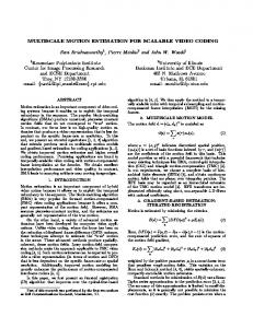
(a)
(b)
(c)
Fig. 1. (a) Vertical and (b) horizontal tag deformation between time frames. By analyzing horizontal and vertical tags independently and setting the velocity component perpendicular to the image encoding to zero (see (1)), the given vectors result. (c) Same patterns, now overlaid. The actual displacement is indicated with the arrow marked (u, v), which results from systems (2), (3). Clearly (u1 , v2 ) 6= (u, v).
and SPAMM data typically suffers from tag fading due to the spin-lattice relaxation (expressed by T1 ), our proposed method incorporates the Harmonic Phase (HARP) technique [7]. Thus, tag-phase information is used instead of brightness information. However, we take the sine of the tag-phase rather than the phase itself, to avoid spatial discontinuities in gray values. This paper is organized as follows. Section 2 briefly summarizes previous work. Section 3 explains our proposed method, Section 4 presents tracking and rotation extraction results. Finally, Section 5, discusses the method and results.
2
Background
In the late 1980s, Zerhouni et al. introduced “tagging” for visualising intramyocardial motion by MR imaging [8], later refined to SPAMM [2]. SPAMM patterns are inherent in the tissue, allowing analysis of the local dynamic behaviour of the LV, while imaging of the cardiac surfaces (e.g., with Cine MRI) focuses on wall thickening. SPAMM data typically suffers from tag fading due to the spin-lattice relaxation (with time-constant T1 ). However, due to velocity errors that occur with PCMRI mentioned above, and the inherent difficulties to transform PCMRI data to strain analysis, motion and deformation analysis with SPAMM imaging data is an active field of research. Suinesiaputra et al. [9] applied the multi-scale generalisation [6] of the OFCE to track human hearts. Their method suffers from the fact that flow components tangential to iso-surfaces cannot be retrieved from data evidence, which was formalised in a “normal flow constraint”. Dougherty et al. [10] also applied optical flow. They estimate global and local cardiac motion in a coarse-to-fine modelbased technique. This technique encompasses a Laplacian filter to compensate for intensity and contrast loss in myocardial tags. Prince and McVeigh [11] developed an optical flow based method which requires extensive prior knowledge
of the relaxation times T1 , T2 and the proton density D0 of the myocardium. The HARP technique, which employs tagging combined with spectral filtering in Fourier space, overcomes tag fading by directly measuring phase information of the MR signal [7]. Thus, the tracking algorithm uses the tag-phase information instead of tag-brightness information. For a review of MRI motion analysis protocols, the reader is referred to [12]. Our method extracts cardiac motion from image sequences with mutually perpendicular encodings. Some methods, however, find the flow vectors’ components by analysis of the perpendicularly encoded images separately, followed by vector addition of the resulting 1D displacement vectors. The displacement components are implicitly assumed to be zero perpendicular to the original encoding direction, and thus to be independent. Although the separate observations should be independent, the displacement components are not (see figure 1). We therefore propose to extract the motion field by simultaneous analysis of the perpendicularly encoded image sequences, with equal 2D motion in both.
3
Methods
The usual way to circumvent the aperture problem is to complement data evidence with prior knowledge, or by stipulating some smoothness hypothesis about the true motion field. However, there is no guarantee that a regularised solution is everywhere close to the physical motion field. If the physical motion field exhibits strong variations at some locations, these will not be retrieved correctly, as they are precluded a priori. We therefore aim for a regularisation free solution, but one that is not hampered by missing data evidence (aperture problem). Florack et al. [6] proposed a multi-scale generalisation of the classical OFCE [5], emphasizing the intrinsic aspects. By imposing conditions reflecting known facts about simulated object/scene dynamics they were able to obtain very good performance by virtue of exploiting the spatial and temporal scale degrees of freedom (dofs) of (Gaussian) derivative filters. Their method’s weakness is that it only improves the way of handling the intrinsic dofs of the OFCE by incorporating scale in a slick way, but it does not handle the aperture problem realistically. It would be desirable if the tangential flow could be retrieved by adding further intrinsic evidence to the existing evidence, obviating the need for regularisation altogether. This is possible if one is in possession of a second independent recording of the same spatiotemporal region of interest. This can be achieved with the help of suitably chosen MR tagging patterns. 3.1
Zeroth Order Polynomial Expansion of the OFCE
Following this new rationale we exploit the strength of the multi-scale OFCE by Florack et al. [6], while at the same time removing its shortcomings. The operational scheme for optical flow extraction makes use of a local polynomial expansion of the flow field (at each point).
Let f be shorthand for f (x, y, t; σ, τ ), the scalar spatiotemporal image sequence as a function of position (x, y), time t, isotropic spatial scale σ > 0, and temporal scale τ > 0. We denote its partial derivatives with respect to x, y, and t by self-explanatory subscripts. These are obtained by convolving the raw image sequence f 0 (x, y, t) = f (x, y, t; 0, 0) with a corresponding derivative of a normalised Gaussian, � 2 � 1 x + y2 1 t2 √ φ(x, y, t; σ, τ ) = exp − − 2 . 2πσ 2 2πτ 2 2σ 2 2τ It has been conjectured in the literature that horizontal and vertical components of the motion field can be retrieved separately from the vertically, respectively horizontally initialized tagging sequences. For a zeroth order polynomial expansion scheme this corresponds to the following system of equations: ( ( fx u1 + fy v1 + ft = 0 gx u2 + gy v2 + gt = 0 and (1) v1 =0 u2 =0 with f and g the perpendicularly encoded MR tagging image sequences. The assumption here is that the true motion field, (u∗ , v ∗ ) say, is given by superposition of the solutions, i.e. (u∗ , v ∗ ) = (u1 , 0) + (0, v2 ). Recall Fig. 1. However, this superposition based argument is incorrect, cf. Fig. 1. Instead of the above systems (1),we must consider the following single system for both components of the physical motion field (u, v) simultaneously. ( fx u+ fy v+ ft = 0 (2) gx u+ gy v+ gt = 0 It is evident from (1) and (2) (besides Fig. 1), that the solutions are indeed fundamentally different. 3.2
First Order Polynomial Expansion of the OFCE
We propose to use a 1st order polynomial expansion scheme, where U (x, y, t) = u + ux x + uy y + ut t respectively V (x, y, t) = v + vx x + vy y + vt t , in which u, ux , uy , ut , v, vx , vy , vt are eight local parameters of the horizontal, respectively vertical local optical flow field approximation U (x, y, t) and V (x, y, t).4 The relevant 1st order OFCE is then given by the following linear system (see [13, 14]). Collecting the unknowns (u, ux , uy , ut , v, vx , vy , vt ) in an 8-entry column vector v, and indicating the 8×8 coefficient matrix by A, and the inhomogeneous term by the 8-entry column vector a, we have Av = a, 4
(3)
The coordinates (x, y, t) are to be understood as local coordinates within the tangent space of a fixed base point in the image sequence. The global motion field, i.e. regarded as a function of this base point, is of course not a simple polynomial.
A= 2 fx 6 fxt 6 6fxx 6 6fxy 6 6 gx 6 6 gxt 6 4gxx gxy
3 fy fxt τ 2 fyt τ 2 fxx σ 2 fxy σ 2 fxy σ 2 fyy σ 2 fyt fx +fxtt τ 2 fy +fytt τ 2 fxxt σ 2 fxyt σ 2 fxyt σ 2 fyyt σ 2 7 7 fxy fxxt τ 2 fxyt τ 2 fx +fxxx σ 2 fy +fxxy σ 2 fxxy σ 2 fxyy σ 2 7 7 2 2 2 2 2 27 fyy fxyt τ fyyt τ fxxy σ fxyy σ fx +fxyy σ fy +fyyy σ 7 , gy gxt τ 2 gyt τ 2 gxx σ 2 gxy σ 2 gxy σ 2 gyy σ 2 7 7 2 2 2 2 2 2 7 gyt gx +gxtt τ gy +gyttτ gxxt σ gxyt σ gxyt σ gyyt σ 7 gxy gxxt τ 2 gxyt τ 2 gx +gxxx σ 2 gy +gxxy σ 2 gxxy σ 2 gxyy σ 2 5 gyy gxyt τ 2 gyyt τ 2 gxxy σ 2 gxyy σ 2 gx +gxyy σ 2 gy +gyyy σ 2
�T � v = u v ut vt ux vx uy vy
�T � . and a = − ft ftt fxt fyt gt gtt gxt gyt
If one would perform separate analysis of the perpendicularly encoded images (which we do not do), this yields two systems to be solved. These can be produced from (3) by replacing the last respectively the first four equations with v1 = 0 , v1,x = 0 , v1,y = 0 , v1,t = 0 ,
(4)
and u2 = 0 , u2,x = 0 , u2,y = 0 , u2,t = 0 ,
(5)
again assuming (incorrectly) the true motion field to be given by (u , v ) = (u1 , 0)+(0, v2 ). It has to be noted that this is not the same as imposing a “normal flow” constraint. Equations (4), (5) assume horizontal respectively vertical flow, while normal flow depends on image structure. ∗
3.3
∗
Calculation of Rotation From a Flow Field
We invoke the generalized Stokes’ theorem: If R is an oriented piecewise smooth n-dimensional manifold (in our case n = 2), with oriented boundary ∂R, and ω is a smooth (n − 1)-form on R, then Z I dω = ω. (6) R
∂R
Take ω = udx + vdy, with (u, v) the motion field, i.e. dω = (vx − uy ) dx ∧ dy (∧ being the wedge product). Take R to be a ring, i.e. the interior of two concentric circles ∂R = ∂Rint ∪ ∂Rext , the orientation of which is deduced from the outward normal of the region R (viz. leftward if you are inside R looking across its boundary). Stokes’ theorem then reduces to the so-called Green’s theorem: I Z udx + vdy . (7) (vx − uy ) dxdy = ∂R
R
For our disconnected boundary parts this yields I I Z (vx − uy ) dxdy = udx + vdy − R
∂Rext
udx + vdy .
(8)
∂Rint
The interpretation of this result is net rotation of the vector field (u, v) inside region R, or equivalently net circulation of the vector field along its boundary.
0.
0.
0.1
0.5
0.2
1.
0.4
1.5
0.8
2.
Fig. 2. Phases shown are {3, 5, 7, 9, 11} (systole), basal slice. Row 1,2: Vertical and horizontal tagging sequences. Row 3: 1st order multi-scale optical flow field from (3). Vectors are colour-coded for direction. Row 4: Results from separate analysis of rows 1 and 2 followed by vector addition from (3) modified with (4) and (5). Row 5, 6: Angle and relative norm differences between rows 3 and 4.
Normalised rotation
Normalised rotation
0.04
0.04
0.02
0.02
0.00
5
10
15
20
Frame
0.00
Normalised rotation 0.04 0.02
5
10
15
20
Frame
0.00
-0.02
-0.02
-0.02
-0.04
-0.04
-0.04
5
10
15
20
Frame
Fig. 3. Rotation plots for three volunteers in three slices (basal: solid, mid slice: dashed, apex: dotted). The first plot is taken from the same subject as presented in figure 2.
4
Experiments and Results
Short-axis MR tagging image data were acquired with a Philips Intera 1.5T scanner (Philips Medical Systems, Best, Netherlands), from 3 volunteers in a basal, a mid-ventricular and an apical slice. For the MR Tagging sequences, a 2D multi-shot gradient-echo with Echo Planar Imaging (EPI factor 9) with breath-holding in end-expiration was used. The following scan parameters were used: TE 4.4ms, TR 19ms, flip angle 10◦ , field-of-view: 300 mm, scan matrix 128, acquisition voxel size 2.34×2.68×8 mm3 reconstructed into 1.17×1.17×8 mm3 . Spacing between the taglines was 8 mm. The LV epi- and endocardial contours were manually indicated by fitting ellipsoids to the image data. The part of the flow field in between the contours (see Fig. 2 bottom row) was used for LV rotation analysis throughout systole, cf. next section for details. Additionally, to demonstrate the differences to our method, motion fields for one subject were calculated by (3) modified with (4), (5) followed by vector addition (see Fig. 2). Differences in vector direction and relative differences in vector L2 -norm are expressed in colour maps. Resulting rotation as a function of time (during systole) is shown in figure 3.
5
Discussion
We presented a multi-scale optical flow-based method for tracking the cardiac LV myocardium from MR tagging images. It is physically well-founded, and does not include any assumptions about the flow field or its representation like, e.g. normal flow, flow perpendicular to original encoding directions, and regularisation or smoothness constraints. This paper showed that the concept works in practice on real data, acquired with clinical protocols, yielding a dense motion field. Employing a multi-scale approach requires (automatic) scale-selection. In our method, automatic scale selection is performed by optimizing for the condition number of the coefficient matrix A in equation (3). The advantages of the 1st order system emerged while calculating LV rotation, requiring uy and vx . Both uy and vx are part of the 8-entry column vector v (see (3)). Thus, calculation of the heart rotation reduces to a normalised summation of (vx − uy ) over the contributing pixels between the contours.
Deformation and strain (another clinically relevant parameter) also involve first order parameters already present in v. Extension of our method for calculation of those parameters is a topic of ongoing research.
References 1. Mirsky, I., Pfeffer, J.M., Pfeffer, M.A., Braunwald, E.: The contractile state as the major determinant in the evolution of left ventricular dysfunction in the spontaneously hypertensive rat. Circ Res 53 (1983) 767–778 2. Axel, L., Dougherty, L.: MR Imaging of Motion with Spatial Modulation of Magnetization. Radiology 171(3) (1989) 841–845 3. Delhaas, T., Kotte, J., van der Toorn, A., Snoep, G., Prinzen, F.W., Arts, T.: Increase in Left Ventricular Torsion-to-Shortening Ratio in Children With Valvular Aorta Stenosis. Magn Reson Med 51 (2004) 135–139 4. Gotte, M.J., van Rossum, A.C., Twisk, J.W.R., et al.: Quantification of regional contractile function after infarction: strain analysis superior to wall thickening analysis in discriminating infarct from remote myocardium. J Am Coll Cardiol 37 (2001) 808–817 5. Horn, B.K.P., Schunk, B.G.: Determining Optical Flow. Artif Intell 17 (1981) 185–203 6. Florack, L., Niessen, W., Nielsen, M.: The Intrinsic Structure of Optic Flow Incorporating Measurement Duality. Int J Comput Vision 27(3) (1998) 263–286 7. Osman, N.F., Kerwin, W.S., McVeigh, E.R., Prince, J.L.: Cardiac Motion Tracking Using CINE Harmonic Phase (HARP) Magnetic Resonance Imaging. Magn Reson Med 42(6) (1999) 1048–1060 8. Zerhouni, E.A., Parish, D.M., Rogers, W.J., Yang, A., Shapiro, E.P.: Human heart: tagging with MR imaging – a method for noninvasive assessment of myocardial motion. Radiology 169(1) (1988) 59–63 9. Suinesiaputra, A., Florack, L.M.J., Westenberg, J.J.M., ter Haar Romeny, B.M., Reiber, J.H.C., Lelieveldt, B.P.F.: Optic flow computation from cardiac MR tagging using a multiscale differential method. In Ellis, R., Peters, T., eds.: Proc MICCAI. Volume 2878 of Lect Notes Comput Sc., Berlin, Springer Verlag (2003) 483–490 10. Dougherty, L., Asmuth, J.C., Blom, A.S., Axel, L., Kumar, R.: Validation of an Optical Flow Method for Tag Displacement Estimation. IEEE T Med Imaging 18(4) (1999) 359–363 11. Prince, J.L., McVeigh, E.R.: Motion estimation from tagged MR image sequences. IEEE T Med Imaging 11(2) (1992) 238–249 12. Axel, L., Montillo, A., Kim, D.: Tagged magnetic resonance imaging of the heart: a survey. Med Image Anal 9(4) (2005) 376–393 13. Florack, L.M.J., van Assen, H.C., Suinesiaputra, A.: Dense Multiscale Motion Extraction from Cardiac Cine MR Tagging using HARP Technology. In Niessen, W.J., Nielsen, M., eds.: Proc. MMBIA. Proc. 11th IEEE ICCV. (2007) 14. van Assen, H.C., Florack, L.M.J., Suinesiaputra, A., Westenberg, J.J.M., ter Haar Romeny, B.M.: Purely Evidence Based Multiscale Cardiac Tracking Using Optic Flow. In Miller, K., Paulsen, K.D., Young, A.A., Nielsen, P.M.F., eds.: Proc. MICCAI 2007 workshop Comput Biomech Med II. (2007) 84–93 Available online: http://cbm2007.mech.uwa.edu.au.