Dec 6, 2007 - case had papulopustular and erythematous cutaneous lesions but first were evaluated as exhibiting a drug reaction. Knowl- edge of clinical ...
Jpn. J. Infect. Dis., 61, 130-132, 2008
Short Communication
Two Possible Cases of Trichosporon Infections in Bone-Marrow-Transplanted Children: the First Case of T. japonicum Isolated from Clinical Specimens Handan A˘gırbas¸lı*, Hulya Bilgen1, Sema Keceli Özcan2, Baris Otlu3, Gulce Sinik4, Nilgün C¸erikc¸io˘glu4, Riza Durmaz3, Emine Can, Nevin Yalman, Gunduz Gediko˘glu and Takashi Sugita5 Our-Children Leukemia Foundation; 1Istanbul University Cerrahpas¸a Medical Faculty Blood Center; 4 Marmara University Medical Faculty, Department of Microbiology, Istanbul; 2Kocaeli University Medical Faculty, Department of Microbiology, Kocaeli; 3Inonu University Medical Faculty, Department of Microbiology, Malatya, Turkey; and 5Department of Microbiology, Meiji Pharmaceutical University, Tokyo, Japan (Received May 16, 2007. Accepted December 6, 2007) SUMMARY: Trichosporon spp. are emerging as opportunistic agents that cause systemic diseases in immunocompromised hosts. Trichosporonosis carries a poor prognosis in neutropenic patients. Trichosporon japonicum was isolated from the air and named by Sugita et al. Here we present the first case of T. japonicum isolated from a clinical specimen. Two cases of acute myeloid leukemia who had Trichosporon isolates are discussed because of their rarity and growing importance. T. asahii was isolated from the throat, feces and urine of the first patient. T. japonicum was isolated from the sputum of the second patient. Both cases produced high MICs to itraconazole, and low MICs to fluconazole and voriconazole. In virulance factor investigations there was (++) biofilm formation in T. japonicum but not in T. asahii. Conventional mycological studies were not adequate for the identification of the isolate at the species level. In our second case as in the first one, the isolate was identified as T. asahii with 99.9% accuracy by API 20C AUX. Although two T. asahii isolates from the same patient yielded identical typing profiles by arbitrary primed-PCR, the isolates of the two different patients showed different arbitrary primedPCR typing profiles. However, the genetic identification of the other patient’s strain gave the result of T. japonicum. Case 1. A 22-month-old boy with acute myeloid leukemia (AML) was admitted for autologous bone marrow transplantation (BMT) in July 2000. The patient was given fluconazole (FLU) prophylaxis according to routine protocol. His absolute neutrophil count (ANC) was below 500/mm3 until day +53 and he had several febrile neutropenic attacks. Due to fever-of-unknown-origin treatment protocol in neutropenic patients, he was given multiple antibiotics and liposomal amphotericin B (AMB). His hemocultures were all sterile. His physical examination revealed numerous squamous and hyperkeratotic lesions. He had icterus and diarrhea. Yeast was isolated in his throat, urine and stool cultures, and identified as Trichosporon asahii. Urine cultures yielded yeast colonies (900 cfu/ml) that lasted 4 days. This time he developed hematuria and proteinuria. Cultures of the catheter exit site and sputum were negative. One of four blood cultures at different times yielded methicillin-resistant coagulase-negative Staphylococci (MRCNS) and the others were negative. Itraconazole (ITRA) was included in the therapy. Fortunately, the patient’s ANC increased to >1,000/mm3 at the 2nd day after identification of T. asahii, and his fever resolved after combination therapy with liposomal AMB (4 mg/kg) + ITRA (100 mg/d ) + FLU (400 mg/d). But he had high C-reactive protein (CRP) values, ranging between 38.3 to 228.2 mg/l, during his follow-up. The combination therapy was continued and he was discharged from the BMT unit at day +103. He continued to recieve liposomal AMB and ITRA for 6 more
months in the outpatient clinic and he recovered. At +11 month he experienced a relapse. He succumbed to death in deep neutropenia and relapse 12 months after BMT without T. asahii isolation. Case 2. An 8-year-old girl with AML was admitted for allogenic BMT in January, 2000. She was discharged from the BMT unit without any problem. She developed central nervous system relapse followed by bone marrow relapse and was hospitalized. Idarubicin (IDA) treatment regimen was applied (day +210 post BMT). When she was in deep neutropenia (ANC, 200/mm3), she developed a high fever with CRP of 200 mg/l. She was given multiple antibiotics and liposomal AMB (5 mg/kg). No improvement was observed. On her physical examination, she had secretory ralles and was under respiratory distress. Liposomal AMB treatment (5 mg/kg) was started; later, the dose was increased. Sputum cultures taken many times during 11 days yielded as follows: Aspergillus fumigatus/yeast (unidentified) + Pseudomonas spp./yeast/yeast + A. fumigatus/Trichosporon japonicum (identified after death) + MRCNS (122 days after BMT)/ Candida famata + Candida tropicalis + MRCNS. Physical examination of the pulmonary system revealed harsh bronchial sounds and crepitant rales. She had a fever that lasted for a week. ITRA (100 mg/d) was added to the treatment. The stool culture yielded C. tropicalis, and the throat culture yielded C. famata. Later, the general condition of the patient deteriorated. On physical exam, she had secretory rales and was experiencing respiratory distress. Then, wheezy breathing started and continous respiratory support with oxygen was applied. After she intubated, she was sent to the intensive care unit. Two days later, she died, 8 months after BMT.
*Corresponding author: Mailing address: Zeytinoglu Cad. Selcuklar Sok. 25/5 Akatlar, Besiktas, Istanbul, Turkey. Tel: +90-212-6311310, -631-1311, Fax: +90-212-631-1312, E-mail: agirbaslihandan @hotmail.com 130
tration of glucose was inoculated with the test strain and incubated at 35°C for 48 h. After washing and staining with 1% safranin the intensity of the film layer formed inside the plastic tubes was evaluated as negative, weakly positive (+), moderately positive (++) or strongly positive (+++) activity. ATCC 35984 Staphylococcus aureus strain was used as positive control (8-10). Isolate from Case 1 (stool): phospholipase (–), acid proteinase (–), esterase (+), biofilm (–). Isolate from Case 2 (sputum): phospholipase (–), acid proteinase (–), esterase (+), biofilm (++). Molecular typing of the isolates was performed by arbitrary primed (AP)-PCR with M13 primer. For AP-PCR analysis, genomic DNA was extracted using a QIAamp DNA mini kit (Qiagen, Valencia, Calif., USA). The primer used in the PCR reaction was the core sequence of phage M13 (5´-GAGG GTGGCGGTTCT-3´). PCR amplification was performed in 50 μl master mix containing approximately 100 ng of DNA as template, 1× amplification buffer, 0.4 mM of dNTP mix, 4 mM MgCl2 and 2.5 U Taq DNA polymerase (11). The reaction mixture was amplified with a Thermal Cycler (Corbett Research Palm-Cycler; Corbett Life Science, Sydney, Australia) according to the following conditions: 2 cycles, each consisting of 5 min at 94°C, 5 min at 40°C, and 5 min at 72°C; then 40 cycles, each consisting of 1 min at 94°C, 1 min at 40°C, and 2 min at 72ºC. Amplification products were electrophoresed by 2% agarose gel; after staining they were visualised and photographed under UV illumination. AP-PCR typing showed that the T. asahii isolates from the same patient were identical (lanes 1 and 2, Figure 1); however, the isolates from the two patients were different (lanes 1 and 2 versus lane 3, Figure 1), clonally unrelated (Figure 1). A yeast strain isolated from the air in Japan was found to represent a new species, and was named T. japonicum (1998) (12). Summer-type hypersensitivity pneumonitis (SHP) is a type 3 or 4 allergy developed by repeated inhalation of arthroconidia of Trichosporon spp. One of these Trichosporon spp. was T. japonicum (13,14). We have isolated T. japonicum from the sputum of a patient, as it is isolated from the air of SHP patients’ houses. Further analysis of the yeast isolate could be performed after the patients had died. In order to investigate retrospectively the epidemiological relationship of T. asahii strains in two different patients at the same dates, we did a molecular typing study on these strains, which had been stored in a freezer since 2000. The yeast isolates were sent to Japan for the genetical confirmation of identification. The molecular typing results revealed that these three Trichosporon strains were two different species but with only the genetic study the identification was made as T. japonicum in 2006. Our case is the first isolation of T. japonicum from a clinical specimen (2000). In the first case excellent T. asahii identification profiles were obtained by API 20C AUX, as in several other studies. In our second case as in the first case, the isolate was identified as T. asahii with 99.9% accuracy by API 20C AUX; however, the genetic identification of this strain gave a different result, T. japonicum. At first studies Trichosporon asteroides, which was the cause of the described blood-stream infections, could not be correctly identified using ID32 C strips. The recent studies of fungal 26S rDNA and ITS region sequence data facilitated the earlier and more reliable identification than phenotypic methods. Unfortunately, these sophisticated molecular methods are performed only in reference centers. Some identifica-
The fungi were identified based on morphology and using API 20C AUX (bioMérieux, Marcy l’Etoile, France). In both cases excellent T. asahii identification profiles were obtained. The intergenic spacer region (IGS) of the rRNA gene was sequenced directly from the polymerase chain reaction (PCR) products with the primer pairs 26SF (ATCCTTTGCAGAC GACTTGA) and 5SR (AGCTTGACTTCGCAGATCGG). The PCR products were sequenced with an ABI 310 DNA sequence and the BigDye Terminator Cycle Sequencing Ready kit (Perkin-Elmer Applied Biosystems, Foster City, Calif., USA) according to the manufacturer’s instructions. The sequence data were analyzed with the National Center for Biotechnology Information (Bethesda, Md., USA) BLAST system (http://www.ncbi.nlm.nih.gov/BLAST/). Ribosomal DNA sequence analysis of throat and stool isolates of Case 1 showed identical sequences. Their sequences matched the IGS of T. asahii (GenBank accession no. AB066396). The IGS sequences of Case 2 sputum isolate corresponded to that of T. japonicum (GenBank accession no. AB066426). The antifungal susceptibilities were determined with the E test system (AB Biodisk, Solna, Sweden). According to the results of reference laboratories, MIC breakpoints for voriconazole (VOR) are >4 μg/ml for resistance and 32, 0.75 and 0.047 μg/ml, respectively; for Case 2, MIC values were 0.032, 1.0, 0.75 and 0.032 μg/ml, respectively. Both cases produced high MICs to ITRA; the T. ashaii isolate had a higher MIC of AMB than T. japonicum. Both Trichosporon isolates produced low MICs to FLU and VOR. The phospholipase test was performed on egg yolk containing agar medium (pH 4.3). The test strain was inoculated onto medium by means of a calibrated loop (0.01 ml) and incubated at 37°C for 4 days. At the end of this period, the precipitation zone formed around the colony was measured and the strength of the reaction was detected by calculating the ratio between colony diameter and the diameter of the precipitation zone including the colony size. A value of 1 showed a negative reaction while values smaller than this indicated +1, +2, +3 or +4, according to the millimetric measurements. Candida albicans sc 5314 was used as the positive control (4,5). For secretory acid proteinase asssay, agar medium with 1% bovine serum albumin (pH 5.0) was used. From each test strain grown in yeast extract-peptone-dextrose broth with a turbidity of 0.5 McFarland, a 10-μl sample was inoculated onto a paper disk placed on the agar medium and incubated at 30°C for 6 days. An opaque precipitation was formed around the disk and was enhanced by ongoing growth. On the 3rd or 4th days the lysis zone around the disk (1 mm, +; 3 - 5 mm, ++; no lysis, –) was measured to determine the proteolytic activity of the strain. The CBS 2730 C. albicans strain was used as a positive control (4,6). The esterase test was performed on Tween 80-containing agar medium (pH 6.8). Each strain inoculated onto the medium in a ring form was examined for the formation of opaque precipitates of opaque crystal resulting from the hydrolysis of Tween 80 and binding of fatty acids with calcium (7). For slime test, Sabouraud broth with an 8% final concen131
saving. Diagnosis relies on clinical suspicion with microbiological confirmation (17). In our cases although we could not isolate the strains from the blood culture, this may have been due to the difficulty of isolation. These findings do not directly correlate with the Trichosporon spp. infections in our cases. In conclusion, difficulties in the identification of Trichosporon spp. cause the delay of a more radical treatment and increase the mortality rate. Indeed, the mortality rate in these patients is estimated to be approximately 78% despite AMB therapy (19). In order to prevent such undesirable results, it would be more proper to insist on taking samples from suspected febrile neutropenic patients. However, clinical suspicion is essential for earlier usage of antifungal therapy to improve the outcome. Clinicians should be aware that Trichosporon fungemia may develop in patients under antifungal therapy.
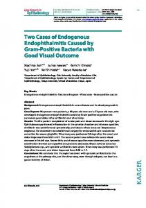
Fig. 1. (A) Agarose gel electrophoresis of the amplified products in AP-PCR with M13 primers. Lane1, Case 1 throat; lane 2, Case 1 stool; lane 3, Case 2 sputum isolates. Lane M is 100-bp DNA ladder. (B) A dendrogram of the DNA fingerprinting patterns of two T. asahii isolates (no. 1 and 2) and one T. japonicum isolate (no. 3).
REFERENCES
tion system such as ID32 C and API Candida kits have been commonly used to identify pathogenic yeasts in clinical microbiology laboratories. Moreover, T. asteroides has not been included in the ID32 C system or other systems. Therefore, it seems impossible to identify clinical isolates of T. asteroides to the species level by their biochemical characteristics used in these systems (15). T. japonicum is also not included in the identification list of commercial kits. For this reason, in order to submit data for epidemiological studies, further genetic studies should be done for isolated Trichosporon spp. This should also include the reevaluation of past isolated Trichosporon spp. If we continue, we think that it will be possible to find more T. japonicum isolates. Careful examination is required in diagnosing Trichosporon infection in immunocompromised hosts, because the infection can be invasive or unusual in appearance (16). Our first case had papulopustular and erythematous cutaneous lesions but first were evaluated as exhibiting a drug reaction. Knowledge of clinical manifestation of cutaneous infections with Trichosporon spp. should lead to earlier diagnosis and institution of appropriate treatment. The two cases reported here were profoundly immunosuppressed and neutropenic at the time of isolation. Response to antifungal agents is poor, mortality is high, and immunological recovery is the most important factor for a favorable outcome in patients with trichosporonosis (17). We believe that in the first patient the immune reconstitution was the major determinant of possible trichosporonosis. Few cases with normal neutrophil counts who recovered from infection without antifungal therapy have been reported in the literature (18). Despite the poor clinical condition of the second patient, the exact contribution of T. japonicum to the patient’s death is difficult to estimate. However, it would be very naive to assume that these pathogenic Trichosporon strains were not the causative pathogens in these patients. In the two patients combination therapy was used because of the severtiy of the neutropenia and high CRP, which was thought to be a good indicator of infection severity in our patients. Trichosporon spp. may colonize and proliferate in different parts of the human body such as the oral cavity, gastrointestinal system, urinary tract or skin. Therefore, the presence of Trichosporon spp. in the sputum, feces and urine specimens does not indicate true infection (15). But in immunocompromised patients it may be life-threatening with a poor prognosis. Early effective treatment may be life-
1. Pfaller, M.A., Diekema, D.J., Rex, J.H., et al. (2006): Correlation of MIC with outcome for Candida species tested against voriconazole: analysis and proposal for interpretive breakpoints. J. Clin. Microbiol., 44, 819-826. 2. Clinical Laboratory Standarts Institute (2002): Reference method for broth dilution antifungal susceptibility testing of yeast. Approved standart. Document M27 A2. 2nd ed. Clinical Laboratory Standarts Institute, Wayne, Pa., USA. 3. Wolf, D.G., Falk, R., Hacham, M., et al. (2001): Multidrug-resistant Trichosporon asahii infection of nongranulocytopenic patients in three intensive care units. J. Clin. Microbiol., 39, 4420-4425. 4. Dagdeviren, M., Cerikcioglu, N. and Karavus, M. (2003): Virulence factors of Candida parapsilosis strains isolated from hospitalized fungemic patients. J. Turkish Microbiol. Soc., 33, . 5. Lane, T. and Garcia, J.R. (1991): Phospholipase production in morphological variants of Candida albicans. Mycoses, 34, 217-220. 6. Cerikcioglu, N. and Alacam, R. (1993): Kandida suslarında salgisal asit proteinaz enzim varlıginin proteinli agar besiyerinde gosterilmesi. Mikrobiol. Bult., 27, 344-351. 7. Rudek, W. (1978): Esterase activity in Candida species. J. Clin. Microbiol., 8, 756-759. 8. Branchini, M.L., Pfaller, M.A., Rhine-Chalberg, J., et al. (1994): Genotypic variation and slime production among blood and catheter isolates of Candida parapsilosis. J. Clin. Microbiol., 32, 452-456. 9. Dolapci, I˙. and Tekeli, A. (2002): Cesitli Candida turlerinde slime faktoru yapiminin arastirilmasi. Mikrobiol. Bult., 36, 323-328. 10. Yucesoy, M. and Karaman, M. (2004): Candida turlerinin biyofilm uretimi ve antifungal duyarlılık paternleri. Mikrobiol. Bult., 38, 91-98. 11. Ayan, M., Durmaz, R., Aktas, E., et al. (2003): Bacteriological, clinical and epidemiological characteristics of hospital-acquired Acinetobacter baumannii infection in a teaching hospital. J. Hosp. Infect., 54, 39-45. 12. Sugita, T. and Nakase, T. (l998): Trichosporon japonicum sp. nov. isolated from the air. Int. J. Syst. Bacteriol., 48, 1425-1429. 13. Sugita, T., Nakajima, M., Ikeda, R., et al. (2002): Sequence analysis of the ribosomal DNA intergenic spacer 1 regions of Thichosporon species. J. Clin. Microbiol., 40, 1826-1830. 14. Sugita, T., Ikeda, R. and Nishikawa, A. (2004): Analysis of Trichosporon isolates obtained from the houses of patients with summer-type hypersensitivity pneumonitis. J. Clin. Microbiol., 42, 5467-5471. 15. Kustimur, S., Kalkanci, A., Caglar, K., et al. (2002): Nosocomial fungemia due to Trichosporon asteroides: firstly described bloodstream infection. Diagn. Microbiol. Infect. Dis., 43,167-170. 16. Erer, M., Galimberti, G., Lucarelli, C., et al. (2005): Trichosporon beigelii: a life thereating pathogen in immunocompromised hosts. Bone Marrow Transplant., 25, 745-749. 17. Nakagawa, T., Nakashima, K., Takaiwa, T., et al. (2000): Trichosporon cutaneum (Trichosporon asahii) infection mimicking hand eczema in a patient with leukemia. J. Am. Acad. Dermatol., 42, 929-931. 18. Hoy, J., Hsu, K.C., Rolston, K., et al. (1986): Trichosporon beigelii infection: a review. Rev. Infect. Dis., 8, 959-967. 19. Fournier, S., Pavageau, W., Feuillhade, M., et al. (2002): Use of voriconazole to successfully treat disseminated Trichosporon asahii infection in a patient with acute myeloid leukaemia. Eur. J. Clin. Microbiol. Infect. Dis., 21, 892-896.
132