CASE REPORT
European Journal of Cardio-Thoracic Surgery 41 (2012) e62–e64 doi:10.1093/ejcts/ezr332 Advance Access publication 9 February 2012
Two separate thoroscopic segmentectomies with vessel sealing system† Atsushi Watanabe*, Masahiro Miyajima, Nobuyoshi Kawaharada and Tetsuya Higami Department of Thoracic and Cardiovascular Surgery, Sapporo Medical University School of Medicine, Sapporo, Hokkaido, Japan * Corresponding author. Department of Thoracic and Cardiovascular Surgery, Sapporo Medical University School of Medicine, South 1, West 16, Chuo-ku, Sapporo 060-8543, Japan. Tel: +81-11-611-2111 (ext. 3312); fax: 81-11-613-7318; e-mail:
[email protected] (A. Watanabe). Received 2 September 2011; received in revised form 24 November 2011; accepted 7 December 2011
Abstract Total thoracoscopic segmentectomy is an appealing concept in terms of providing a parenchyma-sparing treatment. We describe our technique of two seperate total thoracoscopic segmentectomies by using a vessel sealing system (VSS). A 76-year old female with four gradually enlarging ground glass lesions on the right segment 2 (10 and 3 mm) and segment 6 (8 and 3 mm) was admitted to our institute for surgical diagnosis and treatment. Preoperative three-dimensional computed tomography showed that the A2 is composed of descending A2 and ascending A2, branch of V6 coursed to V2 and B1a originated from B2 and B1b from B3. Two separate segmentectomies were subsequently scheduled. Pulmonary vessel division was performed with VSS after proximal ligation. Intersegmental division by VSS and electrocautery was performed with the use of inflation–deflation demarcation line and the pulmonary veins along the intersegmental plane (V2a and V2c for segment 2 and V6b and V6c for segment 6) as guides to confirm the intersegmental plane after pulmonary artery and bronchial divisions. The intraoperative frozen-section examinations revealed adenocarcinoma in situ. Two separate segmentectomies were successfully completed, with a total operative time of 240 min and blood loss of 30 ml. VSS is a very useful and safe device for intersegmental division and pulmonary vessel division. Keywords: Early lung cancer • Thoracoscopic segmentectomy • Vessel sealing system
INTRODUCTION Recently, the vessel sealing system (VSS; LigaSure™ V, Covidien, Mansfield, USA) is gradually being used by surgeons in different fields in order to perform vessel division [1, 2]. Additionally, the application of three-dimensional computed tomography (3D-CT) is gaining popularity in the simulation of the approach method and detection of the course of the vessels. Most recently, thoracoscopic segmentectomy of the lung has been gradually adapted for early non-small-cell lung cancer [3]. In segmentectomy, precise detection of the pulmonary vessels and identification and division of intersegmental plane are very essential issues. Therefore, preoperative 3D-CT enables easy and precise detection of the pulmonary vessels, and VSS provides easy vessel division and safe intersegmental division.
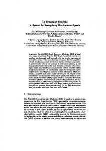
CASE A 76-year old female with four gradually enlarging ground glass lesions on the right segment 2 (10 and 3 mm) and segment 6 (8 and 3 mm) was admitted to our institute for histological diagnosis and treatment. Preoperative 3D-CT showed the following: A2 was composed of descending A2 and ascending A2, small branch of V6 coursed to V2 and B1a originated from B2 and B1b from B3 † Presented at the 25th Annual Meeting of the European Association for Cardio-Thoracic Surgery, Lisbon, Portugal, 1–5 October 2011.
(Fig. 1). Two separate segmentectomies were subsequently scheduled. Two trocar and one 30 mm window incisions were made. A 30° thoracoscope was inserted through the sixth intercostal port at the anterior axillary line. Pulmonary vessel division was performed with VSS (after proximal suture ligation).
S2 segmentectomy First, interlobar dissection between the right upper lobe and lower lobe was performed. After that, the arterial branches were dissected (ascending and descending A2) and node sampling was performed. Secondly, to make the inflation and deflation demarcation line, the segmental bronchus (B2) was ligated at the level just distal to the origin of B1a after inflation of the right lung followed by single ventilation of the left lung. The bronchus was divided with an endoscopic stapler. Outer intersegmental division by electrocautery was performed with the use of inflation–deflation demarcation line, and inner intersegmental division was done with VSS along the intersegmental pulmonary veins (V2a and V2c) as guides to confirm the intersegmental plane (Fig. 2A and B).
S6 segmentectomy First, the arterial branches were dissected (A6) during anticipation of the appearance of the clear demarcation line around S2.
© The Author 2012. Published by Oxford University Press on behalf of the European Association for Cardio-Thoracic Surgery. All rights reserved.
A. Watanabe et al. / European Journal of Cardio-Thoracic Surgery
e63
In order to make the inflation and deflation demarcation line, the segmental bronchus (B6) was ligated after inflation of both lungs. The bronchus was divided with an endoscopic stapler. The intersegmental division was performed along inflation–deflation demarcation line and intersegmental pulmonary veins (V6b and V6c) by the same method as S2 segmentectomy (Fig. 2C and D). Nodes of #11s, #11i, #12U and #12L were sampled during the procedures. The intraoperative frozen-section examinations of two larger lesions in S2 and S6 revealed adenocarcinoma in situ (AIS). Sampled nodes did not manifest the carcinoma. Two separate thoracoscopic segmentectomies were successfully completed with a total operative time of 240 min and blood loss of 30 ml. The postoperative pathological examination of the lesions revealed the 10 mm lesion in segment 6 and 8 mm lesion in segment 6 to be AIS, and the other tiny lesions to be atypical adenomatous hyperplasia. The chest tube was removed on postoperative day 2 and she was uneventfully discharged on postoperative day 6.
DISCUSSION Total thoracoscopic pulmonary segmentectomy is not yet popular with thoracic surgeons. The factors that influence the quality of thoracoscopic pulmonary segmentectomy are intersegmental determination and difficulty in dissection.
Visualization of the inflation–deflation lines is reportedly useful for intersegmental determination and dissection, and the procedure is now applied for less-invasive surgery [4]. Oizumi et al. [5] reported that obtaining an adequate anatomical interpretation of respective cases for dissection of the parenchyma by the use of 3D-CT for preoperative or intraoperative simulation of the pulmonary vessels led to a safe and radical procedure of thoracoscopic segmentectomy. The simulation technique was named SAMURAI (Segmentectomy Achieved by MDCT for Use in Respective Anatomical Interpretation). Intersegmental plane detection in the outer ( peripheral) side employs the demarcation between inflation and deflation segments, in contrast to the use of the intersegmental veins in the inner (central) side. VSS utilizes a new bipolar technology for vascular sealing with a higher current and lower voltage (180 V) than conventional electrocautery. The technique has the ability to regulate output based on the particular kind of tissue and to stop automatically in order to minimize the effect on surrounding tissues [6]. VSS has mainly been used for vessel sealing as an alternative to sutures, haemoclips, staplers and ultrasonic coagulators. VSS can seal the vessel up to 7 mm in diameter and has been used in surgery [7, 8]. Recent experimental research reveals that VSS seems less harmful than the ultrasonic scalpel on the lung tissue immediately after resection. On the other hand, there is no difference in terms of the air tightness effect of the two methods [7].
CASE REPORTS
Figure 1: Preoperative high-resolution CT of the chest shows 10 mm ground glass lesion with a solid component of right segment 2a ( posterior subsegment of posterior segment of right upper lobe) (A), 8 mm ground glass lesion with a solid component of right segment 6a (apical subsegment of apical segment of right lower lobe) (B). Preoperative 3D-CT shows that A2 is composed of descending A2 and ascending A2 (C), and B1a originates from B2 and B1b from B3 (D).
e64
A. Watanabe et al. / European Journal of Cardio-Thoracic Surgery
Furthermore, the device is very useful for lung dissection in order to expose the resected peripheral arteries and bronchi. In conclusion, VSS is a very useful and safe device for intersegmental division and pulmonary vessel division. Conflict of interest: none declared.
REFERENCES [1] Schuchert MJ, Abbas G, Pettiford BL, Luketich JD, Landreneau RJ. Preliminary results of anatomic lung resection using energy-based tissue and vessel coagulative fusion technology. J Thorac Cardiovasc Surg 2010; 140:1168–73. [2] Romano F, Franciosi C, Caprotti R, Uggeri F, Uggeri F. Hepatic surgery using the Ligasure vessel sealing system. World J Surg 2005;29:110–2. [3] Oizumi H, Kanauchi N, Kato H, Endoh M, Suzuki J, Fukaya K et al. Anatomic thoracoscopic pulmonary segmentectomy under 3-dimensional multidetector computed tomography simulation: a report of 52 consecutive cases. J Thorac Cardiovasc Surg 2011;141:678–82. [4] Okada M, Mimura T, Ikegaki J, Katoh H, Itoh H, Tsubota N. A novel videoassisted anatomic segmentectomy technique: selective segmental inflation via bronchofiberoptic jet followed by cautery cutting. J Thorac Cardiovasc Surg 2007;133:753–8. [5] Oizumi H, Kanauchi N, Kato H, Endoh M, Takeda S, Suzuki J et al. Total thoracoscopic pulmonary segmentectomy. Eur J Cardiothorac Surg 2009; 36:374–7. [6] Kennedy JS, Stranahan PL, Taylar KD. High-burst strength, feedbackcontrolled bipolar vessel sealing. Surg Endosc 1998;12:867–8. [7] Campagnacci R, de Sanctis A, Baldarelli M, Rimini M, Lezoche G, Guerrieri M. Electrothermal bipolar vessel sealing device vs. ultrasonic coagulating shears in laparoscopic colectomies: a comparative study. Surg Endosc 2007;21:1526–31. [8] Shigemura N, Akashi A, Nakagiri T, Ohta M, Matsuda H. A new tissuesealing technique using the Ligasure system for nonanatomical pulmonary resection: preliminary results of sutureless and stapleless thoracoscopic surgery. Ann Thorac Surg 2004;77:1415–9.
Figure 2: Thoracoscopic view. (A) Division of intersegmental plane between RS2 and RS1 + 3 by the use of electrocautery (70 W). (B) Dissection of descending A2 with VSS (#: descending A2; ¥: V2c; $: V2a + b). (C) Division of intersegmental plane between RS6 and RS9 + 10 with VSS (#: pulmonary artery to basal segment of right lower lobe; $: A6 stump). (D) The intersegmental plane of RS6 (#: pulmonary artery to basal segment of right lower lobe; $: V6b; ¥: V6c).