Key words: ultrasonography; nec- ... imaging systems are costly and may not be readily available in an .... phone at 517
1448
Yen et al. • ULTRASONOGRAPHY IN NECROTIZING FASCIITIS
Ultrasonographic Screening of Clinically-suspected Necrotizing Fasciitis Zui-Shen Yen, MD, MPH, Hsiu-Po Wang, MD, Huei-Ming Ma, MD, PhD, Shyr-Chyr Chen, MD, Wen-Jone Chen, MD, PhD Abstract Objective: To determine the accuracy of ultrasonography for the diagnosis of necrotizing fasciitis. Methods: This study was a prospective observational review of patients with clinically-suspected necrotizing fasciitis presenting to the emergency department of an urban (Taipei) medical center between October 1996 and May 1998. All patients underwent ultrasonographic examination, with the ultrasonographic diagnosis of necrotizing fasciitis based on the criterion of a diffuse thickening of the subcutaneous tissue accompanied by a layer of fluid accumulation more than 4 millimeters in depth along the deep fascial layer, when compared with the contralateral position on the corresponding normal limb. The final diagnosis of necrotizing fasciitis was determined by path-
ological findings for patients who underwent fasciotomy or biopsy results for patients managed nonoperatively. Results: Data were collected for 62 patients, of whom 17 (27.4%) were considered to suffer from necrotizing fasciitis. Ultrasonography revealed a sensitivity of 88.2%, a specificity of 93.3%, a positive predictive value of 83.3%, a negative predictive value of 95.4%, and an accuracy of 91.9% as regards the diagnosis of necrotizing fasciitis. Conclusions: Ultrasonography can provide accurate information for emergency physicians for the diagnosis of necrotizing fasciitis. Key words: ultrasonography; necrotizing fasciitis; diagnosis. ACADEMIC EMERGENCY MEDICINE 2002; 9:1448–1451.
Necrotizing fasciitis is a serious soft-tissue infection that is rather uncommon but potentially fatal. Common organisms identified in necrotizing fasciitis are streptococci, staphylococci, and anaerobes.1,2 The progression of the disease is usually fulminant and the mortality from this disease can be as high as 76%.2 Early and adequate surgical debridement of affected tissue and fasciotomy have previously been associated with improved patient survival.2,3 Owing to the relative paucity of disease-characteristic skin findings early in the disease’s course, accurate diagnosis can often be extremely difficult, and often relies upon a high index of suspicion.1 Computed tomography (CT)4 and magnetic resonance imaging (MRI)5 have frequently been used to aid in the diagnosis of necrotizing fasciitis, but such imaging systems are costly and may not be readily available in an emergent setting. Frozen-section biopsy6 of the affected site can provide direct evidence of necrotizing fasciitis, but it is an invasive procedure. Ultrasonography, a convenient and non-
invasive tool, has been widely used to evaluate patients in the emergency department (ED). However, the role of ultrasonography in managing necrotizing fasciitis is still unclear. The purpose of this study was to determine, prospectively, the accuracy of ultrasonography for the diagnosis of necrotizing fasciitis.
From the Department of Emergency Medicine, College of Medicine, National Taiwan University, Taipei, Taiwan (ZSY, HPW, HMM, SCC, WJC). Address for correspondence and reprints: Shyr-Chyr Chen, MD, Department of Emergency Medicine, National Taiwan University Hospital, No. 7, Chung-Shan South Road, Taipei 10016, Taiwan. Fax: 886-2-23223150; e-mail:
[email protected]. Received May 8, 2002; revision received July 30, 2002; accepted July 30, 2002.
METHODS Study Design. This was a prospective observational study of patients who were clinically suspected of suffering from necrotizing fasciitis. Ultrasonography examination in the National Taiwan University Hospital was exempt from approval of the medical center review board. Routine informed consent procedures were used. Study Setting and Population. The study was conducted in the ED of the National Taiwan University Hospital, a tertiary-care, university-based medical center receiving approximately 110,000 ED visits annually. All adult patients suffering from clinically-suspected necrotizing fasciitis on limbs and who visited the ED between October 1996 and May 1998 were prospectively identified. Those patients experiencing the rapid development of erythematous, tender, swollen, and hot area(s) on one limb, accompanied by fever (a body temperature >38.5⬚C) or leukocytosis (a white blood cell count
1449
ACAD EMERG MED • December 2002, Vol. 9, No. 12 • www.aemj.org
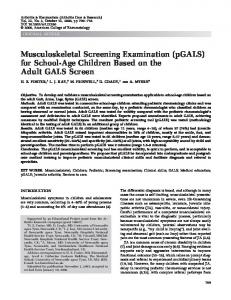
>12,000/mm3), were considered as clinically-suspected cases of necrotizing fasciitis. Patients having suspected necrotizing fasciitis on areas other than limbs, such as the scrotum and neck, were excluded from this study. Study Protocol. When patients presented at the ED, they were first evaluated by one of the emergency physicians, typically at the mid-resident level. All patients suffering from clinically-suspected necrotizing fasciitis underwent ED ultrasonographic examination, performed by the first author (Z-SY). The first author had previously made the (sonographic) diagnosis of more than 50 cases of softtissue diseases. The sonography was performed before the results of all other diagnostic tests were available. The decision to order further diagnostic studies was left to the discretion of the treating physicians. Ultrasonography was performed with a 7.5-MHz linear-array transducer for both longitudinal and transverse imaging. The ultrasonographic diagnosis of necrotizing fasciitis was based on the criterion of a diffuse thickening of the subcutaneous tissue, accompanied by a layer of fluid accumulation more than 4 millimeters in depth along the deep fascial layer (Fig. 1) when compared with the contralateral position on the corresponding normal limb.7 The final diagnosis of necrotizing fasciitis was determined by pathological findings for patients who underwent fasciotomy or biopsy results for patients who were managed nonoperatively. Specific details for all patients were recorded, including the following: patient age, gender, associated medical ill-
nesses, causative organisms, discharge diagnoses, and medical outcomes. Data Analysis. Descriptive statistics were used to evaluate the data set. Measures of test validity, including sensitivity, specificity, positive predictive value, negative predictive value, and accuracy with 95% confidence intervals (95% CIs), were calculated for ultrasonographic examination.
RESULTS Sixty-two patients were evaluated for necrotizing fasciitis during the 20-month study period. All patients were admitted to our hospital. The mean age of the study cohort was 52.7 years (range, 28 to 85 years), and 40 patients (64.5%) were male. Necrotizing fasciitis was demonstrated pathologically for 17 patients. Among the 45 patients without necrotizing fasciitis, 39 had cellulitis, four had deep-vein thrombosis, and two had myositis. Fasciotomy was performed for 21 patients, 16 of whom had necrotizing fasciitis and five of whom had cellulitis only. Four patients died as a result of the progression of necrotizing fasciitis. Diagnostic results from the performed ultrasonography are shown in Table 1. Ultrasonography revealed a sensitivity of 88.2%, a specificity of 93.3%, a positive predictive value of 83.3%, a negative predictive value of 95.4%, and an accuracy of 91.9%. The two false-negative patients survived and were discharged. The three false-positive patients were found to have cellulitis at discharge and they all survived. Of 17 patients suffering from necrotizing fasciitis,
Figure 1. Ultrasonography showing abnormal fluid accumulated in the deep fascial layer.
1450
Yen et al. • ULTRASONOGRAPHY IN NECROTIZING FASCIITIS
TABLE 1. Ultrasonographic Findings for 62 Patients Suspected of Suffering from Necrotizing Fasciitis
tially-toxic contrast medium. Furthermore, at this time of cost-conscious medicine, ultrasonography would clearly appear to be more cost-effective than CT and MRI. The ultrasonographic findings of necrotizing fasciitis are compatible with its pathological process. As the disease progresses, extensive tissue necrosis develops as a result of the action of bacterial enzymes, toxins, or endogenous cytokines.1,10 This causes the abnormal fluid accumulating in the deep fascial layer and the thickening of the subcutaneous layer that can be visualized by ultrasonography. Schmid et al.5 (1998) questioned the accuracy of ultrasonography for detecting the presence of deep fascial fluid, but, from our experience, we believe ultrasonography is able to detect deep fascial fluid without any great difficulty.
Ultrasonographic Findings Positive Negative
Necrotizing Fasciitis
Non–Necrotizing Fasciitis
15 2
3 42
Sensitivity: 88.2% (63.6% to 98.5%)*, specificity: 93.3% (81.7% to 98.6%), positive predictive value: 83.3% (58.6% to 96.4%), negative predictive value: 95.4% (84.5% to 99.4%), accuracy: 91.9% (82.2% to 97.3%). * Numbers in parentheses are 95% confidence intervals.
seven (41.2%) suffered from diabetes. Nine patients with necrotizing fasciitis had positive culture results, with Staphylococcus aureus identified in three cases and streptococcal species in two cases. One patient with necrotizing fasciitis did not undergo a fasciotomy, due to her poor clinical condition. Her final diagnosis was based on MRI and a biopsy result.
DISCUSSION The early diagnosis of necrotizing fasciitis is often difficult.1,6,8 The necessity for rapid lifesaving surgical intervention, however, requires early recognition of the disease. Reliable and convenient tools allowing for the rapid establishment of an accurate diagnosis are essential. Our results have shown that ultrasonography is a good diagnostic tool for this purpose, demonstrating a sensitivity of 88.2% and a specificity of 93.3% for the diagnosis of necrotizing fasciitis in limbs. Different radiological methods have been used previously for the diagnosis of necrotizing fasciitis. Plain radiographs9 can provide information such as soft-tissue thickening and internal gas formation within limbs, although plain radiography typically shows no specific abnormality until the necrotizing process is well advanced, and anaerobic conditions and/or diabetes appear necessary for the production of clinically-detectable quantities of gas.1,9 Computed tomography4 and MRI5 facilitate the detection of fascial thickening, focal fluid collection, and soft-tissue gas production and retention. Both of these imaging modalities have sensitivities between 80% and 100%; however, both CT and MRI may not be readily available in an emergent setting, and there are risks associated with interdepartmental transfer of critically ill patients from the ED to the radiography department. In addition, these imaging studies require exposure to somewhat toxic contrast media. In comparison, ultrasonography can be performed at the bedside with no concern about radioactivity or patient contact with a poten-
LIMITATIONS We acknowledge that our study does have some limitations. First, the location of necrotizing fasciitis in our study patients was limited to limbs only. Second, the impact of ultrasonography on outcomes for patients suffering from necrotizing fasciitis was not investigated. Finally, this investigation was carried out at only one institution in Taiwan, and its generalizability to the situations at other institutions remains unknown. Further studies are needed to determine the clinical role of ultrasonography and its role in the management of necrotizing fasciitis.
CONCLUSIONS We believe that ultrasonography, a convenient tool with good sensitivity and specificity for such an application, can provide accurate information for emergency physicians for the diagnosis of necrotizing fasciitis. References 1. 2.
3.
4.
5.
6.
7.
Green RJ, Dafoe DC, Raffin TA. Necrotizing fasciitis. Chest. 1996; 110:219–29. McHenry CR, Piotrowski JJ, Petrinic D, Malangoni MA. Determinants of mortality for necrotizing soft-tissue infections. Ann Surg. 1995; 221:558–65. Voros D, Pissiotic C, Georgantas D, Katsaragakis D, Antoniou S, Papadimitriou J. Role of early and extensive surgery in the treatment of severe necrotizing soft tissue infections. Br J Surg. 1993; 80:1190–1. Wysoki MG, Santora TA, Shah RM, Friedman AC. Necrotizing fasciitis: CT characteristics. Radiology. 1997; 203: 859–63. Schmid MR, Kossmann T, Duewell S. Differentiation of necrotizing fasciitis and cellulitis using MR imaging. AJR. 1998; 170:615–20. Stamenkovic I, Lew PD. Early recognition of potentially fatal necrotizing fasciitis. The use of frozen-section biopsy. N Engl J Med. 1984; 310:1689–93. Tsai CC, Lai CS, Yu ML, Chou CK, Lin SD. Early diagno-
1451
ACAD EMERG MED • December 2002, Vol. 9, No. 12 • www.aemj.org
8.
sis of necrotizing fasciitis by utilization of ultrasonography. Kaohsiung J Med Sci. 1996; 12:235–40. Lille ST, Sato TT, Engrav LH, Foy H, Jurkovich GJ. Necrotizing soft tissue infections: obstacles in diagnosis. J Am Coll Surg. 1996; 182:7–11.
9.
10.
Fisher JR, Conway MJ, Takeshita RT, Sandoval R. Necrotizing fasciitis: importance of roentgenographic studies for soft-tissue gas. JAMA. 1979; 241:803–6. Stevens DL. Invasive group A streptococcal infections. Clin Infect Dis. 1992; 14:2–13.
䢇
Where to Find AEM Instructions for Authors For complete instructions for authors, see the January or July issue of Academic Emergency Medicine; visit the SAEM web site at www.saem.org/ inform/autinstr.htm; or contact SAEM via e-mail at
[email protected], via phone at 517-485-5484, or via fax at 517-485-0801.