software framework to help real-time monitoring system for acoustic ablation therapy. Figure 1 shows the ... For TCP/IP networking, all the modules have network ...
Ultrasound Imaging Software Framework for Real-Time Monitoring of Acoustic Ablation Therapy Hyun-Jae Kanga, Nishikant P. Deshmukha, Philipp Stolkaa, E. Clif Burdettec, Emad M. Boctora,b a
b
Dept. of Computer Science, Johns Hopkins University, Baltimore, MD, USA Dept. of Radiation Oncology, Johns Hopkins University, Baltimore, MD, USA c Acoustic MedSystems, Inc., Champaign, IL, USA ABSTRACT
The concern with interstitial ablative therapy for a treatment of hepatic tumors has been growing. In spite of advances in these therapies, there are several technical challenges due to tissue deformation and target motion: localization of the tumor and monitoring for ablator’s tip and thermal dose in heated tissue. In the previous work[1], a steerable acoustic ablator, called ACUSITT, for targeting of ablation tip accurately into tumor area has been developed. However, real-time monitoring techniques for providing image feedback of the ablation tip positioning and thermal dose deposited in the tissue by heating are still needed. In this paper, a new software framework for real-time monitoring ablative therapy during pre- and intra-operation is presented. The software framework provides ultrasound Brightness Mode (B-Mode) image and elastography simultaneously and with real-time. A position of ablator’s tip and a region of heated tissue are monitored on B-Mode image, because the image represents tissue morphology. Furthermore, ultrasound elasticity image is used for finding a boundary and region of tumor on pre-ablation, and monitoring thermal dose in tissue during ablation. By providing B-Mode image and elastography at the same time, reliable information for monitoring thermal therapy can be offered. 1.
INTRODUCTION
The concern with interstitial ablative therapy for the treatment of hepatic tumors has been growing. There are several methods in this therapy: chemical ablation, cryoablation [2], radiation therapy [3], and heat ablative therapies using different energy sources such as radiofrequency (RF) [4], laser, microwave [5] or focused ultrasound [6]. In spite of advances in these therapies, there are several technical challenges due to tissue deformation and target motion: localization of the tumor and monitoring for ablator’s tip and thermal dose in heated tissue. Moreover, current ablative systems are highly dependent on operator skill. In the previous work, a steerable acoustic ablator, called ACUSITT, for targeting of ablation tip accurately into tumor area has been developed[1]. However, real-time monitoring techniques for providing image feedback of the ablation tip positioning and thermal dose deposited in the tissue by heating are still needed. In this paper, we report a new ultrasound software framework to help real-time monitoring system for acoustic ablation therapy. Figure 1 shows the block diagram of this new real-time monitoring system. Ultrasound imaging software framework affords a real-time ultrasound Brightness Mode (B-Mode) image and elastography simultaneously. B-Mode images represent tissue-morphology by collecting and displaying the intensity information of reflected ultrasound wave, therefore this imaging modality is mainly used for diagnose and monitoring. On B-Mode image, a location of the ablation tip can be tracked in real-time by a needle detection algorithm which is one of the image processing algorithms. On the other hand, ultrasound elastography represents the information of tissues’s stiffness and its elastic module. Since the abnormal tissue of tumor area has different stiffness compared to normal tissue of surrounding, the area and boundary of tumor can be recognized in ultrasound elastography. Moreover, ultrasound thermal image, heat-induced echo-strain image, is very similar to ultrasound elastography. During the ablation, the speed of sound of heated tissue is higher than non-heated tissue. By tracking shifts in the backscattered RF signals, heat-induced echo-strain image is generated using a similar time-delay estimation algorithm of ultrasound elastography. In heat-induced echo-strain image, low and high strains correspond to constant and higher temperature respectively. Thus, thermal dose in heated tissue can be estimated by using ultrasound elasticity image. This paper is organized as follows: Section 2 describes the software architecture with detail explanation of each software
Medical Imaging 2012: Ultrasonic Imaging, Tomography, and Therapy, edited by Johan G. Bosch, Marvin M. Doyley, Proc. of SPIE Vol. 8320, 83201E · © 2012 SPIE CCC code: 1605-7422/12/$18 · doi: 10.1117/12.912362 Proc. of SPIE Vol. 8320 83201E-1 Downloaded From: http://proceedings.spiedigitallibrary.org/ on 04/20/2014 Terms of Use: http://spiedl.org/terms
module and our experimental testbed. In Section 3, we present results of our development and ex-vivo tissue experiment with a discussion. ID LOPS
yconej
2re
VPIS 10 1
ycona b0MGL GGUGL
IGWb
MW XE
Cfl DV
Figure 1 Block diagram of monitoring system for acoustic ablative therapy 2.
METHODS
As seen in the Figure 1, the software framework is composed of several specialized executable modules: RF-Server2.0, Elasticity-Imaging (EI), ImageViewer, Multi-Image Viewer and Framechooser modules. Ultrasound data such as RF data, elastography, and B-Mode Image from or into each software module are communicated between software modules with an extended version of OpenIGTLink, called OpenIGTLinkMUSiiC [7]. For TCP/IP networking, all the modules have network classes of OpenIGTLinkMUSiiC (MUSiiCServerT, MUSiiCClientT, or both) that support multiple-client connections via TCP/IP network simultaneously [7]. Ultrasound software framework and acoustic ablator system are synchronized by a Transistor-Transistor Logic (TTL) control signal of the controller switch box (see Figure 1). Figure 2 shows the time-sequence diagram of ultrasound data acquisition and processing the data according to TTL signal. Before ablation, a position of ablator’s tool tip is tracked on B-Mode image streams and a boundary and region of tumor are identified on ultrasound elasticity images in real-time. During acoustic ablative therapy, ultrasound software framework is externally triggered by TTL control signal from our controller switch box for anti-synchronization (see Figure 1, 2). While N-frames of RF data are collected in RFServer2.0, ultrasound B-Mode and elastography streams are generated in the software framework simultaneously. These data are used for monitoring of thermal dose in the heated tissue during acoustic ablation therapy.
Proc. of SPIE Vol. 8320 83201E-2 Downloaded From: http://proceedings.spiedigitallibrary.org/ on 04/20/2014 Terms of Use: http://spiedl.org/terms
Pre- Ablation
Ablation Trigger Signal
Trigger Signal
Collect RF-data (N-Frames) 0
Collect RF-data (N-Frames)
Ablation/Water Pump 2
10
Ablation/Water Pump
12
20
Time (second)
B-Mode Image
Tracking a tip of ablator
Monitoring Ablation
Monitoring Ablation
Detecting a boundary and region of tumor
Monitoring thermal dose
Monitoring thermal dose
Elastography
Figure 2 Time-sequence diagram for triggered monitoring of acoustic ablation 2.1 RF-Server 2.0 and Elasticity Image modules 2GLAGL
EI92OL
Figure 3 Block diagram of: (a) RF-Server 2.0, (b) Elasticity Image Module (EIM) We developed a software module, RF-Server1.0, for collecting ultrasound RF data using Texo Software Developing Kit (SDK) in our previous research[8]. But, the software can collect only ultrasound RF data without any filter for the data. On the other side, ultrasound RF data and B-Mode image streams are needed for real-time monitoring of acoustic ablation therapy. A new data-collection software module, RF-Server2.0, has been built by changing the hardware interface API with Port SDK (Ultrasonix. Co.)[9] in this research. This module allows high-performance ultrasound data collection with Field-programmable gate array (FPGA)-based filtering (three-band finite impulse response (FIR) filter for depth-dependent image quality improvement) and real-time B-Mode image generation simultaneously (refer Figure 3). As the diagram indicates, RF-Server2.0 has two set of MUSiiCQueue and MUSiiCServerT classes of
Proc. of SPIE Vol. 8320 83201E-3 Downloaded From: http://proceedings.spiedigitallibrary.org/ on 04/20/2014 Terms of Use: http://spiedl.org/terms
OpenIGTLinkMUSiiC [7] to send ultrasound RF data and B-Mode image streams to other client programs. The block diagram of the Elasticity Imaging module is shown in Figure 3 (b). Because the algorithm of ultrasound elastography needs high computation power, we implemented our algorithm on GPU-EI-Engine based on Graphics processing unit (GPU) programming technique [10] as like Figure 3. Elasticity Image module receives ultrasound RF data stream from RF-Server2.0 via TCP/IP network, generates elastography streams from consecutive RF data frames, and then sends out the results over TCP/IP. 2.2 ImageViewer, Multi-Image Viewer and Framechooser modules The block diagrams of Image Viewer, Multi-Image Viewer and Framechooser are in Figure 4. As seen in the figure, the basic structures of these three modules are similar due to their basic function, receiving ultrasound data from network and displaying the data with real-time. Image Viewer module only provides the basic functionality including simple image processing such as zoom in/out, adjusting brightness and contrast of image. While, Multi-image Viewer has two set of MUSiiCClientT and MusiiCQueue for receiving different image data (B-Mode and Elastography) and its own specific class (MUSiiCImageMerging) to display multiple images on one screen. Moreover, post-processing is applied on Framechooser module to choose the best available strain images. In this module, strain images can be filtered and selected and sent to other module for further processing of data with the results from the computation of GPU-EI-Engine. a
Graphic User Interface TCP/IP Network MUSiiCClientT
MUSiiCQueue
MUSiiC ImageDisplay
b
Control Parameters
Graphic User Interface
B‐Mode Data
TCP/IP Network MUSiiC ClientT
MUSiiCQueue
MUSiiC ClientT
MUSiiCQueue
MUSiiC Image Merging
Elastography
MUSiiC ImageDisplay
Image Data
c
Graphic User Interface TCP/IP Network
TCP/IP Network
MUSiiCClientT
MUSiiCServerT
MUSiiCQueue
MUSiiCQueue
MUSiiC ImageDisplay
Filtering Frame
Figure 4 Block diagram of: (a) Image Viewer, (b) Multi-Image Viewer, (c) Framchooser module
Proc. of SPIE Vol. 8320 83201E-4 Downloaded From: http://proceedings.spiedigitallibrary.org/ on 04/20/2014 Terms of Use: http://spiedl.org/terms
2.3 Experimental Testbed
Ultrasound Machine
Acoustic Ablator
Controller Switch Box Workstation Acoustic Abalation System
Ex-Vivo Tissue (Turkey Breast)
Figure 5 System setup for real-time monitoring of acoustic ablation Therapy Figure 5 shows the system setup for our ex-vivo tissue experiment of real-time monitoring of acoustic ablation therapy. In our experiment, we used a turkey breast tissue as an ex-vivo tissue. For collecting ultrasound RF data and B-Mode Image, our system setup used SONIX-CEP machine (UltraSonix Medical Co.) equipped with 3D linear array transducer (model 4DL14-5/38). For real-time monitoring of thermal dose in a tissue, our controller switch box is connected ultrasound machine (SONIX-CEP) and Acoustic power generator with Bayonet Neil-Concelman (BNC) cables to provide the anti-synchronization that is based on the time-sequence (Figure 2) with TTL signals. 3.
RESULTS
In our software framework, RF-Server2.0 collects ultrasound RF data and B-Mode image and provides the data to other client modules (Elasticity Image, Image Viewer, Multi-image Viewer and Framechooser module) in real time. So, the performance of this program is important for real-time monitoring of acoustic ablation system. To verify the capacity of RF-Server2.0, we collected RF data and B-Mode image streams on different depth and sampling frequency. Also, we tested this module with/ without network connections. The result of our test is in table 1. As the table, data-collecting performance depends on the depth of ultrasound data or image. But, there is little difference in the performance of RFServer2.0 between different sampling frequencies. Moreover RF-Server2.0 has similar frame rates per second with/ without TCP/IP network connection, because it has an independent network class (MUSiiCServerT).
Depth(cm) 3 4 5 6
Sampling Frequency 20 MHz Sampling Frequency 40 MHz RF B B+N RF B B+N 70.922 70.922 70.922 71.429 70.922 64.103 58.140 58.480 58.140 58.140 58.140 58.140 49.020 49.261 49.261 49.261 49.261 49.020 42.735 42.553 42.735 42.735 42.553 42.553
RF
Collecting RF Data
B
Collecting B-Mode Image
B+N
Collecting B-Mode Image with TCP/IP Network connection
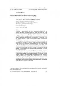
Table 1 The performance of RF-Server 2.0 (fps) Figure 6 shows gross pathology image (a), ultrasound B-Mode Image (b) and ultrasound elastography (c). As the histopathology image, we can see two lesions (L1 and L2) on both ultrasound B-Mode Image and elastography.
Proc. of SPIE Vol. 8320 83201E-5 Downloaded From: http://proceedings.spiedigitallibrary.org/ on 04/20/2014 Terms of Use: http://spiedl.org/terms
Moreover, strain results of ultrasound elastography are in good correspondence with the histopathlogy.
a
b L1
L2
L1
Figure 6 Intraoperative thermal monitoring of acoustic ablation therapy. (a) Gross pathology Image, (b) Ultrasound B-Mode Image, (c) Ultrasound Elastography. 4.
CONCLUSION
In this research, we developed a software framework which is composed of specialized executable modules: RFServer2.0, Elasticity Image, Image Viewer, Multi-Image Viewer and Framechooser modules. The software framework is based on network distributed system and multithreaded architecture. That is, each module of the software framework has its own independent task-thread for improving its performance with multithreaded programming. Moreover, one or two MUSiiCQueue class, thread-safety queue for multithreaded program, and network classes of OpenIGTLinkMUSiiC (MUSiiCServerT, MUSiiCClinetT, or both) are implemented in all modules for TCP/IP network communication. Using our software framework, we can monitor the location of ablation tip (with B-Mode Image), tumor area (with Elastography), and distribution of thermal dose in tissue (with Elastography). Our software framework is based on the component of ultrasound data processing, therefore, we see that this software framework can be applied other ultrasound research. 5. [1] [2] [3] [4] [5] [6] [7] [8] [9] [10]
REFERENCES E. M. Boctor, P. Stolka, H. J. Kang et al., "Precisely shaped acoustic ablation of tumors utilizing steerable needle and 3D ultrasound image guidance." 7625, 91. B. S. Kuszyk, J. K. Boitnott, M. A. Choti et al., “Local Tumor Recurrence Following Hepatic Cryoablation: Radiologic-histopathologic Correlation in a Rabbit Model1,” Radiology, 217(2), 477 (2000). L. G. Koniaris, D. Y. Chan, C. Magee et al., “Focal hepatic ablation using interstitial photon radiation energy1,” Journal of the American College of Surgeons, 191(2), 164-174 (2000). M. A. Choti, “Surgical management of hepatocellular carcinoma: resection and ablation,” Journal of vascular and interventional radiology, 13(9), S197-S203 (2002). N. Izumi, Y. Asahina, O. Noguchi et al., “Risk factors for distant recurrence of hepatocellular carcinoma in the liver after complete coagulation by microwave or radiofrequency ablation,” Cancer, 91(5), 949-956 (2001). G. Ter Haar, “Ultrasound focal beam surgery,” Ultrasound in medicine & biology, 21(9), 1089-1100 (1995). H.-J. Kang, P. J. Stolka, and M. B. Emad, “OpenITGLinkMUSiiC: A Standard Communications Protocol for Advanced Ultrasound Research,” MIDAS Journal, (2011). P. J. Stolka, H.-J. Kang, and M. B. Emad, “The MUSiiC toolkit: Modular Real-Time Toolkit for Advanced Ultrasound Research,” MIDAS Journal, (2010). http://www.ultrasonix.com/ N. Deshmukh, H. Rivaz, and E. Boctor, "GPU-Based Elasticity Imaging Algorithms." 45-54.
Proc. of SPIE Vol. 8320 83201E-6 Downloaded From: http://proceedings.spiedigitallibrary.org/ on 04/20/2014 Terms of Use: http://spiedl.org/terms