Int J CARS (2015) 10:1493–1503 DOI 10.1007/s11548-014-1133-6
ORIGINAL ARTICLE
Ultrasound texture-based CAD system for detecting neuromuscular diseases Tim König · Johannes Steffen · Marko Rak · Grit Neumann · Ludwig von Rohden · Klaus D. Tönnies
Received: 31 July 2014 / Accepted: 18 November 2014 / Published online: 2 December 2014 © CARS 2014
Abstract Purpose Diagnosis of neuromuscular diseases in ultrasonography is a challenging task since experts are often unable to discriminate between healthy and pathological cases. A computer-aided diagnosis (CAD) system for skeletal muscle ultrasonography was developed and tested for myositis detection in ultrasound images of biceps brachii. Methods Several types of features were extracted from rectangular and polygonal image regions-of-interest (ROIs), including first-order statistics, wavelet-based features, and Haralick’s features. Features were chosen that are sensitive to the change in contrast and structure for pathological ultrasound images of neuromuscular diseases. The number of features was reduced by applying different sequential feature selection strategies followed by a supervised principal component analysis. For classification, two linear approaches were investigated: Fisher’s classifier and the linear support vector machine (SVM) as well as the nonlinear k-nearest neighbor approach. The CAD system was benchmarked on datasets of 18 subjects, seven of which were healthy, while 11 were affected by myositis. Three expert radiologists provided pre-classification and testing interpretations. Results Leave-one-out cross-validation on the training data revealed that the linear SVM was best suited for discriminating healthy and pathological muscle tissue, achieving 85/87 % accuracy, 90 % sensitivity, and 83/85 % specificity, depending on the radiologist. Conclusion A muscle ultrasonography CAD system was T. König (B) · J. Steffen · M. Rak · K. D. Tönnies Department of Simulation and Graphics, Otto von Guericke University, Universitätsplatz 2, 39106 Magdeburg, Germany e-mail:
[email protected] G. Neumann · L. von Rohden Department of Radiology and Nuclear Medicine, Otto von Guericke University, Leipziger Straße 44, 39120 Magdeburg, Germany
developed, allowing a classification of an ultrasound image by one-click positioning of rectangular ROIs with minimal user effort. The applicability of the system was demonstrated with the challenging example of myositis detection, showing highly accurate results that were robust to imprecise user input.
Keywords Texture analysis · Computer-aided diagnosis · Ultrasound imaging · Neuromuscular diseases · Classification
Introduction Neuromuscular diseases, e.g., progressive spinal muscular atrophy (Werdnig–Hoffmann disease), Duchenne muscular dystrophy, or myositis are common neurological disorders. Incidence rates are 10/100,000 for progressive spinal muscular atrophy, 29/100,000 for Duchenne muscular dystrophy, and 1/100,000 for myositis. Ultrasonography is a convenient technique to visualize healthy and pathological skeletal muscle tissue, providing high resolution scans with noninvasive real-time image acquisition. Neuromuscular disorders, like myositis, often cause structural muscle changes that can be observed in ultrasound images. It is assumed that the replacement of muscle tissue by fat and an increase or a thickening of fibrous connected tissue is the main reason for an increased muscle echo intensity in pathological tissue, since the number of reflections increases within the muscle resulting in partially higher intensities in the ultrasound image [29]. Hence, only few reflections occur in healthy muscle tissue during image acquisition, resulting in a low echo intensity and thus in rather dark images with more evenly distributed high-intensity peaks. As
123
1494
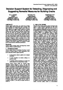
a consequence, texture of healthy muscle tissue seems to be more oriented and structured due to higher contrast changes, while pathological muscle tissue appears to be diffuse and unstructured due to its smaller contrast changes. We treat the issue of detection of neuromuscular diseases in ultrasound images at the challenging example of myositis detection. Myositis, or inflammatory myopathy, is a rare disease in which the immune system chronically inflames the body’s muscle tissue. Persistent inflammation progressively weakens or destroys the muscle tissue and is commonly accompanied by pain. Over time, this process may also lead to a loss of muscle mass. Myositis most often appears in childhood, at the age of 5–15, and in adulthood, at the age of 40–60, although it might occur at any age. In contrast to other neuromuscular disorders like progressive spinal muscular atrophy or Duchenne muscular dystrophy, a texture analysis of myositis is more difficult, since the disease may only occur focally and brightness changes in pathological cases are often small [28,30]. For this reason, we developed a CAD system for detecting myositis in ultrasound images, which can be easily adapted to detect similar neuromuscular disorders. Under the assumption that pathological muscle tissue appears to be diffuse and unstructured, a texture-based analysis seems adequate for detecting myositis in ultrasound images. A computer-aided approach serves several purposes: It provides an independent opinion, which can be taken into consideration, and it saves time for radiologists. Moreover, it helps young professionals with less experience to identify myositis in ultrasound images. A well-trained process may also be capable to support an early diagnosis. Diagnosis from visual inspection can be difficult for the radiologist in early stages of neuromuscular diseases or if myopathies cause only little structural changes in muscle groups. Figure 1 shows ultrasound images of pathological muscle tissue with different degree of severity of myositis. Discrimination between healthy and strongly affected muscle tissue in Fig. 1a, d seems to be rather intuitive, compared to the more difficult discrimination between healthy and little affected muscle tissue in Fig. 1a, b, or little and medium affected muscle tissue in Fig. 1b, c. The latter distinctions are challenging tasks even for automated classification.
Int J CARS (2015) 10:1493–1503
Related work Texture features are well established in many medical applications incorporating ultrasound imaging. Most problems require a combination of statistical and spectral features, since they represent the nature of deterministic tissue-related variation and non-deterministic influences from image generation ,respectively, [16,25,31,35,38]. These problems include diagnosis and classification of, for example, diffuse, chronic, alcoholic, fatty liver diseases, and liver cancer in ultrasound images [4,15,19, 20,24,26]. Such methods often require texture measures that rely on spatial gray level dependence, gray level cooccurrence, fractal dimension, Fourier transform, Law’s texture energy, discrete wavelet transform, or wavelet packet decomposition features combined with, for example, probabilistic neural networks or support vector machines as classifiers. More recent methods also include an automatic ROI detection or segmentation of the liver [22, 32]. Another important research branch of texture analysis in ultrasound imaging is the detection of breast cancer. Earlier work uses first-order statistics or the Fourier transform to extract texture features [12,13]. Autocorrelation [6], morphological [5], auto-covariance [16], or Haralick’s [21] features of the breast tissue have also been used. More recently, approaches combine morphological features, generated by active contour or level set segmentation, with texture features derived from autocorrelation functions, first-order statistics, or spatial gray level dependences [34,39] as well as shearlet transform [40], which have proven to produce good and stable classification rates. There are several other research areas of texture analysis in ultrasound imaging, e.g., the diagnosis of prostate cancer in transrectal images. Here, methods use, e.g., combinations of features derived from gray level co-occurrence matrices, Gabor transform, or discrete wavelet transform [3,25,33]. To detect carotid atherosclerotic plaque in ultrasound images, different approaches exist, which for instance use features based on first- and second-order statistics, fractal dimensions, discrete wavelet transform, wavelet package decomposition,
Fig. 1 Exemplary stages of myositis disease on ultrasound images of biceps brachii showing healthy (a) and pathological muscle tissue with different degree of severity: b little, c medium, and d strongly affected muscle tissue
123
Int J CARS (2015) 10:1493–1503
gray level co-occurrence matrix, Law’s filter masks, and the Fourier transform [1,2,8,35,37]. Some geometrical, Gabor, and statistical features as well as combinations of these have also been used for Parkinson’s disease diagnostics in transcranial sonography [7,17,31]. Regarding skeletal muscle ultrasound, Pohle et al. [28] proposed an approach for examining a number of different neuromuscular diseases. Although they obtained good classification rates, they did not use a standardized examination procedure. Scan and ROI estimation were performed subjectively by one person for already preselected images. Moreover, it was necessary to record digital ultrasound images on analog S-VHS cassettes during that time. A re-digitization, which is often a source of error, was required for a computeraided evaluation. However, it was shown that a texture-based analysis of ultrasound images may work well for detecting progressive spinal muscular atrophy and Duchenne muscular dystrophy. Pillen and van Alfen [27] also investigated neuromuscular diseases in ultrasound images. They proposed a quantitative gray scale analysis using first-order histogram-based statistical features, e.g., the mean intensity values of the ROIs. This approach performs well for diseases that cause a regular, increased echo intensity like Duchenne muscular dystrophy. However, methods using only first-order statistics most likely fail for irregular or focally located diseases, as in some cases of myositis. König et al. [18] proposed a classification approach for detecting myositis with an overall accuracy of ≥85 %. They used a combination of two statistical and two wavelet features, measuring both, brightness and structural changes of pathological tissue in ultrasound images. Moreover, they demonstrated that too small ROI sizes (