Asian J. Nursing Edu. and Research 3(3): July-September 2013
www.anvpublication.org
ISSN-2231-1149
RESEARCH ARTICLE
Understanding of Urodynamics Testing Mrs. Malar Kodi Aathi1, Ms. Akojiam Sangita Devi2, Mr. Gaurav Kohli3 1
Assistant Professor, Dept of Child Health Nursing, M.M Institute of Nursing, M.M University Mullana, Ambala Haryana.133207 2 Dept of Medical Surgical Nursing, M.M Institute of Nursing, M.M University Mullana, Ambala Haryana.133207 3 Dept of Community Health Nursing, M.M Institute of Nursing, M.M University Mullana, Ambala Haryana.133207 *Corresponding Author Email:
[email protected]
ABSTRACT: A systematic investigation of urodynamics will provide an explanation and comprehension of various invasive and non invasive investigations involved in urodynamics such as uroflowmetry, post void residual measurement, cystometric test, leak point pressure measurement, pressure flow study, electromyography, video Urodynamic tests. This simple, painless study is a series of tests that allows your physician to evaluate any problems your bladder may have storing or emptying urine. Reasons for these tests may include incontinent (leak urine), bladder may not empty completely, uncomfortable symptoms, such as the frequent need to urinate or a constant urgent need to urinate, urine stream may be intermittent or weak, Client may have persistent urinary tract infections. Bladder and abdominal pressure is measured during filling and voiding. Urodynamic equipment calculates detrusor pressure by subtracting abdominal from pelvic. This pressure measures during filling and voiding of urine. The terminology in this abstract has comply with cooper measure guide, International Continence Society and department of urodynamics standard and quality control.
KEY WORDS: Comprehension, Urodynamics, Uroflowmetry, post void residual measurement, cystometry, leak point pressure measurement, pressure flow study, electromyography, video Urodynamic tests, incontinence.
INTRODUCTION: Lower Urinary Tract Symptoms and no neurologic diseases and conditions. These findings have important diagnostic and management implications. Continued improvement in Urodynamic testing will help support advancements in understanding these associations. Participants in clinical trials can play a more active role in our own health care, gain access to new research treatments before they are and help others by contributing to health The National Institute of Diabetes and Digestive and widely available, 1 Kidney Diseases (NIDDK) sponsors studies of diagnostic research. Urodynamic methods and devices. The NIDDK also sponsors programs aimed at understanding, diagnosing, and Urodynamics Urodynamics is the term that encompasses a treating lower urinary tract problems. NIDDK-funded number of tests used in the investigation of women with lower urinary tract symptoms (LUTS). Some are non studies have shown associations between (LUTS) invasive, such as flows studies, but the majority are non invasive, requiring urethral catherization and placement of Received on 04.04.2013 Modified on 25.04.2013 an abdominal pressure catheter in the vagina, rectum or Accepted on 01.05.2013 © A&V Publication all right reserved stoma. The International Continence Society (ICS) sets In recent years, researchers have learned much about the causes and treatment of lower urinary tract problems. Researchers are looking into ways to more accurately assess LUTS and diagnose lower urinary tract problems. Research focuses on designing and developing more accurate and reliable diagnostic tests and tools.2
Asian J. Nur. Edu. and Research 3(3): July-Sept., 2013; Page 171-176
171
Asian J. Nursing Edu. and Research 3(3): July-September 2013
definitions and standards for urodynamics investigation. It publishes standardization reports that are available online and should be adhered to. Non invasive urodynamics tests, such as Uroflowmetry, do not have any associated morbidity. Invasive tests involving urethral catherization have a recognized risk of urinary tract infections of between 1 and 10%. Most centers do not advocate the prophylactic use of antibiotics for cystometry apart from in specific indication, for example renal transplant patients.6 What is urodynamics? Urodynamic tests range from simple observation to precise measurements using sophisticated instruments.
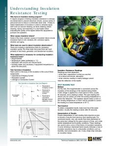
Uroflowmetry Purpose: Uroflowmetry is the measurement of urine speed and volume. Special equipment automatically measures the amount of urine and the flow rate how fast the urine comes out. Procedure: Uroflowmetry equipment includes a device for catching and measuring urine and a computer to record the data. During a Uroflowmetry test, the person urinates privately into a special toilet or funnel that has a container for collecting the urine and a scale. The equipment creates a graph that shows changes in flow rate from second to second so the health care provider can see when the flow rate is the highest and how many seconds it takes to get there. Results of this test will be abnormal if the bladder muscles are weak or urine flow is blocked. Another approach to measuring flow rate is to record the time it takes to urinate into a special container that accurately measures the volume of urine.4 Uroflowmetry measurements are performed in a health care provider’s office; no anesthesia is needed. Figure: 1.
Most Urodynamic tests focus on the bladder’s ability to hold urine and empty steadily and completely. Urodynamic tests can also show whether the bladder is having involuntary contractions that cause urine leakage.7 Urodynamic tests range from simple observation to precise measurements using sophisticated instruments. For simple observation, a health care provider may record the length of time it takes a person to produce a urinary stream, note the volume of urine produced, and record the ability or inability to stop the urine flow in midstream. For precise Time required: measurements, imaging equipment takes pictures of the 1-2 minutes bladder filling and emptying, pressure monitors record the pressures inside the bladder, and sensors record muscle and nerve activity. The health care provider will decide the type of Urodynamic test based on the person’s health information, physical exam, and LUTS (lower urinary tract symptoms). The Urodynamic tests results help diagnose the cause and nature of a lower urinary tract problem. Urodynamic tests include Uroflowmetry, post void residual measurement, cystometric test, leak point pressure measurement, pressure flow study, electromyography, video Urodynamic tests.5 Types of Urodynamic tests
Fig. 1. Normal Uroflowmetry study demonstrating a normal bellshaped pattern ( A) and an abnormal screening Uroflowmetry study ( B) in a Valsalva voider with an interrupted, intermittent flow pattern
172
Asian J. Nursing Edu. and Research 3(3): July-September 2013
Posts void Residual Measurement: Purpose: This Urodynamic test measures the amount of urine left in the bladder after urination. The remaining urine is called the post void residual.
Purpose: A cystometric test measures how much urine the bladder can hold, how much pressure builds up inside the bladder as it stores urine, and how full it is when the urge to urinate begins.
Procedure: Post void residual can be measured with ultrasound equipment that uses harmless sound waves to create a picture of the bladder. Bladder ultrasounds are performed in a health care provider’s office, radiology center, or hospital by a specially trained technician and interpreted by a doctor, usually a radiologist. Anesthesia is not needed. Post void residual can also be measured using a catheter a thin flexible tube. A health care provider inserts the catheter through the urethra up into the bladder to remove and measure the amount of remaining urine. A post void residual of 100 milliliters or more is a sign that the bladder is not emptying completely. Catheter measurements are performed in a health care provider’s office, clinic, or hospital with local anesthesia. (Figure: 2)
Procedure: A catheter is used to empty the bladder completely. Then a special, smaller catheter is placed in the bladder. This catheter has a pressure-measuring device called a manometer. Another catheter may be placed in the rectum to record pressure there.
Time required: Will take up to 30 minutes
Once the bladder is emptied completely, the bladder is filled slowly with warm water. During this time, the person is asked to describe how the bladder feels and indicate when the need to urinate arises. When the urge to urinate occurs, the volume of water and the bladder pressure are recorded. The person may be asked to cough or strain during this procedure to see if the bladder pressure changes. A cystometric test can also identify involuntary bladder contractions. Cystometric tests are performed in a health care provider’s office, clinic, or hospital with local anesthesia.(figure:3) Time required: 10-20 minutes
Figure: 2 Posts void Residual Measurement
Cystometric Test:
Figure:3 Cystometry
173
Asian J. Nursing Edu. and Research 3(3): July-September 2013
common in women but can occur with a cystocele or, Leak Point Pressure Measurement: rarely, after a surgical procedure for urinary incontinence. Purpose: This Urodynamic test measures pressure at the point of Pressure flow studies are performed in a health care leakage during a cystometric test. provider’s office, clinic, or hospital with local anesthesia. (Figure:4) Time required: 1-5 minutes Procedure: While the bladder is being filled for the cystometric test, it may suddenly contract and squeeze some water out without Electromyography: warning. The manometer measures the pressure inside the Purpose: Electromyography uses special sensors to bladder when this leakage occurs. This reading may provide measure the electrical activity of the muscles and nerves in information about the kind of bladder problem that exists. and around the bladder and the sphincters. If the health care The person may be asked to apply abdominal pressure to provider thinks the urinary problem is related to nerve or the bladder by coughing, shifting position, or trying to muscle damage, the person may be given an exhale while holding the nose and mouth. These actions electromyography. help the health care provider evaluate the sphincters. Procedure: The sensors are placed on the skin near the Time required: 30 minutes urethra and rectum or on a urethral or rectal catheter. Muscle and nerve activity is recorded on a machine. The Pressure Flow Study: patterns of the nerve impulses show whether the messages Purpose: A pressure flow study measures the bladder pressure sent to the bladder and sphincters are coordinated correctly. required to urinate and the flow rate a given pressure Electromyography is performed by a specially trained generates. technician in a health care provider’s office, outpatient clinic, or hospital. Anesthesia is not needed if sensors are placed on the skin. Local anesthesia is needed if sensors are Procedure: After the cystometric test, the person empties the bladder, placed on a urethral or rectal catheter. during which time a manometer is used to measure bladder pressure and flow rate. This pressure flow study helps Time required: 30 minute identify bladder outlet blockage that men may experience with prostate enlargement. Bladder outlet blockage is less
Figure:4 Normal pressure flow studies.
174
Asian J. Nursing Edu. and Research 3(3): July-September 2013
Eyeball Cystometrogram: A simple "eyeball Cystometrogram (CMG)" can be performed at the bedside or in the office. The patient is placed in the supine position and catheterized with an 18 Fr catheter. Post-void residual urine is measured. The plunger is removed from a 50-60 mL catheter tip syringe and the barrel of the syringe is connected directly to the end of the catheter. Water or saline is infused into the bladder by pouring into the open end of the syringe. The height of the barrel is raised or lowered until there is steady flow. The patient is instructed to neither try to void nor to inhibit micturition, but rather to report their sensations to the clinician. If the rate of infusion begins to slow down or if the fluid begins to back up, the barrel of the syringe is raised or lowered until flow just stops. The height of the meniscus, above the symphysis, is a measure of vesical pressure. If the pressure does rise, it may be due to an involuntary detrusor contraction, to an increase in abdominal pressure or to low bladder compliance. Increases in intra-abdominal pressure are usually detectable by visual observation or palpation of the abdomen, but low bladder compliance may be difficult to distinguish from an involuntary detrusor contraction. An involuntary detrusor contraction is characterized by a sudden increase in pressure which is not volitional. In most neurologically normal patients the involuntary detrusor contraction is perceived by the patient as an urge to void, but some patient may be completely unaware of it. If involuntary detrusor contractions are suspected, but not demonstrated, the examination is repeated in the upright position. The patient is asked to cough or strain and the pressure response observed to see if this stimulates an involuntary detrusor contraction, which will be apparent as a sustained increase in pressure that persists long after the increase in abdominal pressure has abated.3 Video Urodynamic Tests: Purpose: Video Urodynamic tests take pictures and videos of the bladder during filling and emptying. Procedure: The imaging equipment may use x rays or ultrasound. If x-ray equipment is used, the bladder will be filled with a special fluid, called contrast medium, that shows up on x rays. X rays are performed by an x-ray technician in a health care provider’s office, outpatient facility, or hospital; anesthesia is not needed. If ultrasound equipment is used, the bladder is filled with warm water and harmless sound waves are used to create a picture of the bladder. The pictures and videos show the size and shape of the bladder and help the health care provider understand the problem. Bladder ultrasounds are performed in a health care provider’s office, radiology center, or hospital by a specially trained technician and interpreted by a doctor, usually a radiologist. Although anesthesia is not needed for the ultrasound, local anesthesia is needed to insert the catheter to fill the bladder.
Nursing responsibilities: A careful history, examination and completion of frequency volume chart should be maintained firstly Relevant past medical history, including surgery and neurological disease should be include as well as parity and obstetric history Client’s drug history should b taken prior to urodynamics investigation as some medication, for example, diuretics can affect urinary symptoms such as urine frequency. Frequency/volume chart will contain information about voiding frequency, as well as voiding volumes. On its own it can be used as diagnosing tool Its nurses responsibility to give an information leaflet about what to expect during testing and any questions should be answered Before embarking on urodynamics question should be identified, i.e. what are the symptoms required to provide diagnosis will be determined by the urodynamics questions Degree of incontinence and the number of pads should be noted, as should any co existing difficulties8
SUMMARY: Urodynamic as an essential part of the investigation and its measures nerve and muscle function, pressures around and in the bladder, flow rates and other factors which might help to explain a person's incontinence. Some people find these tests embarrassing and uncomfortable. Urodynamic studies are a set of investigation that define underlying pathophysiology and facilitates better treatment of symptoms. Urodynamic studies are the best diagnostic tool in the management of patients with LUTS. Being invasive and time consuming, it is unnecessary to perform Urodynamic tests in each and every patient with LUTS. However, in patients undergoing any surgical procedure designed to modify the function of the lower urinary tract, an objective assessment by Urodynamic evaluation is mandatory. Patients with recurrent LUTS after initial medical treatment or patients with persistent symptoms after adequate treatment should be referred for Urodynamic studies before undertaking further treatment.
ACKNOWLEDGEMENTS: With profound gratitude we express my heartfelt veneration toward my esteemed; the keen and invaluable family members and friends for enlightening guidance, interest, valuable suggestions and consistent encouragement at all stages of work. We deeply appreciate their untiring and outstanding contribution, encouraging words for compiling the narrative review paper.
REFERENCES: 1.
Time required: Approximately one half to three-quarters 2. of an hour
175
Berkow, R., and Fletcher, A. J. (Ed.). (2006).The management of urinary incontinence in women guidelines. (2nd edition) National Institute for Health and Clinical Excellence.CG40. Balivas and Groutz. (2000) Bladder outlet obstruction nomogram for women with lower urinary tract symptomatology. Neurourol urodyn . 19: 553-64.
Asian J. Nursing Edu. and Research 3(3): July-September 2013
3.
4.
5.
6.
7.
8.
Bates et al. (1976) First report on the standardization of terminology of lower urinary tract function. Urinary incontinence:. British Journal of Urology 48: 39. Schafer, Abrahams and Liao (2002). Good Urodynamics Practices: Uroflowmetry filling cystometry, and pressure flow studies. Neurourol Urodyn ,23:261-74 Cooper, Fletter, Zaszczurynski and Damaser.( 2011) Comparison of air charged and water filled urodynamic pressure measurement catheters. Neurourol Urodyn. 30: 329-34 Latthe, Foon and Toozs. (2008) Prophylactic antibiotics in urodynamic: a systematic review of effectiveness and safety. Neurourol Urodyn . 27:167-73. Rosenzweig and Bhatia. (1992). Temporal separation of coughinduced urethral and bladder pressure spikes in women with urinary incontinence. International Journal of Urology 39: 165. Ouslander, Kane and Abrass. (1982) Urinary incontinence in elderly nursing home patients. JAMA. 248: 1194.
176