Bulletin of the NYU Hospital for Joint Diseases 2010;68(2):147-8
147
Image Erosive Spinal Tophus in a Patient with Gout and Back Pain Jonathan Samuels, M.D., Robert T. Keenan, M.D., M.P.H., Rena Yu, M.D., Michael H. Pillinger, M.D., and Tibor Bescke, M.D.
A
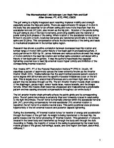
75-year-old male developed intractable low-back pain, right-sided radiculopathy, and bilateral groin pain that severely limited his ability to stand for a sustained period of time, even long enough to take a shower. His previous medical history included bladder cancer, morbid obesity, diabetes, hypertension, chronic renal insufficiency, obstructive sleep apnea, atherosclerosis with coronary stent placement, carpal tunnel syndrome, severe osteoarthritis, and gout. A magnetic resonance image of the spine revealed a lobulated destructive mass eroding into bone and destroying the L5-S1 joint space, with foraminal narrowing and encroachment on the nerve root (Fig. 1, arrows). An abdominal computed tomography scan performed as follow-up for the bladder cancer revealed thickening of the bladder wall and hypertrophy of the prostate, and confirmed the expansile mass in the right posterior L5 vertebral body. Given the patient’s history of malignancy, a CT-guided aspiration biopsy was performed. Histopathological examination showed abundant, negatively birefringent needle-shaped crystals (Fig. 2, inset on right with magnified cytology smear), consistent with a gouty tophus. The patient received epidural steroid injections and urate-lowering allopurinol therapy, requiring 750 mg
Jonathan Samuels, M.D., Assistant Professor of Medicine, Michael H. Pillinger, M.D., Associate Professor of Medicine and Pharmacology, and Robert T. Keenan, M.D., M.P.H., are within the Division of Rheumatology, Department of Medicine; Tibor Bescke, M.D., Clinical Assistant Professor of Neurology, is from Interventional Neuroradiology, Department of Neurology; and Rena Yu, M.D., is from the Department of Pathology, NYU Langone Medical Center, New York, New York. Correspondence: Jonathan Samuels, M.D., Peter D. Seligman Center for Advanced Therapeutics, NYU Hospital for Joint Diseases, 246 E. 20th Street, New York, New York 10003; jonathan.
[email protected].
Figure 1 MR images, 1.5 Tesla, T2 with gadolinium. Left, axial image at L5; Right, saggital image.
Figure 2 Biopsy of mass at T5-S1 joint stained with nonaqueous alcoholic eosin. Magnification not known.
daily to achieve a target serum uric acid level less than 6.0 mg/dL. With this regimen, the patient experienced dramatic clinical improvement. Gout rarely involves the spine,1-7 but should be considered in patients with high uric acid and back pain or radiculopathy.
References 1.
2.
Fontenot A, Harris P, Macasa A, et al. An initial presentation of polyarticular gout with spinal involvement. J Clin Rheumatol. 2008 Jun;14(3):188-9. Konatalapalli RM, Demarco PJ, Jelinek JS, et al. Gout in the
Samuels J, Keenan RT, Yu R, Pillinger MH, Bescke T. Erosive spinal tophus in a patient with gout and back pain. Bull NYU Hosp Jt Dis. 2010;68(2):147-8.
148
3.
4.
5.
Bulletin of the NYU Hospital for Joint Diseases 2010;68(2):147-8 axial skeleton. J Rheumatol. 2009;36(3):609-13. Kuo YJ, Chiang CJ, Tsuang YH. Gouty arthropathy of the cervical spine in a young adult. J Chin Med Assoc. 2007;70(4):180-2. Lam HY, Cheung KY, Law SW, Fung KY. Crystal Arthropathy of the lumbar spine: a report of 4 cases. J Orthop Surg, 2007;15(1):94-101. Mahmud T, Basu D, Dyson PH. Crystal arthropathy of the
6. 7.
lumbar spine: a series of six cases and a review of the literature. J Bone Joint Surg Br. 2005;87(4):513-7. Nygaard HB, Shenoi S, Shukla S. Lower back pain caused by tophaceous gout of the spine. Neurology. 2009;73(5):404. Souza AW, Fontenele S, Carrete H Jr, et al. Involvement of the thoracic spine in tophaceous gout. A case report. Clin Exp Rheumatol. 2002;20(2):228-30.