Letter to the Editor
Clinics in Orthopedic Surgery 2013;5:87-88 • http://dx.doi.org/10.4055/cios.2013.5.1.87
Usage of a Curved Chisel When Resecting Osteochondroma in the Long Bone Akio Sakamoto, MD Department of Orthopaedic Surgery, Graduate School of Medical Sciences, Kyushu University, Fukuoka and Department of Orthopaedic Surgery, National Hospital Organization Kokura Medical Center, Kitakyushu, Japan
To the Editor: Osteochondroma, which is the most common benign bone tumor, is an exophytic bone tumor covered by a cap of cartilage. It exists in two clinical settings, as a solitary lesion (solitary osteochondroma) and as a multiple lesion (multiple hereditary exostoses). The most common affected site is the metaphysis around the knee, especially in the distal femur. When pain or neurologic symptoms develop due to compression, resection of the tumor is recommended. The resection line is supposed to be at the base of the tumor, namely at the border with the normal bone. The
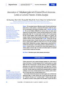
recurrence of osteochondroma is rare, but it does recur in an estimated 2% of cases.1) Moreover, the development of osteosarcoma at the initial site of a treated osteochondroma has been reported.2) In addition, osteosarcoma has been reported within the stalk of an osteochondroma.3) The most common resection tools are a flat chisel, a bone saw or a wire-saw. However, these tools make complete resection of an osteochondroma in the metaphysis of a lone bone difficult, due to continuity of the osteochondroma and the host bone, and because of the anatomical concave shape of the metaphysis of the host bone. In contrast, usage of a curved chisel (radius, 40 or 45 mm; length, 48.5 or 65 mm; width, 15 or 20 mm; Sonne Co. Tokyo, Japan) (Fig. 1) for transposition osteotomy of the acetabulum4) with or without an intraoperative radiograph makes complete resection of osteochondroma in the long bone easier, thereby minimizing the chance of its recurrence and reducing the possibility of the subsequent development of a malignant bone tumor (Fig. 2).
Fig. 1. Lateral views of a curved chisels.
CONFLICT OF INTEREST No potential conflict of interest relevant to this article was reported.
REFERENCES 1. Florez B, Monckeberg J, Castillo G, Beguiristain J. Solitary osteochondroma long-term follow-up. J Pediatr Orthop B. 2008;17(2):91-4. 2. Gomez-Brouchet A, Accadbled F, Rubie H, et al. Rapid development of an osteosarcoma after surgical resection of an osteochondroma. J Pediatr Orthop. 2007;27(6):640-2.
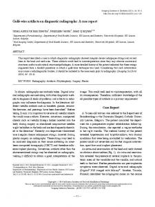
Fig. 2. Plain radiographs of osteochondroma in the distal femur before (A) and after (B) the operation.
3. Bovee JV, Sakkers RJ, Geirnaerdt MJ, Taminiau AH, Hogendoorn PC. Intermediate grade osteosarcoma and chondrosarcoma arising in an osteochondroma: a case report of a patient with hereditary multiple exostoses. J Clin Pathol. 2002;55(3):226-9.
Copyright © 2013 by The Korean Orthopaedic Association
This is an Open Access article distributed under the terms of the Creative Commons Attribution Non-Commercial License (http://creativecommons.org/licenses/by-nc/3.0) which permits unrestricted non-commercial use, distribution, and reproduction in any medium, provided the original work is properly cited.
Clinics in Orthopedic Surgery • pISSN 2005-291X eISSN 2005-4408
88 Letter to the Editor Clinics in Orthopedic Surgery • Vol. 5, No. 1, 2013 • www.ecios.org
4. Matsuo A, Jingushi S, Nakashima Y, et al. Transposition osteotomy of the acetabulum for advanced-stage osteoarthritis of the hips. J Orthop Sci. 2009;14(3):266-73.
Received July 7, 2012; Accepted August 17, 2012 Correspondence to: Akio Sakamoto, MD Department of Orthopaedic Surgery, Graduate School of Medical Sciences, Kyushu University, 3-1-1 Maidashi, Higashi-ku, Fukuoka 812-8582, Japan Tel: +81-92-642-5488, Fax: +81-92-642-5507 E-mail:
[email protected]