Molecular Human Reproduction Vol.8, No.4 pp. 399–408, 2002
Use of cDNA arrays to generate differential expression profiles for inflammatory genes in human gestational membranes delivered at term and preterm K.W.Marvin1, J.A.Keelan, R.L.Eykholt, T.A.Sato and M.D.Mitchell Liggins Institute and Division of Pharmacology & Clinical Pharmacology, Faculty of Medical and Health Sciences, University of Auckland, 85 Park Road, Grafton, Auckland, New Zealand 1To
whom correspondence should be addressed at: Liggins Institute, Faculty of Medical and Health Sciences, University of Auckland, Private Bag 92019, Auckland, New Zealand. E-mail:
[email protected]
Inflammatory processes are implicated in preterm labour (PTL). To identify potential novel markers for PTL, we have used commercial cDNA arrays to generate profiles of differential expression of inflammation-associated genes in gestational membranes with term and PTL. RNA for cDNA probe synthesis was isolated from reflected human amnion and choriodecidua membranes delivered following Caesarean section at term before the onset of labour (TNL, n ⍧ 4), spontaneous labour at term (TSL, n ⍧ 4), and PTL with and without chorioamnionitis (PTL⍣INF and PTL–INF respectively, n ⍧ 4 each). Profiles were displayed relative to TNL and statistical comparisons of TSL versus TNL and PTL⍣INF versus PTL–INF were performed. Elevated expression of chemokines macrophage inflammatory protein 1β(MIP–1β) and pulmonary and activation-regulated chemokine (PARC) was observed in PTL⍣INF compared to PTL–INF amnion and choriodecidua respectively (P ⍧ 0.03). Likewise, the cytokines oncostatinM and pre-B cell enhancing factor (PBEF) were more highly expressed in PTL⍣INF compared with PTL–INF and in TSL compared with TNL respectively (P ⍧ 0.03). Conversely, inhibin A, tissue inhibitors of matrix metalloproteinase (TIMP)-3 and TIMP-4 were all significantly elevated in PTL–INF compared with PTL⍣INF (P ⍧ 0.03). Furthermore, differential expression patterns of classes of genes, grouped according to function (e.g. chemokines), were noted. The cDNA array approach holds promise for identification of new candidate markers or combinations thereof for prediction or diagnosis of PTL, as well as for increasing our understanding of the particular aetiologies involved. Key words: amnion/chorioamnionitis/cytokines/labour/preterm labour
Introduction Despite decades of research and the widespread introduction and use of a variety of tocolytic therapies, preterm birth rates have remained relatively static for decades. Shortened gestation results in most of the perinatal deaths and the need for neonatal intensive care in industrialized countries (Morrison, 1990; Copper et al., 1993; Mercer and Lewis, 1997; Lockwood and Kuczynski, 1999). Costs attributable to preterm delivery are $US 5.4 billion and £200 million per year in the USA and the UK respectively (Morrison, 1990; Keirse, 1995; Hall et al., 1997). Inflammatory processes have long been implicated in the mechanisms of parturition (Liggins, 1981). In particular, cervical ripening involves recruitment of macrophages and neutrophils by chemokines, such as IL-8, and release of collagen-degrading enzymes which facilitate remodelling (Barclay et al., 1993; Osmers et al., 1995a,b, Bokstrom et al., 1997; Sennstrom et al., 1997; McLaren et al., 2000). In © European Society of Human Reproduction and Embryology
cervicovaginal fluid, many investigators have measured chemokines, cytokines and tissue matrix proteins and reported correlations between levels of these markers and cervical dilation and/or parturition (Dudley et al., 1994; Inglis et al., 1994; Burrus et al., 1995; Sagawa et al., 1996; Winkler and Rath, 1996; Rizzo et al., 1997, 1998a,b; Ogawa et al., 1998; Winkler et al., 1999; Marvin et al., 2000a). Leukocytosis also occurs in both the uterus and gestational membranes. Histological studies have indicated that prior to the onset of labour, there is an increased presence of inflammatory cells (Hillier et al., 1993; Bokstrom et al., 1997; Papadogiannakis, 1997; Thomson et al., 1999; Keski-Nisula et al., 2000), presumably recruited by chemokines and other chemoattractant molecules (Osmers et al., 1995a,b; Cohen et al., 1996; Maehara et al., 1996; Keelan et al., 1997b; Denison et al., 1998; Laham et al., 1999; Athayde et al., 2000) and accompanied by local expression of cell adhesion molecules (Marvin et al., 1999, 2000b) and metalloproteinases (Denison et al., 1999). 399
K.W.Marvin et al.
While many of these studies suggest a role for inflammatory reactions in the mechanism of normal parturition at term, studies of abnormal pregnancies provide strong evidence that inflammatory reactions within gestational tissues as a result of intrauterine infection play a substantial role in the mechanisms of preterm labour and birth. Elevated levels of cytokines and matrix metalloproteases in amniotic fluid (Greig et al., 1993; Romero et al., 1993; Dudley et al., 1994; Gomez et al., 1994; Halgunset et al., 1994; Romero et al., 1994a,b; Stallmach et al., 1995; Olah et al., 1996; Hoskins et al., 1997; Hsu et al., 1998a,b; Fortunato et al., 1999) and cervicovaginal fluid (Rechberger and Woessner, 1993; Inglis et al., 1994; Rizzo et al., 1996; Sato et al., 1996; Tanaka et al., 1998; Lenhart et al., 1999; Watari et al., 1999; Sugano et al., 2000) have been measured in pregnancies delivered preterm, particularly those shown to be positive for chorioamnionitis or microbial infiltration of the amniotic cavity (Cherouny et al., 1993; Greig et al., 1993; Hillier et al., 1993; Gomez et al., 1994; Dudley et al., 1996a; Putz et al., 1996; Maeda et al., 1997; Arntzen et al., 1998; Rizzo et al., 1998a,b). These substances appear to be produced both by infiltrating leukocytes and by resident cells of the gestational membranes (Fidel et al., 1994; Jones et al., 1995; Dudley et al., 1996b; Keelan et al., 1997b). Many of the cytokines found associated with preterm pregnancies have been found to stimulate the local production of uterotonins such as prostaglandins (Romero et al., 1989; Bry et al., 1994; Dudley et al., 1996c; Ishihara et al., 1996; Pollard and Mitchell, 1996), providing at least one possible pathway through which an acute inflammatory response could bring about the onset of labour (Dudley, 1997; Gomez et al., 1997; Keelan et al., 1997). The majority of studies to date on term and preterm parturition have focused on substances with well-established roles as inflammatory mediators. Until recently, technical and economic limitations have severely restricted the number of components of the inflammatory process which may be evaluated in a single study. Recently, with the advent of cDNA array technology, it has become possible to examine simultaneously the expression of sets of genes encoding multiple classes of proteins, and hence assess global changes in gene expression in response to a given condition (Young, 2000). In order to characterize inflammatory changes associated with parturition from a broad perspective, unbiased by prior selection based on previous studies, we have applied a commercially available medium density array system to RNA isolated from gestational membranes derived from pregnancies delivered before and after labour at term and preterm. This system, unlike some micro-array systems, is targeted to the analysis of genes known to be, or that are potentially, associated with the inflammatory process. Furthermore, its use requires no dedicated array preparation or analysis equipment.
Materials and methods Placental tissue bank The establishment of a placental tissue bank comprising protein, RNA and tissue samples from term and preterm amnion, choriodecidual and villous tissues has been described previously (Marvin et al., 1999,
400
2000b). Briefly, placentae were obtained: at term following elective Caesarean section before the onset of labour (indications: previous Caesarean or malpresentation) (TNL, n ⫽ 15); after spontaneous labour and uncomplicated vaginal delivery at term (TSL, n ⫽ 15); and after preterm labour and delivery (PTL, n ⫽ 31). The amnion and choriodecidua were manually separated and samples of these tissues and also of villous placental tissue were rinsed and then frozen in liquid nitrogen within 1 h of delivery. Consent for the collection of placental tissues was obtained from all women according to the guidelines of the Auckland Human Ethics Committee. Extraction for analysis of RNA Total RNA was isolated as previously described (Marvin et al., 2000b). RNA quality was assessed electrophoretically. Trace DNA contamination was removed from 10 µg aliquots of the samples by repeating the guanidine isothiocyanate–acid phenol–chloroform extraction and isopropanol precipitation of the RNA. These samples were redissolved in MilliQ (Millipore, Bedford, MA, USA)-filtered, diethylpyrocarbonate (Sigma Chemical Co., St Louis, MO, USA)treated water before use. Assessment of evidence of infection and leukocyte infiltration The preterm labour (PTL) subgroups, PTL with (PTL⫹INF, n ⫽ 12) or without (PTL–INF, n ⫽ 14) evidence of intrauterine infection, have been previously defined (Marvin et al., 2000b). These include samples obtained from women following the start of spontaneous preterm labour, but exclude complication by pre-eclampsia or intrauterine growth restriction. Evidence of intrauterine infection was defined as histological report (if available) indicating chorioamnionitis, definite or probable neonatal sepsis, or two or more of: maternal fever, maternal tachycardia, fetal tachycardia, uterine tenderness and foul smelling liquor. Histological reports, unfortunately, were not available for all PTL samples. Therefore, leukocyte infiltration of the gestational membranes was additionally characterized immunohistochemically. The extent of cells in the gestational membranes staining positive for CD45 with anti-CD45 leukocyte common antigen (Dako Corp., Carpinteria, CA, USA) were reported (Marvin et al., 1999) according to the grades (0 to 4 for degree of infiltration of the gestational membranes ⫹ or – for each of the fetal membranes) (Salafia et al., 1989). For the present study, only samples from singleton pregnancies were chosen. One of the seven PTL–INF and four of the nine PTL⫹INF group were excluded for discordant data on infection (clinical signs, neonatal sepsis or CD45⫹ fetal membranes in PTL–INF, and negative histology or CD45– fetal membranes in PTL⫹INF). From the remaining samples in each group, four amnion and four choriodecidual samples that produced good quality RNA in sufficient quantity to avoid compromise of the full tissue bank sample set for future studies were selected for the present study. cDNA probe preparation Radiolabelled first strand cDNA was prepared using a cocktail of primers for each of the 384 genes on the expression arrays (Human Cytokine-specific Primers; R&D Systems, Minneapolis, MN, USA) in accordance with the manufacturer’s instructions. Briefly, 2 µg of total RNA was denatured for 2 min at 90°C and annealed with 4 µg of primer cocktail by cooling to 42°C over 24 min in a Progene (Techne, Cambridge, UK) thermocycler. This was followed by a 2.5 h incubation at 42°C in the presence of 333 µmol/l each dATP, dGTP and dTTP (Roche Diagnostics Ltd, Auckland, New Zealand) 1.67 µmol/l dCTP (Roche), 20 IU RNasin (RNA guard, Amersham Pharmacia Biotechnology, Auckland, New Zealand) 20 µCi [33P]dCTP (Amersham Pharmacia), 50 IU AMV reverse transcriptase (Roche)
Inflammatory gene activation with labour in the buffer supplied with the enzyme. Unincorporated nucleotides were removed by gel filtration (NucTrap Push Columns; Statagene, La Jolla, CA, USA). Hybridization of array membranes with cDNA probes Four pairs of Human Cytokine Expression Arrays (R&D Systems) from a single lot were used in this study. Each array was probed successively with four labelled first strand cDNA probes from the four different amnion or choriodecidual samples in one of the four delivery groupings. The blots were stripped [30 min at 100°C in 10 mmol/l Tris–HCl, pH 8.0, 1 mmol/l EDTA, 1% sodium dodecyl sulphate (SDS; Life Technologies Ltd, Auckland, New Zealand)] between successive probings. A pair of blots was used for one tissue (amnion or choriodecidua) and one timing of delivery (term or preterm) so that the variable between two blots in a pair was either the presence or not of labour (for term samples) or of infection (for preterm samples). The blots were pre-hybridized for 2 h at 65°C in 5 ml of: 5⫻SSPE [900 mmol/l NaCl, 50 mmol/l sodium phosphate, pH 7.7, 5 mmol/l EDTA], 2% SDS, 5⫻Denhardt’s reagent [0.1% Ficoll (mol. wt 400 000; Sigma), 0.1% polyvinylpyrrolidone (Sigma), 0.1% bovine serum albumin (Life Technologies)] and 100 µg/ml denatured fish sperm DNA (Roche). They were then hybridized with heat-denatured cDNA in 3 ml of this solution for 16–18 h. Equal activities of probe were utilized for all four blots used for either term or preterm samples. The blots were washed three times with 0.5⫻SSPE, 1% SDS at room temperature and three times for 20 min each with 0.1⫻SSPE, 1% SDS at 65°C. The membranes were sealed in plastic and exposed to general purpose phosphorimager screens for 7 days. The screens were scanned at 50 µm resolution in a Storm 860 Instrument (Molecular Dynamics Inc., Sunnyvale, CA, USA) and the spots were quantitated as volumes (density⫻area) with ImageQuaNT (Molecular Dynamics). Expression of data and statistical analyses Volumes arising from the two dots representing a gene on each image of an array were averaged, corrected for background (bkg), and normalized for housekeeping gene (hk) expression. Background was determined at 12 unspotted locations and averaged. The normalization factors were the ratios of the mean of the bkg subtracted values for eight hk across all 32 cDNA samples tested to the mean of the bkg subtracted values for these hk in a single sample. A ninth hk, hypoxanthine phosphoribosyltransferase, was excluded for lack of signal. Six negative controls were also included on the blots. The limit of detection for each probing was defined as either the mean of the volumes of the negative controls in that probing or 2 SD of bkg above the bkg, whichever was greater. Volumes less than this were replaced with the limit of detection before the data were normalized and were assigned a tied lowest rank during non-parametric statistical analyses. Genes exhibiting ratios of median normalized expression of 5:1 between compared groups are considered to be candidate differentially expressed genes. Wilcoxon tests were, in addition, used to evaluate probabilities of equal expression between groups of each of the genes on the array. The two term groups were compared with each other. Likewise, the two preterm groups were compared with each other. P 艋 0.05 was considered to indicate a significant differential expression. (With n ⫽ 4, this is essentially equivalent to non-overlapping ranges for expression of a gene between two conditions being compared.) Familywise error rates were not controlled in this analysis because only comparisons intended prior to the start of the array experiments were made, and the purpose of these experiments was to profile candidate genes for further analysis, which is, therefore, inclusively defined as an expression ratio of 5:1 or P 艋 0.05.
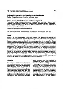
Results The median gestational age at delivery for the term groups, TNL and TSL, was 38.5 and 39.5 weeks respectively (n ⫽ 4 per group). All were uncomplicated by evidence of infection. The clinical details of the PTL–INF and PTL⫹INF are noted in Table I. All 384 genes in the array exceeded the threshold of detection in at least one of the 32 samples tested, while 369 exceeded it in two samples in at least one group. Of these, 301 were detected in all four samples of at least one group in at least one tissue. Sixty-three genes were detected in all 32 samples. Figures 1 and 2 summarize the differential expression observed in these blots for 90 genes that have relevance to inflammatory responses in the tissues. These genes are listed in Table II along with their GenBank accession numbers and a number representing their placement in Figures 1 and 2. Table III lists the candidate genes for differential expression from Figures 1 and 2. Information regarding the expression of genes less overtly associated with inflammation will be published elsewhere. The profiles of chemokine expression exhibit elevated expression of several of these genes in the leukocyte-infiltrated (PTL⫹INF) tissue samples compared with the PTL–INF and at TSL compared with TNL (Figures 1 and 2). MIP-1β in the amnion was 8-fold (P ⫽ 0.03) higher in the PTL⫹INF than in the PTL–INF group (Figure 1B,C, number 13 and Table III). Pulmonary and activation-regulated chemokine (PARC) expression was likewise elevated (P ⫽ 0.03) in the PTL⫹INF choriodecidua (Figure 2B,C, number 16). Several others including GRO-α, GRO-β, I-1309, IL-8, MCP-1, MIP-1α and MIP3α were also candidates for differential expression, particularly in the amnion, with high expression in PTL⫹INF compared with PTL–INF (Table III and Figures 1 and 2, numbers 4, 5, 7, 8, 10, 12 and 14 respectively). Notably, in none of the TNL amnion samples did expression of IL-8, MIP-1α, or MIP-1β exceed the limit of detection. Furthermore, GRO-α and GRO-β were both detectable in only one TNL amnion sample. Expression of each of these five genes was detected in all four PTL⫹INF amnion samples and also in at least three of the four PTL–INF amnion samples. GRO-β, GRO-γ and IL-8 were also detectable in all four TSL amnion samples where they were significantly (P ⫽ 0.03) more highly expressed than in TNL samples. Expression of chemokines in the preterm choriodecidua was generally reflective of that in the amnion. At term, no changes in expression of GRO-β, GRO-γ and IL-8 were evident in the choriodecidua. Among the pro-inflammatory cytokines, IL-1β, oncostatin M and pre-B cell enhancing factor (PBEF) were the most notable candidates for greater expression with infection (Figures 1C and 2C, numbers 19, 29 and 30 respectively). IL-1β was much more highly expressed in three of the four PTL⫹INF samples relative to the other groups in both the amnion and choriodecidua. The weakly expressing PTL⫹INF sample for the two tissue types, however, originated from different placentae. Oncostatin M expression differed significantly (P ⫽ 0.03) in choriodecidua between PTL⫹INF and PTL–INF, and in both amnion and choriodecidua was always 401
K.W.Marvin et al.
Figure 1. Profiles of differential inflammation-related gene expression in amnion samples obtained after (A) term spontaneous labour (TSL) or preterm labour (B) without (PTL–INF) or (C) with (PTL⫹INF) infection compared with that in samples from Caesarean section at term without labour (TNL). The results are represented as ratios of median normalized expression between groups (left ordinate): bars above the x-axis, TSL:TNL, PTL–INF:TNL or PTL⫹INF:TNL; bars below the x-axis, TNL:TSL, TNL:PTL–INF or TNL:PTL⫹INF (ordinate labelled in italics). Where expression of a gene failed to exceed the threshold of detection (TD) in more than one sample in a group, the median TD for the TNL group (TDTNL) was substituted in calculations of the ratios. Also shown in A (—, right ordinate) are ratios of median normalized expression with TNL to TDTNL to represent the degree of expression exhibited by that group. Each Human Cytokine Expression Array was probed successively with four independent 33P-labelled first strand cDNAs derived from the four amnion samples used for one of the four groups. One array was used for each group. Bars 1–90 correspond to genes 1–90 shown in Table II.
Table I. Clinical details for preterm deliveries after the start of labour (PTL) Sample at delivery (weeks) PTL–INF 1 2 3 4 PTL⫹INF 1 2 3d 4e 5
Gestational age (g)
Birthweight infiltrationa
Leukocyte infection
Clinical details
Delivery
32 32 34 31
1750 2800 2360 1850
2 1 1 1
(–)b (–)b (–) (–)
– – – –
V CS V, PROM CS, PROM
27 31 25 29 26
800 1590 890 1190 705
3 3 4 4 3
(⫹) (⫹) (⫹) (⫹)c (⫹)
FT, NS FT, MT, NS UT, NS BS, NS NS
CS V, PROM V, PROM V, PROM V
⫾ INF ⫽ with or without infection; FT ⫽ fetal tachycardia; MT ⫽ maternal tachycardia; UT ⫽ uterine tenderness; BS ⫽ positive placental bacterial swab; NS ⫽ neonatal sepsis; V ⫽ vaginal (delivery); CS ⫽ Caesarean section; PROM ⫽ premature rupture of membranes. aScore, 0–4, for CD45⫹ cells in the gestational membranes (⫹) or (–), presence or absence of infiltration of the fetal membranes respectively. b,cPathologist reports available: bnegative or cpositive for chorioamnionits. dChoriodecidua sample only. eAmnion sample only.
greater with PTL⫹INF than following TNL. PBEF expression was greater in all samples delivered with labour than in the TNL samples. Furthermore, the increase in the amnion with labour at term (P ⫽ 0.03) was significant. Interestingly, instead of being increased in infection or labour, the expression of 402
TNF-α was highest in three of the four TNL amnion samples compared to all other samples. Among anti-inflammatory cytokines, elevated expression of inhibin α in the PTL–INF amnion was most notable (Figure 1B, number 35). All four samples of PTL–INF amnion exhibited
Inflammatory gene activation with labour
Figure 2. Profiles of differential gene expression in choriodecidual samples obtained after preterm and term labour relative to Caesarean section at term without labour. Details are described for Figure 1 but for choriodecidual samples.
greater expression of inhibin α than did any of the other amnion samples, and the 3-fold difference between PTL–INF and PTL⫹INF was significant (P ⫽ 0.03). This was not accompanied by a large increase in expression of activin βA subunit (Figure 1B, number 34). Conversely, in the term choriodecidua, activin βA, which was expressed in all choriodecidual samples, increased significantly (P ⫽ 0.03) with labour (Figure 2A, number 34), while inhibin did not. Another TGF-β family member, MIC-1 (Figure 2A, number 36), also increased significantly in choriodecidua with term labour (P ⫽ 0.03). Changes in responsiveness to modulators of inflammation may be mediated through availability of receptors. Expression of the naturally occurring IL-1 receptor antagonist (IL-1ra) (Figure 1 and 2, number 31) was higher in three PTL⫹INF choriodecidual samples than in any other samples but was undetectable in the fourth PTL⫹INF choriodecidual sample. Several chemokine and cytokine receptors are candidates for greater expression with PTL⫹INF and TSL (Figures 1 and 2, Table III). Moreover, the data suggest a trend toward a modestly increased expression of a large number of cytokine receptor genes in the amnion for either PTL (Figure 1) group compared to term. Chemokine receptor CXCR-2 (Figure 1, number 46) was significantly more highly expressed in the amnion with PTL⫹INF than with PTL–INF (P ⫽ 0.03), while CXCR-4 (Figure 1, number 47) expression with PTL⫹INF was more variable. It was undetectable in one PTL⫹INF choriodecidual and two PTL⫹INF amnion samples, but highly elevated in the remaining PTL⫹INF samples. With TSL (versus TNL), CXCR-1 (Figure 2, number 45) was significantly elevated in the choriodecidua (P ⫽ 0.03). Among cytokine receptors, TGF-β receptor III
was significantly (P ⫽ 0.03) more highly expressed in TSL than in TNL choriodecidual samples and G-CSF receptor was highly elevated in three of the four PTL⫹INF choriodecidual relative to the PTL–INF samples (Figure 2, numbers 65 and 66 respectively). In addition, the lipopolysaccharide receptor, CD14, was more highly expressed in this tissue with PTL⫹INF versus PTL–INF (Figures 2B,C, number 68). The profiles of matrix metalloproteinase (MMP) and tissue inhibitor of metalloproteinases (TIMP) also appear to exhibit distinctive patterns with preterm labour. In particular, dramatically higher MMP-14 (Figure 1, number 74) was observed in the amnion for all eight preterm deliveries relative to term. Interestingly, expression of TIMP-3 and TIMP-4 in the amnion was greater with PTL–INF than with PTL⫹INF (P ⫽ 0.03) (Figure 1, numbers 77 and 78 respectively, and Table III). Finally, differences in median expression of cell adhesion molecules with labour at term and with infection preterm were all ⬍5-fold. However, expression of an extracellular matrix receptor subunit, α6-integrin, was significantly lower, and that of a leukocyte associated subunit, β2-integrin, was significantly greater, in choriodecidua with PTL⫹INF than with PTL–INF (P ⫽ 0.03) (Figure 1, numbers 87 and 89 respectively, and Table III).
Discussion Several approaches to the prediction of preterm labour have been described over the years. Studies of biochemical markers, such as serum corticotrophin-releasing hormone, salivary estriol, and cervicovaginal fetal fibronectin, pro-inflammatory cytokines and cell adhesion molecules offer hope that indicators 403
K.W.Marvin et al.
Table II. Inflammation-associated genes assessed in Figures 1 and 2
Table II. continued
No.
Gene name (accession no.a)
No.
Gene name (accession no.a)
1 2 3 4 5 6 7 8 9 10 11 12 13 14 15 16 17 18 19 20 21 22 23 24 25 26 27 28 29 30 31 32 33 34 35 36 37 38 39 40 41 42 43 44 45 46 47 48 49 50 51 52 53 54 55 56 57 58 59 60 61 62 63 64 65 66 67 68 69 70
IP-10 (X02530) ENA-78 (X78686) Fractalkine (U91835) GRO-α (J03561) GRO-β (M36820) GRO-γ (M36821) I-309 (M57502) IL-8 (Y00787) MIP-1δ (AF031587) MCP-1 (S69738) MCP-2 (Y16645) MIP-1α (AF043339) MIP-1β (J04130) MIP-3α (U77035) MIP-3β (AB000887) PARC (AB000221) RANTES (M21121) IL-1α (M28983) IL-1β (M15330) IL-6 (M14584) IL-11 (M57765) IL-16 (M90391) IFN-γ (X13274) TNF-α (M10988) Lymphotoxin[LT] β (L11015) G-CSF (X03438) M-CSF (M64592) MIF (M25639) Oncostatin M (AH001516) PBEF (U02020) IL-1ra (M55646) IL-4 (M13982 IL-10 (M57627) Activin βA (M13436) Inhibin α (M13981) MIC-1 (AF019770) TGF-β1 (M38449) CCR-1 (L10918) CCR-2A (U03882) CCR-4 (X85740) CCR-6 (Z79784) CCR-7 (L31581) CCR-9 (Y12815) CX3CR-1 (U20350) CXCR-1 (L19591) CXCR-2 (M73969) CXCR-4 (X71635) CXCR-5 (X68149) IL-1 RI (X16896) IL-2 Rα (X01057) IL-3 Rα (M74782) IL-4 Rα (X52425) IL-10 Rα (U00672) IL-11 Rα (Z38102) IL-12 Rβ1 (U03187) IL-18 R (U43672) IFN-α/β Rα (J03171) IFN-γ R1 (J03143) IFN-γ R2 (U05875) TNF RI (M63121) TNF RII (M55994) LT-β R (L04270) TGF-β RI (AF054598) TGF-β RII (M85079) TGF-β RIII (L07594) G-CSF R (M59818) Endoglin (X72012) CD14 (X06882) MMP-1 (X05231) MMP-7 (NM002423)
71 72 73 74 75 76 77 78 79 80 81 82 83 84 85 86 87 88 89 90
MMP-8 (NM002424) MMP-9 (J05070) MMP-10 (X07820) MMP-14 (D26512) TIMP-1 (S68252) TIMP-2 (M32304) TIMP-3 (U67195) TIMP-4 (U76456) ICAM-1 (J03132) ICAM-2 (X15606) ICAM-3 (S50015) VCAM-1 (X53051) E-Selectin (M30640) L-Selectin (M25280) P-Selectin (M25322) Integrin-α5 (X06256) Integrin-α6 (X53586) Integrin-β1 (X07979) Integrin-β2 (M15395) Integrin-β4 (X51841)
404
aFor
GenBank sequences used in design of the cDNA amplicons spotted on the arrays.
or combinations thereof may be developed for accurate clinical prediction of preterm labour (Lockwood and Kuczynski, 1999; Lu and Goldenberg, 2000). However, while these methods are promising, they are not yet sufficiently conclusive for routine use. Similarly, development of tocolytic treatments has also met with only limited success (Higby et al., 1993; Hill, 1995). β-Adrenergic sympathomimetics delay labour by only 24–48 h (Higby et al., 1993). Prostaglandin synthesis inhibitors constrict the fetal ductus arteriosus and promote renal insufficiency, oligohydramnios and pulmonary hypertension in the fetus (Higby et al., 1993). Finally, antibiotics do not halt infectionassociated preterm labour. Other agents have been ineffective as tocolytics. Effective treatment of preterm labour will require both early diagnosis and effective intervention in the pathophysiological processes responsible for the disorder. Products of genes exhibiting altered expression in association with particular aetiologies, with labour and with preterm labour, in general are both potential markers of, and potential participants in, the molecular mechanisms underlying preterm labour. As such, their identification may lead not only to new diagnostics but better understanding of these mechanisms, which may in turn lead to design of improved diagnostic and treatment regimes. In the present study, the use of Human Cytokine Expression Arrays has permitted the simultaneous evaluation of the expression in gestational membranes of a large number of genes which are related to the control of inflammatory responses. This has the advantage of revealing the effects of labour at term and of infection preterm on groups of related genes. It may also aid discovery of novel markers of preterm labour with or without chorioamnionitis, revealing candidate genes which have had limited study to date in the gestational tissues. The approach is primarily a survey, assisting in design of subsequent studies of expression focused on a smaller number of genes but having greater statistical power.
Inflammatory gene activation with labour
Table III. Candidate differentially expressed genesa Gene name
No.b
Tissue
Comparedc
Chemokines GRO-α 4 Am Preterm GRO-α 4 CD Preterm GRO-β 5 Am Preterm GRO-β 5 Am Term GRO-γ 6 Am Term I-309 7 Am Preterm IL-8 8 Am Preterm IL-8 8 CD Preterm IL-8 8 Am Term MCP-1 10 CD Preterm MIP-1α 12 Am Preterm MIP-1β 13 Am Preterm MIP-3α 14 Am Preterm PARC 16 CD Preterm Cytokines IL-1β 19 Am Preterm IL-1β 19 CD Preterm TNF-α 24 Am Term Oncostatin M 29 CD Preterm PBEF 30 Am Term PBEF 30 Am Preterm PBEF 30 CD Preterm IL-1ra 31 CD Preterm Activin βA 34 CD Term Inhibin α 35 Am Preterm MIC-1 36 CD Term Chemokine receptors CXCR-1 45 CD Term CXCR-2 46 CD Preterm CXCR-4 47 Am Preterm CXCR-4 47 CD Preterm Cytokine and other receptors TGF-β RIII 65 CD Term G-CSF R 66 CD Preterm CD14 68 CD Preterm Matrix metalloproteinases (inhibitors of) TIMP-3 77 Am Preterm TIMP-4 78 Am Preterm Cell adhesion molecules Integrin-α6 87 CD Preterm Integrin-β2 89 CD Preterm aRatio
Ratio
P
6.0 6.2 5.7 1.5 1.7 12.0 44.8 18.5 3.5 7.2 7.1 7.8 8.3 4.8
0.06 0.11 0.06 0.03 0.03 0.18 0.06 0.31 0.02 0.25 0.06 0.03 0.19 0.03
17.4 7.8 0.07 2.6 6.2 11.2 11.5 9.3 3.6 0.34 2.0
0.06 0.11 0.19 0.03 0.03 0.11 0.11 0.12 0.03 0.03 0.03
2.4 2.3 11.1 6.0
0.03 0.03 0.64 0.18
1.4 8.5 3.3
0.03 0.06 0.03
0.13 0.12
0.03 0.03
0.53 2.4
0.03 0.03
of median expression 艌5 or 艋0.2 or P 艋 0.05.
bNumber from Table II and Figures 1 and 2. cPreterm ⫽ PTL⫹INF:PTL–INF; Term ⫽ TSL:TNL
The array approach generally suffers from three major limitations. First, the size of mRNA is not determined by the method. Thus related genes and regulation of production of active gene products through alternate mRNA splicing may result in false positives or false negatives respectively, for altered gene expression. Second, changes in mRNA expression do not necessarily correlate closely to changes in abundance and activity of gene products. This is a factor in interpretation of all analyses of mRNA expression. Third, the number of replicates per group is small, often n ⫽ 1 sample or pool of samples for each group, permitting no analysis of variability. We have demonstrated that using reprobing of arrays on nylon membranes, it is feasible to increase the number of replicates to n ⫽ 4, permitting limited assessment of variability. Essentially, with the Wilcoxon test and n ⫽ 4, non-overlapping ranges are required for groups to be significantly different, a condition that the variability of gene expression in human gestational membranes, particularly in the PTL groups, often
appeared to preclude. Considerable benefit in statistical power could undoubtedly be obtained from further increases in group size. However, the time and cost to produce this increase would be considerable and would not obviate the need for follow-on experiments. The results obtained with the present arrays, although not necessarily definitive with respect to particular genes, will aid greatly in design of subsequent experiments, both in hypothesis-building regarding differential expression of candidate genes and groups thereof, and in the degree of variation which needs to be accommodated by the design. The association of increased chemokine expression with leukocyte infiltration accords with expectation. GRO-α, -β and -γ and IL-8 all promote chemotaxis of neutrophils. MIP1α and -1β, and PARC attract monocytes, dendritic cells and several lineages of T-cell. Expression of CXCR2 and CXCR4 is characteristic respectively of neutrophils and of these other lineages. Neutrophils are the major component of leukocytes that infiltrate the membranes during chorioamnionitis and the other lineages have also been detected. Clearly, changes in gene expression in these tissues may reflect several mechanisms. Increased expression of a gene may reflect the differential content of infiltrating cells and factors such as mode of delivery, or may either directly or secondarily reflect the underlying inflammation-promoting mechanisms of the resident cells of the tissues. Additionally, advancing gestational age also affects expression of some genes. Thus, the intriguing apparent differential expression of genes between PTL–INF and term labour (e.g. inhibin α, MMP-14 and TIMP-4, Figure 1B, numbers 35, 74 and 78 respectively) may be either associated with gestational age or pathophysiology. Conversely, at term, proinflammatory changes in expression commence prior to labour (Elliott et al., 2001). Further studies, with inclusion of preterm not in labour samples, will be required to distinguish these possibilities. We have previously demonstrated that immunoreactive IL-8 increases significantly with labour either at term or preterm (Keelan et al., 1999). Thus the elevated IL-8 expression after TSL, compared with TNL, reflects our previous findings, and similar results for GRO-β and GRO-γ lend further support to the idea that normal labour has characteristics of an inflammatory response. Less supportive of this model, however, are the results with respect to expression of cytokines. Little change was observed with TSL for IL-1β or IL-6, while TNFα even appears to decline markedly with TSL in the amnion. Expression of oncostatin M, an IL-6 family member, likewise, did not appear to change with TSL although it increased significantly in the choriodecidua with chorioamnionitis. However, expression of the less studied pro-inflammatory cytokine, PBEF, increased significantly with term labour. Recent reports suggest that this may be a consequence of stretch in the gestational membranes (Nemeth et al., 2000a,b). PBEF is, however, also a new potential marker of chorioamnionitis as we and another study (Ognjanovic et al., 2001) have shown. As the major reported role for this factor is as a pre-B cell differentiation factor (Samal et al., 1994), it is unlikely that PBEF would have been quickly noted as worthy of study in the gestational membranes, were it not for such differential 405
K.W.Marvin et al.
screens as subtractive hybridization (Nemeth et al., 2000a,b) or cDNA array analysis. The thick extracellular matrix of the amnion may be quite important for the integrity of the membranes. In this tissue, the high expression of the matrix protease MMP-14 in all preterm deliveries and the decline of matrix protease inhibitors TIMP-3 and TIMP-4, with preterm infection may thus play a role in the high rate of preterm premature spontaneous rupture of membranes. In conclusion, these studies may provide a major step forward for development of new strategies for the prediction, diagnosis and prevention of preterm labour and delivery.
Acknowledgements We gratefully acknowledge Dr M.Coleman and the theatre staff at the National Women’s Hospital, Auckland, New Zealand, for their assistance with the collection of tissues. Mr J.Graham and Ms C.Paton are also thanked for their assistance with the histological characterization of the tissues, as are Drs L.McCowan and M.Batton for their clinical input. We also thank Mr A.W.Stewart, Biostatistician, for his advice on analysis of the data. This study was funded by the Health Research Council of New Zealand, the New Zealand Lottery Health Grants Board and the Royal Society of New Zealand Marsden Fund.
References Arntzen, K.J., Kjollesdal, A.M., Halgunset, J. Vatten, L. and Austgulen, R. (1998) TNF, IL-1, IL-6, IL-8 and soluble TNF receptors in relation to chorioamnionitis and premature labor. J. Perinat. Med., 26, 17–26. Athayde, N., Romero, R., Maymon, E., Gomez, R. Pacora, P., Yoon, B.H. and Edwin, S.S. (2000) Interleukin 16 in pregnancy, parturition, rupture of fetal membranes, and microbial invasion of the amniotic cavity. Am. J. Obstet. Gynecol., 182, 135–141. Barclay, C.G., Brennand, J.E., Kelly, R.W. and Calder, A.A. (1993) Interleukin8 production by the human cervix. Am. J. Obstet. Gynecol., 169, 625–632. Bokstrom, H., Brannstrom, M., Alexandersson, M. and Norstrom, A. (1997) Leukocyte subpopulations in the human uterine cervical stroma at early and term pregnancy. Hum. Reprod., 12, 586–590. Bry, K., Hallman, M. and Lappalainen, U. (1994) Cytokines released by granulocytes and mononuclear cells stimulate amnion cell prostaglandin E2 production. Prostaglandins, 48, 389–399. Burrus, D.R., Ernest, J.M. and Veille, J.C. (1995) Fetal fibronectin, interleukin6, and C-reactive protein are useful in establishing prognostic subcategories of idiopathic preterm labor. Am. J. Obstet. Gynecol., 173, 1258–1262. Cherouny, P.H., Pankuch, G.A., Romero, R., Botti, J.J., Kuhn, D.C., Demeres, L.M. and Appelbaum, P.C. (1993) Neutrophil attractant/activating peptide-1/ interleukin-8: association with histologic chorioamnionitis, preterm delivery, and bioactive amniotic fluid leukoattractants. Am. J. Obstet. Gynecol., 169, 1299–1303. Cohen, J., Ghezzi, F., Romero, R., Ghidini, A., Mazor, M., Tolosa, J.E., Goncalves, L.F. and Gomez, R. (1996) GRO alpha in the fetomaternal and amniotic fluid compartments during pregnancy and parturition. Am. J. Reprod. Immunol., 35, 23–29. Copper, R.L., Goldenberg, R.L., Creasy, R.K., DuBard, M.B., Davis, R.O., Entman, S.S., Iams, J.D. and Cliver, S.P. (1993) A multicenter study of preterm birth weight and gestational age-specific neonatal mortality. Am. J. Obstet. Gynecol., 168,78–84. Denison, F.C., Kelly, R.W., Calder, A.A. and Riley, S.C. (1998) Cytokine secretion by human fetal membranes, decidua and placenta at term. Hum. Reprod., 13, 3560–3565. Denison, F.C., Kelly, R.W., Calder, A.A. and Riley, S.C. (1999) Secretory leukocyte protease inhibitor concentration increases in amniotic fluid with the onset of labour in women: characterization of sites of release within the uterus. J. Endocrinol., 161, 299–306. Dudley, D.J. (1997) Pre-term labor—an intra-uterine inflammatory response syndrome. J. Reprod. Immunol., 36, 93–109.
406
Dudley, D.J., Hunter, C., Mitchell, M.D. and Varner, M.W. (1994) Clinical value of amniotic fluid interleukin-6 determinations in the management of preterm labour. Br. J. Obstet. Gynecol., 101, 592–597. Dudley, D.J., Collmer, D., Mitchell, M.D. and Trautman, M. S. (1996a) Inflammatory cytokine mRNA in human gestational tissues: implications for term and preterm labor. J. Soc. Gynecol. Invest., 3, 328–335. Dudley, D.J., Edwin, S.S., Dangerfield, A., Van Waggoner, J. and Mitchell, M.D. (1996b) Regulation of cultured human chorion cell chemokine production by group B streptococci and purified bacterial products. Am. J. Reprod. Immunol., 36, 264–268. Dudley, D.J., Edwin, S.S. and Mitchell, M.D. (1996c) Macrophage inflammatory protein-1-alpha regulates prostaglandin E2 and interleukin-6 production by human gestational tissues in vitro. J. Soc. Gynecol. Invest., 3, 12–16. Elliott, C.L., Loudon, J.A., Brown, N., Slater, D.M., Bennett, P.R. and Sullivan, M.H. (2001) Expression of IL-1b and IL-8 in human fetal membranes throughout pregnancy. Am. J. Reprod. Immunol., 46, 260–267. Fidel, P.L., Jr, Romero, R., Ramirez, M., Cutright, J., Edwin, S.S., LaMarche, S., Cotton, D.B. and Mitchell, M.D. (1994) Interleukin-1 receptor antagonist (IL-1ra) production by human amnion, chorion, and decidua. Am. J. Reprod. Immunol., 32, 1–7. Fortunato, S.J., Menon, R. and Lombardi, S.J. (1999) MMP/TIMP imbalance in amniotic fluid during PROM: an indirect support for endogenous pathway to membrane rupture. J. Perinat. Med. 27, 362–368. Gomez, R., Romero, R., Galasso, M., Behnice, E., Insunza, A. and Cotton, D.B. (1994) The value of amniotic fluid interleukin-6, white blood cell count, and gram stain in the diagnosis of microbial invasion of the amniotic cavity in patients at term. Am. J. Reprod. Immunol., 32, 200–210. Gomez, R., Romero, R., Edwin, S.S. and David, C. (1997) Pathogenesis of preterm labor and preterm premature rupture of membranes associated with intraamniotic infection. Infect. Dis. Clin. North Am., 11, 135–176. Greig, P.C., Ernest, J.M., Teot, L., Erikson, M. and Talley, R. (1993) Amniotic fluid interleukin-6 levels correlate with histologic chorioamnionitis and amniotic fluid cultures in patients in premature labor with intact membranes. Am. J. Obstet. Gynecol., 169, 1035–1044. Halgunset, J., Johnsen, H., Kjollesdal, A.M., Qvigstad, E., Espevik, T. and Austgulen, R. (1994) Cytokine levels in amniotic fluid and inflammatory changes in the placenta from normal deliveries at term. Eur. J. Obstet. Gynecol. Reprod. Biol., 56, 153–160. Hall, M.H., Danielian, P. and Lamont, R.F. (1997) The importance of preterm birth. In Elder, M.G., Lamont, R.F. and Romero, R., (eds), Preterm Labor. Churchill Livingstone, New York, pp. 1–27. Higby, K., Xenakis, E.M. and Pauerstein, C.J. (1993) Do tocolytic agents stop preterm labor? A critical and comprehensive review of efficacy and safety. Am. J. Obstet. Gynecol., 9, 1256–1259. Hill, W.C. (1995) Risks and complications of tocolysis. Clin. Obstet. Gynecol., 38, 725–745. Hillier, S.L., Witkin, S.S., Krohn, M.A., Watts, D.H., Kiviat, N.B. and Eschenbach, D.A. (1993) The relationship of amniotic fluid cytokines and preterm delivery, amniotic fluid infection, histologic chorioamnionitis, and chorioamnion infection. Obstet. Gynecol., 81, 941–948. Hoskins, I.A., Zandieh, P., Schatz, F. and Lee, C. (1997) Amniotic fluid granulocyte colony stimulating factor levels: a rapid marker for diagnosing chorioamnionitis. Am. J. Reprod. Immunol., 38, 286–288. Hsu, C.D., Meaddough, E., Aversa, K. and Copel, J.A. (1998a) The role of amniotic fluid L-selectin, GRO-alpha, and interleukin-8 in the pathogenesis of intraamniotic infection. Am. J. Obstet. Gynecol., 178, 428–432. Hsu, C.D., Meaddough, E., Aversa, K., Hong, S.F., Lu, L.C., Jones, D.C. and Copel, J.A. (1998b) Elevated amniotic fluid levels of leukemia inhibitory factor, interleukin 6, and interleukin 8 in intra-amniotic infection. Am. J. Obstet. Gynecol., 179, 1267–1270. Inglis, S.R., Jeremias, J., Kuno, K., Lescale, K., Peeper, Q., Chervenak, F.A. and Witkin, S.S. (1994) Detection of tumor necrosis factor-alpha, interleukin-6, and fetal fibronectin in the lower genital tract during pregnancy: relation to outcome. Am. J. Obstet. Gynecol., 171, 5–10. Ishihara, O., Numari, H., Saitoh, M., Arai, Y., Takanashi, H., Kitagawa, H. and Kinoshita, K. (1996) Prostaglandin E2 production by endogenous secretion of interleukin-1 in decidual cells obtained before and after the labor. Prostaglandins, 52, 199–208. Jones, C.A., Williams, K.A., Finlay-Jones, J.J. and Hart, P.H. (1995) Interleukin 4 production by human amnion epithelial cells and regulation of its activity by glycosaminoglycan binding. Biol. Reprod., 52, 839–847. Keelan, J.A., Coleman, M. and Mitchell, M.D. (1997a) The molecular mechanisms of term and preterm labor: recent progress and clinical implications. Clin. Obstet. Gynecol., 40, 460–478.
Inflammatory gene activation with labour Keelan, J.A., Sato, T. and Mitchell, M.D. (1997b) Interleukin (IL)-6 and IL8 production by human amnion: regulation by cytokines, growth factors, glucocorticoids, phorbol esters, and bacterial lipopolysaccharide. Biol. Reprod., 57, 1438–1444. Keelan, J.A., Marvin, K.W., Sato, T.A. Coleman, M., McCowan, L.M. and Mitchell, M.D. (1999) Cytokine abundance in placental tissues: evidence of inflammatory activation in gestational membranes with term and preterm parturition. Am. J. Obstet. Gynecol., 181, 1530–1536. Keirse, M.J. (1995) New perspectives for the effective treatment of preterm labor. Am. J. Obstet. Gynecol., 173, 618–628. Keski-Nisula, L., Aalto, M.L., Katila, M.L. and Kirkinen, P. (2000) Intrauterine inflammation at term: a histopathologic study. Hum. Pathol., 31, 841–846. Laham, N., Brennecke, S.P. and Rice, G.E. (1999) Interleukin-8 release from human gestational tissue explants: effects of gestation, labor, and chorioamnionitis. Biol. Reprod., 61, 823–827. Lenhart, J.A., Ohleth, K.M., Ryan, P.L., Palmer, S.S. and Bagnell, C.A. (1999) Effect of relaxin on tissue inhibitor of metalloproteinase-1 and -2 in the porcine uterus and cervix. Ann. NY Acad. Sci., 878, 565–566. Liggins, G.C. (1981) Cervical ripening as an inflammatory reaction. In Ellwood, D.A. and Anderson, A.B.M. (eds), The Cervix in Pregnancy and Labour, Clinical and Biochemical Investigations. Churchill Livingstone, Edinburgh, pp. 1–9. Lockwood, C.J. and Kuczynski, E. (1999) Markers of risk for preterm delivery. J. Perinat. Med., 27, 5–20. Lu, G.C. and Goldenberg, R.L. (2000) Current concepts on the pathogenesis and markers of preterm births. Clin. Perinatol., 27, 263–83. Maeda, K., Matsuzaki, N., Fuke, S., Mitsuda, N., Shimoya, K., Nakayama, M., Suehara, N. and Aono, T. (1997) Value of the maternal interleukin 6 level for determination of histologic chorioamnionitis in preterm delivery. Gynecol. Obstet. Invest., 43, 225–231. Maehara, K., Kanayama, N., Maradny, E.E., Uezato, T., Fujita, M. and Terao, T. (1996) Mechanical stretching induces interleukin-8 gene expression in fetal membranes—a possible role for the initiation of human parturition. Eur. J. Obstet. Gynecol. Reprod. Biol., 70, 191–196. Marvin, K.W., Keelan, J.A., Sato, T.A., Coleman, M.A., McCowan, L.M. and Mitchell, M.D. (1999) Expression of intercellular adhesion molecule-1 (ICAM-1) in choriodecidua with labour and delivery at term and preterm. Reprod. Fertil. Dev., 11, 255–262. Marvin, K., Keelan, J., Coleman, M.A., McCowan, L.M., Zhou, R.L. and Mitchell, M.D. (2000a) Intercellular adhesion molecule-1 (ICAM-1) in cervicovaginal fluid of women presenting with preterm labor: predictive value for preterm delivery. Am. J. Reprod. Immunol., 43, 264–271. Marvin, K.W., Keelan, J.A., Sato, T.A., Coleman, M.A., McCowan, L.M., Miller, H.C. and Mitchell, M.D. (2000b) Enhanced expression of intercellular adhesion molecule-1 (ICAM-1) in amnion with term and preterm labour. Placenta, 21, 115–121. McLaren, J., Taylor, D.J. and Bell, S.C. (2000) Increased concentration of pro-matrix metalloproteinase 9 in term fetal membranes overlying the cervix before labor: implications for membrane remodeling and rupture. Am. J. Obstet. Gynecol., 182, 409–416. Mercer, B.M. and Lewis, R. (1997) Preterm labor and preterm premature rupture of the membranes. Diagnosis and management. Infect. Dis. Clin. North Am., 11, 177–201. Morrison, J.C. (1990) Preterm birth: a puzzle worth solving. Obstet. Gynecol., 76, 5S–12S. Nemeth, E., Tashima, L.S., Yu, Z. and Bryant-Greenwood, G.D. (2000a) Fetal membrane distention: I. Differentially expressed genes regulated by acute distention in amniotic epithelial (WISH) cells. Am. J. Obstet. Gynecol., 182, 50–59. Nemeth, E., Millar, L.K. and Bryant-Greenwood, G. (2000b) Fetal membrane distention: II. Differentially expressed genes regulated by acute distention in vitro. Am. J. Obstet. Gynecol., 182, 60–67. Ogawa, M., Hirano, H., Tsubaki, H., Kodama, H. and Tanaka, T. (1998) The role of cytokines in cervical ripening|correlations between the concentrations of cytokines and hyaluronic acid in cervical mucus and the induction of hyaluronic acid production by inflammatory cytokines by human cervical fibroblasts. Am. J. Obstet. Gynecol., 179, 105–110. Ognjanovic, S., Bao, S., Yamamoto, S.Y., Garibay-Tupas, J., Samal, B. and Bryant-Greenwood, G.D. (2001) Genomic orginization of the gene coding for human pre-B-cell colony enhancing factor and expression in the human fetal membranes. J. Mol. Endocrinol., 26, 107–117. Olah, K.S., Vince, G.S., Neilson, J.P., Deniz, G. and Johnson, P.M. (1996) Interleukin-6, interferon-gamma, interleukin-8, and granulocytemacrophage colony stimulating factor levels in human amniotic fluid at term. Am. J. Obstet. Gynecol., 32, 89–98.
Osmers, R.G.W., Adelmann-Grill, B.C., Rath, W., Stuhlsatz, H. W. Tschesche, H. and Kuhn, W. (1995a) Biochemical events in cervical ripening dilatation during pregnancy and parturition. J. Obstet. Gynecol., 21, 185–194. Osmers, R.G.W., Blaser, J., Kuhn, W. and Tschesche, H. (1995b) Interleukin8 synthesis and the onset of labor. J. Obstet. Gynecol., 86, 223–229. Papadogiannakis, N. (1997) Traffic of leukocytes through the maternofetal placental interface and its possible consequences. Curr. Top. Microbiol. Immunol., 222, 141–157. Pollard, J.K. and Mitchell, M.D. (1996) Intrauterine infection and the effects of inflammatory mediators on prostaglandin production by myometrial cells from pregnant Women. Am. J. Obstet. Gynecol., 174, 682–686. Putz, I., Wienecke, B., Winkler, M. and Rath, W. (1996) Interleukin-1alpha and interleukin-1beta levels in amniotic fluid during pregnancy: correlation with microbial invasion of the amniotic cavity. Geburtsh. Frauenheilk., 56, 640–644. Rechberger, T. and Woessner, J.F., Jr (1993) Collagenase, its inhibitors, and decorin in the lower uterine segment in pregnant women. Am. J. Obstet. Gynecol., 168, 1598–603. Rizzo, G., Capponi, A., Rinaldo, D., Tedeschi, D., Arduini, D. and Romanini, C. (1996) Interleukin-6 concentrations in cervical secretions identify microbial invasion of the amniotic cavity in patients with preterm labor and intact membranes. Am. J. Obstet. Gynecol., 175, 812–817. Rizzo, G., Capponi, A., Vlachopoulou, A., Angelini, E., Grassi, C. and Romanini, C. (1997) The diagnostic value of interleukin-8 and fetal fibronectin concentrations in cervical secretions in patients with preterm labor and intact membranes. J. Perinatal. Med., 25, 461–468. Rizzo, G., Capponi, A., Vlachopoulou, A., Angelini, E., Grassi, C. and Romanini, C. (1998a) Ultrasonographic assessment of the uterine cervix and interleukin-8 concentrations in cervical secretions predict intrauterine infection in patients with preterm labor and intact membranes. Ultrasound Obstet. Gynecol., 12, 86–92. Rizzo, G., Capponi, A., Vlachopoulou, A., Angelini, E., Grassi, C and Romanini, C. (1998b) Interleukin-6 concentrations in cervical secretions in the prediction of intrauterine infection in preterm premature rupture of the membranes. Gynecol. Obstet. Invest., 46, 91–95. Romero, R., Durum, S., Dinarello, C.A., Oyarzun, E., Hobbins, J.C. and Mitchell, M.D. (1989) Interleukin-1 stimulates prostaglandin biosynthesis by human amnion. Prostaglandins, 37, 13–22. Romero, R., Bo, Hyun, Y., Kenney, J.S., Gomez, R., Allison, A.C. and Sehgal, P.B. (1993) Amniotic fluid interleukin-6 determinations are of diagnostic and prognostic value in preterm labor. Am. J. Reprod. Immunol., 30, 167–183. Romero, R., Gomez, R., Galasso, M., Munoz, H., Acosta, L., Yoon, B.H., Svinarich, D. and Cotton, D.B. (1994a) Macrophage inflammatory protein1alpha in term and preterm parturition: Effect of microbial invasion of the amniotic cavity. Am. J. Reprod. Immunol., 32, 108–113. Romero, R., Gomez, R., Galasso, M., Mazor, M., Berry, S.M., Quintero, R.A. and Cotton, D.B. (1994b) The natural interleukin-1 receptor antagonist in the fetal, maternal, and amniotic fluid compartments: the effect of gestational age, fetal gender, and intrauterine infection. Am. J. Obstet. Gynecol., 171, 912–921. Sagawa, T., Furuta, I., Negishi, H., Kishida, T., Begum, S. and Fujimoto, S. (1996) Cytokines concentrations in the cervical mucus of pregnant women. J. Obstet. Gynecol. Res., 22, 517–522 Salafia, C.M., Weigl, C. and Silberman, L. (1989) The prevalence and distribution of acute placental inflammation in uncomplicated term pregnancies. Obstet. Gynecol., 73, 383–389. Samal, B., Sun, Y., Stearns, G., Xie, C., Suggs, S. and McNiece, I. (1994) Cloning and characterization of the cDNA encoding a novel human preB-cell colony-enhancing factor. Mol. Cell. Biol., 14, 1431–1437. Sato, T., Ito, A., Ogata, Y., Nagase, H. and Mori, Y. (1996) Tumor necrosis factor alpha (TNFalpha) induces pro-matrix metalloproteinase 9 production in human uterine cervical fibroblasts but interleukin 1alpha antagonizes the inductive effect of TNFalpha. FEBS Lett., 392, 175–178. Sennstrom, M.K.B., Brauner, A., Lu, Y., Granstrom, L. M.M., Malmstrom, A.L. and Ekman, G.E. (1997) Interleukin-8 is a mediator of the final cervical ripening in humans. Eur. J. Obstet. Gynecol. Reprod. Biol., 74, 89–92. Stallmach, T., Hebisch, G., Joller-Jemelka, H.I., Orban, P., Schwaller, J. and Engelmann, M. (1995) Cytokine production and visualized effects in the feto-maternal unit: quantitative and topographic data on cytokines during intrauterine disease. Lab. Invest., 73, 384–392.
407
K.W.Marvin et al. Sugano, T., Nasu, K., Narahara, H., Kawano, Y., Nishida, Y. and Miyakawa, I. (2000) Platelet-activating factor induces an imbalance between matrix metalloproteinase-1 and tissue inhibitor of metalloproteinases-1 expression in human uterine cervical fibroblasts. Biol. Reprod., 62, 540–546. Tanaka, Y., Narahara, H., Takai, N., Yoshimatsu, J., Anai, T. and Miyakawa, I. (1998) Interleukin-1-beta andinterleukin-8 in cervicovaginal fluid during pregnancy. Am. J. Obstet. Gynecol., 179, 644–649. Thomson, A.J., Telfer, J.F., Young, A., Campbell, S., Stewart, C.J., Cameron, I.T., Greer, I.A. and Norman, J.E. (1999) Leukocytes infiltrate the myometrium during human parturition: further evidence that labour is an inflammatory process. Hum. Reprod., 14, 229–236. Watari, M., Watari, H., DiSanto, M.E., Chacko, S., Shi, G.P. and Strauss, J.F., III (1999) Pro-inflammatory cytokines induce expression of matrix-
408
metabolizing enzymes in human cervical smooth muscle cells. Am. J. Path., 154, 1755–1762. Winkler, M. and Rath, W. (1996) The role of cytokines in the induction of labor, cervical ripening and rupture of the fetal membranes. Zeitschr. Geburtsh. Perinatol., 200, 1–12. Winkler, M., Oberpichler, A., Tschesche, H., Ruck, P., Fischer, D.C. and Rath, W. (1999) Collagenolysis in the lower uterine segment during parturition at term: correlations with stage of cervical dilatation and duration of labor. Am. J. Obstet. Gynecol., 181, 153–158. Young, R.A. (2000) Biomedical discovery with DNA arrays. Cell, 102, 9–15. Submitted on May 29, 2001; resubmitted on September 28, 2001; accepted on December 19, 2001