2Department of Computer Science and Engineering, School of Engineering and Applied Science, Washington University, St. Louis, Missouri. Stroke incidence ...
Case Report
Use of Computer Games as an Intervention for Stroke Rachel M. Proffitt, BS,1 Gazihan Alankus, MS,2 Caitlin L. Kelleher, PhD,2 and Jack R. Engsberg, PhD1 1
Program in Occupational Therapy, Washington University School of Medicine, St. Louis, Missouri; Department of Computer Science and Engineering, School of Engineering and Applied Science, Washington University, St. Louis, Missouri
2
Current rehabilitation for persons with hemiparesis after stroke requires high numbers of repetitions to be in accordance with contemporary motor learning principles. The motivational characteristics of computer games can be harnessed to create engaging interventions for persons with hemiparesis after stroke that incorporate this high number of repetitions. The purpose of this case report was to test the feasibility of using computer games as a 6-week home therapy intervention to improve upper extremity function for a person with stroke. One person with left upper extremity hemiparesis after stroke participated in a 6-week home therapy computer game intervention. The games were customized to her preferences and abilities and modified weekly. Her performance was tracked and analyzed. Data from pre-, mid-, and postintervention testing using standard upper extremity measures and the Reaching Performance Scale (RPS) were analyzed. After 3 weeks, the participant demonstrated increased upper extremity range of motion at the shoulder and decreased compensatory trunk movements during reaching tasks. After 6 weeks, she showed functional gains in activities of daily living (ADLs) and instrumental ADLs despite no further improvements on the RPS. Results indicate that computer games have the potential to be a useful intervention for people with stroke. Future work will add additional support to quantify the effectiveness of the games as a home therapy intervention for persons with stroke. Key words: occupational therapy, stroke, upper extremity, video games
S
troke incidence, new or recurrent, is about 795,000 every year, which makes it a leading cause of serious, long-term disability in the United States.1 As the US population ages, this number is expected to rise, leading to an increase in the number of people with a long-term disability. In 1999, more than 1.1 million American adults reported difficulty with functional limitations and activities of daily living (ADLs) or instrumental ADLs resulting from stroke.2 These functional limitations are often a result of hemiparesis, found in at least 50% of survivors 6 months post stroke.3 Hemiparesis is partial paralysis of 1 side of the body and often affects the upper extremities more than the lower extremities. Stroke survivors who experience hemiparesis in the upper extremities have deficits in range of motion, strength, and motor control. These deficits may affect their ability to perform everyday tasks such as bathing, dressing, or cooking.4 Hemiparesis can also affect participation in everyday life. People may avoid certain activities altogether after a stroke because of fear of failure or insecurity with having a disability.5 For people with hemiparesis, regaining function in the upper extremity as early as possible is
important because it can lead to a more complete recovery.6 Fortunately, the brain has been shown to have an inherent capacity to recover some of the lost function after a stroke through neural plasticity.7 Neural plasticity is the concept that the brain can be remodeled and that areas of the brain can take on new functions to make up for areas that may have been damaged. Within the first 3 months after a stroke, many of the gains in function can be attributed to this concept of neural plasticity.8 Even with these spontaneous gains, a person may not recover all function and still have deficits. Rehabilitation services are therefore a necessary and integral part of recovery after a stroke. Therapists can address limitations in the function and participation of people with stroke while taking advantage of neural plasticity by providing interventions to improve physiological function. Contemporary motor learning theories can be
Top Stroke Rehabil 2011;18(4):417–427 © 2011 Thomas Land Publishers, Inc. www.thomasland.com doi: 10.1310/tsr1804-417
417
418
TOPICS IN STROKE REHABILITATION/JULY-AUG 2011
used to guide therapy practice and incorporate the principles of neural plasticity.9 Interventions under this theory often require a high number of repetitions of purposeful, task-directed movements to create lasting motor changes for the client.10,11 The exact number is unknown; however, animal models have shown that the number of repetitions needs to be in the realm of several hundred.10,11 Current practice is often inconsistent with current teachings, and many therapists are unaware of the number of repetitions needed for functional motor changes. In a study that looked at the actual number of repetitions during a typical session, active or passive movements were about 3 times more frequent than purposeful, task-directed movements.12 Out of all of the different types of movements, the highest number of repetitions achieved during a session was around 40.12 As a way to overcome the challenge of incorporating hundreds of repetitions into an hour or halfhour therapy session, many therapists prescribe home exercises. However, many people do not complete these home exercises. Several reasons for lack of completion include lack of adherence to therapy regimen, cognitive factors, lack of motivation, and repeated failures during the attempted movements. If a client does not complete the prescribed exercises, this may lead to an incomplete recovery.6 The motivational characteristics of computer games can be harnessed to create interventions that are both effective and engaging for the client.13 Computer games have the potential to incorporate a high number of repetitions into a relatively short time frame while still remaining fun and motivating to the person playing the game. Other researchers have used these motivational characteristics in the design of their games and systems,14 including TheraDrive15 and Palanca.16 Although participants in those studies showed improvements in function, these systems are limited in what therapeutic motions are involved. In other systems, client preferences can also be taken into account during the design process and allow programmers to create games that are of personal interest to the persons with stroke.17 The purpose of this case report was to test the feasibility of using computer games as a 6-week home therapy intervention to improve upper extremity function for a person with stroke.
Methods Participant
A 62-year-old female with stroke was recruited for this investigation. She had a diagnosis of ischemic cerebral vascular accident 17 years ago that resulted in left side hemiparesis. The active range of motion in her left upper extremity was limited, with less than 90° of shoulder flexion and shoulder abduction. She also had limited elbow extension (-24°) and could only bring her forearm from full pronation to neutral. Her left hand was typically held in a fist with no active extension in her thumb and minimal active finger extension with gravity. She was able to read and understand English at a sixth-grade level and understand all verbal and auditory directions given through a computer medium. She had no aphasia, visual deficits, or cognitive deficits. She had not previously received computer games as therapy. Informed consent was obtained prior to testing and intervention. The Institutional Review Board of Washington University School of Medicine approved the protocol for the study. Intervention
A series of computer games was created using the Looking Glass software.17 All games focused on some aspect of upper extremity movement. The games were then modified to meet the abilities, interests, and therapeutic needs of the participant. The games were controlled by either 1 or 2 Nintendo Wii remotes connected to the computer via Bluetooth or a Webcam and a brightly colored sock worn on the hand. The game setup software allowed the participant to calibrate the controllers each time she played. This calibration feature allowed the participant’s limited range of motion to be translated into full movement of the objects on the screen. For instance, the participant’s range of motion at the wrist from 15° of wrist extension to 20° of wrist flexion made a helicopter fly to the top and the bottom of the screen, respectively. If this range changed the next time she played, the program would calibrate to match. Table 1 provides a summary of the games and the prescribed amount of time for each week of the
Computer Games as Stroke Intervention
Table 1.
Description of games for the intervention and prescribed times Movements involved
Game
Description
Helicopter
Participant controls vertical location of helicopter as it flies forward, trying to avoid buildings and collect fuel cells for points. Same
Helicopter Baseball catch
Pong
419
Three pitchers in the outfield throw baseballs and basketballs to the participant; points are awarded for catching baseballs only. Participant controls the vertical location of a paddle to keep a ball from going past it, playing against the computer.
intervention. All of the games were played from a sitting position in front of a laptop computer (Figure 1). Using the game manager, the research assistant created long-term goals and associated short-term goals for the participant. Games were then attached to each short-term goal. Each week, the participant played the games for 60 to 75 minutes each day, 5 days a week for 6 weeks. After the evaluation and modification of games, the research assistant met with the participant and her husband to set up the computer and additional devices (Wii remote, Webcam). The research assistant then instructed the participant and her husband on how to use the games. This included how to open the games, how to set up the devices, how to load the games, how long to play each game, how to close the games, and what to do when issues associated with running the games arose. On the fifth day of each week, the participant met with the research assistant and computer programmers at the Human Performance Laboratory.
Prescribed times
Shoulder flexion/ extension
20 min/day
Wrist flexion/ extension Shoulder and elbow, all planes
15 min/day (last 4 weeks only) 20 min/day
Elbow flexion/ extension
20 min/day
The participant brought the computer and devices to the meeting. During the meeting, the research assistant and programmers downloaded use data from the computer for the previous week. The use data contained information about the amount of time the participant played the game, total score for each session, and angles/length and width of area used during game play. The research assistant and programmers made changes to the games or software and added additional games as necessary. If any changes were made, the participant was given the necessary instructions for playing and setting up the games. A short, semistructured interview was conducted during each meeting. These interviews included questions about setup and learning time, game playability, enjoyment, and any problems that arose. Triangulation was established by having 3 observers record answers from the interview. All sessions were videotaped to ensure accuracy of data reporting. The participant also kept a journal of her experience playing the games. At the end of the study, member checking was completed with the
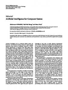
Figure 1. The participant playing 2 of the computer games with screen shots of the games. The game on the left is Helicopter; the participant plays by moving her arm up and down from the shoulder with a Wii remote strapped to it. The game on the right is Baseball Catch; the participant plays by moving her arm around space to control the position of the baseball glove.
420
TOPICS IN STROKE REHABILITATION/JULY-AUG 2011
participant to ensure the accuracy of interview notes and notes from the journal. Outcome assessment data collection
There were 3 outcome assessment data collection sessions. The first session took place prior to the start of the intervention and provided baseline data. The second session took place at the end of the first 3 weeks of the intervention. The third session took place within 1 week after completing the entire 6-week intervention. Data were collected in the Human Performance Laboratory. Each session lasted about 2 hours. Rest periods were provided as needed. Basic descriptive information (eg, height, weight, age, ethnicity, marital status) was collected during the first session. There were 4 outcome measures. The first was the Action Research Arm Test (ARAT) used to assess upper extremity function.12 The ARAT has high validity (r > 0.87) for assessing upper extremity motor function in persons with stroke.18,19 The second was the Activity Card Sort (ACS), which documents a person’s level of activity input in instrumental, leisure, and social activities and changes in activity participation following the
stroke.20 The person sorts 80 cards with various activities into several categories (ie, Do Now, Do Not Do, Gave Up Due to Injury/Illness, Do Less). The sort can be completed before and after an intervention to determine a change in participation. The ACS has high construct validity for persons with stroke and is a better predictor of quality of life than other measures.21 The third was a simple reach to target task, the Reaching Performance Scale (RPS),22 modified for use in our laboratory.4,23 The fourth was simple active range of motion in all planes for all joints of the upper extremity collected using our video motion capture system.24 The ARAT was completed following the standardized instructions and recorded on the accompanying form. The ACS was completed following the standardized instructions, and the numbers of the activities sorted into each category were recorded on a form. At the 3- and 6-week time points, the baseline sort was reviewed with the participant and she was asked to note if her participation had changed for any activity. Changes were noted on a form. For the RPS, 29 reflective surface markers were placed on anatomical landmarks of the participant’s upper extremities, head, and trunk (Figure 2A). A video
Figure 2. (A) Still image of the participant with the reflective surface markers placed on anatomical landmarks of the upper extremity and torso. Three surface markers and the target are highlighted. (B) Still image of the participant during a forward reach with her left upper extremity.
Computer Games as Stroke Intervention
motion capture system25 captured the movement of the surface markers during the trials. To set up the target, a researcher asked the participant to sit on a bench with no back support with her hands in her lap or at rest for her affected arm. Her trunk was stabilized by the researcher, and she was asked to simply bring her arm out in front of her. As the participant kept her arm out in front of her, a target (2.5 cm diameter reflective marker on a tripod) was placed at the height and distance of her index finger. For the simple reach, the participant was instructed to start with her hands at rest and reach forward to touch the target. The test was repeated 3 times. For the extended reach, the target was moved 15 cm forward from the participant, and she was instructed to start with her hands at rest and reach forward to touch the target. This distance was less than what was used in other studies.22 For the participant in this study, 15 cm provided a challenge. The test was repeated 3 times. The whole process, including target setup, was repeated in the coronal plane for a side reach and side extended reach. Both right and left upper extremities were tested (Figure 2B).
421
For active range of motion, the same surface marker placement and video motion capture system were used. The participant was asked to perform active range of motion movements in all planes for each joint. Each motion was performed twice. Both right and left upper extremities were tested. Data processing
The data from the ARAT were processed according to the developer’s guidelines. A subscore for each section and an overall score were calculated. The data from the ACS were processed using the developer’s software and guidelines. Scores were calculated for each activity category as well as a global composite score. For the RPS, the data were tracked and edited to produce a “Tinkertoy” image (Figure 3). The raw 3-dimensional coordinates for each marker as a function of time series were imported into a spreadsheet for analysis. The reach-path ratio was defined as the ratio of the actual path taken to the straight line from the index finger to the target. The data were also imported into MATLAB26 for analysis.
Vertex
R acromion
C7
L acromion Sternal notch
T8 L medial & lateral epicondyle
R medial & lateral epicondyle
Xiphoid process
L radial & ulnar styloid
R radial & ulnar styloid
Figure 3. A “Tinkertoy” image of the marker set used for analysis of the Reaching Performance Scale and active range of motion. Only the markers used in the definitions of the movements for analysis are included. L = left; R = right.
422
TOPICS IN STROKE REHABILITATION/JULY-AUG 2011
The body segments were defined as follows: thorax (C7, T8, sternal notch, xiphoid process), humerus (acromion process, lateral epicondyle, medial epicondyle), and forearm (lateral epicondyle, medial epicondyle, ulnar styloid, radial styloid). The motions used in the RPS analysis were defined from the body segments as detailed in Table 2. Angles were calculated across the time series for each motion during each of the 3 reaches. The maximum, minimum, and range were calculated for each motion during each of the 3 reaches. To identify key variables in this study, we then conducted an exploratory analysis with the maximum, minimum, and range values. Variables indicating changes that were occurring as a consequence of the intervention were used in subsequent analyses. The key variables identified from the RPS were the humerothoracic angle, humerothoracic external rotation, thorax lateral rotation, and thorax extension. For the active range of motion movements, the data were tracked, edited, and processed through MATLAB using the same motions definitions as the RPS (Table 2). The data for each motion of each movement were plotted and visually analyzed for trends over time. Similar to the RPS, key variables indicating changes that were occurring as a consequence of the intervention were identified. The only key variable included in data processing was the humerothoracic plane angle. Descriptive statistics
For each motion of the RPS, the calculated angles (minimum, maximum, and range) for each of the 3 reaches were plotted at baseline.
The mean line and 2 standard deviation bands for the baseline points were plotted as well. The 3 calculated angles from the 3-week and 6-week outcome assessment periods were plotted on the same graph. The data points indicated noticeable improvement if 2 or more consecutive points fell outside of the 2 standard deviation band.27 This process was completed for each reach of the RPS as well as the reach-path ratio data. Results Playing the games
The use data that were downloaded at the end of each week showed that the participant played up to 4 games each day as prescribed by the research assistant. On average, the participant completed around 250 repetitions for each game per day. This varied slightly from game to game and amounted to around 1,000 repetitions per day. Reaching Performance Scale
After 3 weeks of the intervention, the participant demonstrated increased left humerothoracic elevation range during the forward extended reach on the RPS (Figure 4A). All 3 of the data points at 3 weeks were outside of the baseline 2 standard deviation band. At 6 weeks, all of the data points had returned to within the band, indicating return to baseline. At 6 weeks, the participant demonstrated increased humeral internal/external range during both the forward reach and the forward extended reach on the RPS (Figure 4B).
Table 2. Motions used for the Reaching Performance Scale and active range of motion analysis Motion
Description
Humerothoracic plane Humerothoracic angle
Angle of humerus within the transverse plane Angle of the humerus relative to the thorax; can occur anywhere within the transverse plane (For the forward reaches, the angle is mostly shoulder flexion. For the side reaches, the angle is mostly shoulder abduction.) Rotation of the humerus along its long axis Anterior/posterior flexion of the thorax Lateral flexion of the thorax Rotation of the thorax about the vertical axis Angle of forearm to the humerus Axial rotation of the forearm with respect to the humerus
Humerothoracic external rotation Thorax extension Thorax lateral rotation Thorax axial rotation Elbow flexion Elbow pronation
Computer Games as Stroke Intervention
(a)
Left humerothoracic angle range
(b)
34 Degrees
Degrees
32
28 26 24 22 Baseline
(c)
3 weeks
Left internal/external rotation range 35 33 31 29 27 25 23 21 19 17 15
36
30
6 weeks
Left lateral trunk bending range
423
Baseline
(d)
3 weeks
6 weeks
Left sagittal trunk bending range 25
4 3.5
20
3 2.5
15
2 10
1.5 1
5
0.5 0
0 Baseline
3 weeks
6 weeks
Baseline
3 weeks
6 weeks
Figure 4. (A) Humerothoracic angle range during forward extended reach at baseline, 3 weeks, and 6 weeks. (B) Humerothoracic internal/external rotation range during forward extended reach at baseline, 3 weeks, and 6 weeks. (C) Lateral trunk bending range during forward reach at baseline, 3 weeks, and 6 weeks. (D) Sagittal trunk bending range during forward extended reach at baseline, 3 weeks, and 6 weeks.
Active range of motion
After a visual analysis of the active range of motion movements, the participant demonstrated a trend toward shoulder flexion occurring more in a sagittal plane (where flexion should normally occur) as opposed to more in a coronal plane after
3 weeks. After 6 weeks, shoulder flexion occurred more in a sagittal plane but was slightly less than at 3 weeks (Figure 5). Action Research Arm Test
The participant demonstrated no change in score on the ARAT for her left upper extremity after 3 weeks of the intervention. After 6 weeks, her score increased from 6 to 8. This increase in score
Left humerothoracic plane during shoulder flexion 60 55 50 Degrees
Again, all 3 of the data points were outside the baseline 2 standard deviation band. At 3 weeks, 2 of the data points remained within the band, with 1 data point falling outside the band but indicating a slight decrease in range. The participant also demonstrated decreased lateral trunk bending during the forward reach on the RPS after 3 weeks of the intervention but no difference from baseline after 6 weeks (Figure 4C). After 6 weeks, she demonstrated decreased sagittal trunk bending during the forward extended reach on the RPS with no change after only 3 weeks of the intervention (Figure 4D). The reach-path ratio data showed no noticeable improvement from baseline to 3 weeks and 6 weeks.
45 40 35 30 25 20
Baseline
3 weeks
6 weeks
Figure 5. The angle of the humerus in the transverse plane during active shoulder flexion.
424
TOPICS IN STROKE REHABILITATION/JULY-AUG 2011
was because the participant was able to complete the first task of the grip subsection: pouring water from 1 glass to another. She received 2 points for completing the task but with great difficulty (she spilled water on the towel on the table). This increase in score is not clinically significant (> 6 points) and falls within the margin of error.
The participant shared several other stories, such as being able to move her toothpaste tube on the sink counter before it fell off, using her left arm for balance against a wall while bending over, using her left arm to hold plates on the counter while using her right hand to wash them, and using her left arm to raise the covers up in bed while getting adjusted underneath.
Activity card sort
The participant demonstrated no change in participation level after 3 and 6 weeks of the intervention for all activity subcategories as well as overall participation. Subjective increases in function
The qualitative data from the weekly interviews revealed several subjective increases in function. During the third week of the intervention, the participant stated that she was getting out a crock pot for cooking dinner and was holding it in her right arm. Without thinking, she used her left arm to guide the power cord onto the counter in front of the electrical outlet, allowing her to later plug it in. She stated that she had not been able to move the cord like that in the past 17 years since her stroke. During the fifth week of the intervention, the participant stated that she was able to bathe completely independently. For the past 17 years since her stroke, she had either had to have someone hold up her left arm so she could wash under it or had to prop her left arm up on something to wash under it. She was now able to hold her left arm up by herself and wash underneath it. The participant’s general comments about increased movement abilities included statements such as the following: “My arm moves a whole lot faster.” “After 1 week and adding an extra game, I can do something I haven’t been able to do for a year – get something to move where I want.” “I can envision discovering more things to do.” “Not as much work is needed to extend my fingers.” “It’s easy to do.” [holding left arm up in shower] “It [left arm] does what I want it to do without thinking about it too much.”
Discussion The purpose of this case report was to test the feasibility of using computer games as a 6-week home therapy intervention to improve upper extremity function for a person with stroke. It is clear that the 6-week home therapy intervention had an impact not only on the range of motion and motor control for the participant with stroke but also on her overall function. Many of her reported subjective increases in function were unprompted and were generally spontaneously reported during the weekly meetings. What is most interesting is that many of her subjective increases in function were not directly correlated with any of the games. Some of her new gains in function, such as holding her arm up in the shower, are clearly the same movement being used to play the Helicopter game. However, other functional movements, such as moving her toothpaste on the sink or holding onto the wall for balance, are made up of several movements that she used to play the games. The lack of change in participation, as shown by the ACS results, may be due to the short time frame of an intervention that is focused primarily on improving upper extremity function and not necessarily activity participation. Although not as compelling as the subjective i n c re a s e s i n f u n c t i o n , t h e p a r t i c i p a n t demonstrated noticeable improvements in her upper extremity range of motion as well as subjective increases in motor control. As shown in several of the graphs in Figure 4, she improved her upper extremity range of motion and trunk stability at 3 weeks but returned to her baseline level at 6 weeks. One potential explanation of this result is the day-to-day variability that may exist within the participant. Factors responsible for this variability include
Computer Games as Stroke Intervention
levels of fatigue, motivation, and muscle tone. Additional participant testing may have clarified this issue; however, further testing was beyond the scope of the study. Completing the protocol with additional participants as well as some reliability testing should resolve this issue. The increases that occurred throughout the 6 weeks provide good evidence for motor learning theories. The high number of repetitions the participant performed every day while playing the games is far greater than what is typically received in outpatient therapy12 and is comparable with therapies such as constraintinduced movement therapy. 28 Other virtual reality and computerized games/systems are more limited in the number of repetitions that can be achieved during game play (generally no more than 150 per hour).29-31 The games were also motivating and fun for the participant to play while still allowing her to achieve a high number of repetitions per day. She went as far as to set personal goals for herself for each game when she played (ie, try to get all of the fuel cells in between the buildings). This supports the idea that computer-based and virtual reality systems, such as this one, contain motivating characteristics necessary for rehabilitation similar to other systems.13-16 This report does have several limitations. First, this was only a case report with 1 person with stroke. The physical, cognitive, and emotional outcomes from a stroke can vary widely from person to person. It is therefore difficult to generalize the results to the broader stroke population. Also, the participant was 17 years post stroke. These games need to be tested with persons with stroke at several stages post stroke, including very early stages. Second, the methodology for the RPS may have influenced the results. The methods described in previous studies23 may not be applicable for persons with stroke and other upper extremity limitations. Each time the assessment was performed, the target was placed at a distance that was comfortable for the person with stroke instead of at a fixed distance. This makes it more difficult to compare results across time as well as calculate the time required for the reach. For future studies, the target should be placed at a fixed distance from the participant
425
instead of being variable across time. This fixed distance may depend on the severity of stroke of each participant. Third, the outcome measures described in this study limited the ability to quantify small increases in upper extremity function. The ACS measures participation on a larger scale than the changes that were seen in this study. A measure such as the Canadian Occupational Performance Measure or the FIMTM* may allow for easier quantification of increases in function. Fourth, the mixed methodology used in this study was not clearly defined from the beginning. This could have affected the rigor and trustworthiness of the qualitative data that were collected. The qualitative methods need to be more clearly defined in future studies and possibly include various methods of data collection as well as better integration with the quantitative data.32 Clinically, this case report provides a compelling example of how computer games can be used as an intervention for a person with stroke. In an outpatient rehabilitation setting, many therapists prescribe home exercises.33 These games may be a feasible, motivating alternative to home exercises and allow the person with stroke to achieve a high number of repetitions. Although this report did not include outpatient therapy as an intervention, the potential for these games to complement the treatment effects of outpatient therapy is high. Future work should be done in several stages. First, a controlled study to test the effectiveness of the computer games as an intervention should be done with more persons with stroke. This may also permit the researchers to conduct a focus group, thus strengthening the qualitative results. The limitations of the RPS discussed in this case report should be taken into consideration when choosing outcome measures. As in this report, a home-based setting would still be ideal for a controlled study. After a controlled study is completed, the games can be tested in a variety of settings, including outpatient, inpatient, and acute care rehabilitation settings.
*
FIM TM is a trademark of Uniform Data System for Medical Rehabilitation, a division of UB Foundation Activities, Inc.
426
TOPICS IN STROKE REHABILITATION/JULY-AUG 2011
Conclusion The results of this case report indicate that computer games have the potential to be a useful intervention for people with stroke. Although we only had 1 participant, she demonstrated many increases in upper extremity function despite being 17 years post stroke. Future work with these games will be able to provide additional support to the effectiveness of these games as
a home therapy intervention for persons with stroke. Acknowledgments This work was supported by a grant from the Clinical and Translational Research Funding Program through Barnes-Jewish Hospital Foundation and Washington University Institute of Clinical and Translational Sciences.
REFERENCES 1. American Heart Association. Heart disease and stroke statistics 2010 update: a report from the American Heart Association. Circulation. 2010;121:e46–e215. 2. Rosamond W, Flegal K, Furie K, et al. Heart disease and stroke statistics – 2008 update: a report from the American Heart Association Statistics Committee and Stroke Statistics Subcommittee. Circulation. 2008;177:e26–e146. 3. Kelly-Hayes M, Beiser A, Kase CS, Scaramucci A, D’Agostino RB, Wolf PA. The influence of gender and age on disability following ischemic stroke: the Framingham study. J Stroke Cerebrovasc Dis. 2003;12(3):119–126. 4. Wagner JM, Lang CE, Sahrmann SA, Edwards DF, Dromerick AW. Sensorimotor impairments and reaching performance in subjects with posttroke hemiparesis during the first few months of recovery. Phys Ther. 2006;87(6):751–765. 5. Lai SM, Studenski S, Duncan PW, Perera S. Persisting consequences of stroke measured by the Stroke Impact Scale. Stroke. 2002;33:1840–1844. 6. Lotze M, Cohen LG. Volition and imagery in neurorehabilitation. Cogn Behav Neurol. 2006;19:135–140. 7. Govender P, Kalra L. Benefits of occupational therapy in stroke rehabilitation. Expert Rev Neurother. 2007;7(8):1013–1019. 8. Woodson AM. Stroke. In: Trombly CA, Radomski MV, eds. Occupational Therapy for Physical Dysfunction. Baltimore, MD: Lippincott Williams & Wilkins; 2005:817–853. 9. Sabari JS. Optimizing motor behavior using the Carr and Shepherd approach. In: Trombly CA, Radomski MV, eds. Occupational Therapy for Physical Dysfunction. Baltimore, MD: Lippincott Williams & Wilkins; 2005:501–519. 10. Kleim JA, Jones TA, Schallert T. Motor enrichment and the induction of plasticity before or after brain injury. Neurochem Res. 2003;28(11):1757–1769. 11. Nudo RJ. Functional remodeling of motor cortex: Implications for stroke rehabilitation. Paper presented at: McDonnell Conference, Washington University, St. Louis, MO; 2005.
12. Lang CE, MacDonald JR, Gnip C. Counting repetitions: an observational study of outpatient therapy for people with hemiparesis post-stroke. J Neurol Phys Ther. 2007;31:3–10. 13. Jack D, Boian R, Merrians AS, et al. Virtual realityenhanced stroke rehabilitation. IEEE Trans Neural Syst Rehabil Eng. 2001;9(3):308–318. 14. Broeren J, Claesson L, Goude D, Rydmark M, Sunnerhagen K. Virtual rehabilitation in an activity centre for community-dwelling persons with stroke. Cerebrovasc Dis. 2008;26:289–296. 15. Johnson M, Ramachandran B, Paranjape R, Kosasih J. Feasibility study of TheraDrive: a low-cost gamebased environment for the delivery of upper arm stroke therapy. Conf Proc IEEE Eng Med Biol Soc. 2006;695–698. 16. Bach-y-Rite P, Wood S, Leder R, et al. Computerassisted motivating rehabilitation (CAMR) for institutional, home, and educational late stroke programs. Top Stroke Rehabil. 2002;8(4):1–10. 17. Alankus G, Lazar A, May M, Kelleher C. Towards customizable games for stroke rehabilitation. CHI Conf Proc. 2010;2113–2122. 18. Hseih CL, Hsueh IP, Chiang FM, Lin PH. Inter-rater reliability and validity of the Action Research Arm Test in stroke patients. Age Ageing. 1998;27(2):107–113. 19. Yozbatiran N, Der-Yeghiaian L, Cramer SC. A standardized approach to performing the Action Research Arm Test. Neurorehabil Neural Repair. 2008;22(1):78–90. 20. Baum C, Edwards D. Activity Card Sort. St. Louis, MO: Washington University School of Medicine; 2001. 21. Law M, Baum C, Dunn W. Measuring participation. In: Law M, Baum C, Dunn W, eds. Measuring Occupational Performance: Supporting Best Practice in Occupational Therapy. Thorofare, NJ: SLACK Incorporated; 2001:107–128. 22. Levin MF, Desrosiers J, Beauchemin D, Bergeron N, Rochette A. Development and validation of a scale for rating motor compensations used for reaching in patients with hemiparesis: the Reaching Performance Scale. Phys Ther. 2004;84(1):8–22.
Computer Games as Stroke Intervention
23. Shurtleff TL, Standeven JW, Engsberg JR. Changes in dynamic trunk/head stability and functional reach after hippotherapy. Arch Phys Med Rehabil. 2009;90:1185–1195. 24. Norkin CC, White DJ. Measurement of Joint Motion: A Guide to Goniometry. 3rd ed. Philadelphia, PA: F.A. Davis Company; 2003. 25. Cortex. Version 1.0. Santa Rosa, CA: Motion Analysis Corporation; 2008 26. MATLAB. Version 7. Natick, MA: The Math Works, Inc; 2010. 27. Ottenbacher KJ. Reliability and accuracy of visually analyzing graphed data from single-subject designs. Am J Occup Ther. 1986;40:464–469. 28. Page SJ, Levine P. Modified constraint-induced therapy in patients with chronic stroke exhibiting minimal movement ability in the affected arm. Phys Ther. 2007;87:872–878. 29. Deutsch JE, Merians AS, Adamovich S, Poizner H, Burdea GC. Development and application of virtual reality technology to improve hand use
30.
31.
32.
33.
427
and gait of individuals post-stroke. Restor Neurol Neurosci. 2004;22:371–386. Stewart JC, Yeh SC, Jung Y, et al. Intervention to enhance skilled arm and hand movements after stroke: a feasibility study using new virtual reality system. J Neuroeng Rehabil. 2007;4(21): e1–e6. Viau A, Feldman AG, McFadyen BJ, Levin M. Reaching in reality and virtual reality: a comparison of movement kinematics in health subjects and in adults with hemiparesis. J Neuroeng Rehabil. 2004;1(11):e1–e7. Corcoran MA. Using mixed methods designs to study therapy and its outcomes. In: Creswell JW, Plano VL, eds. Designing and Conducting Mixed Methods Research. Thousand Oaks, CA: Sage Publications; 2007:411–419. Werner RA, Kessler S. Effectiveness of an intensive outpatient rehabilitation program for postacute stroke patients. Am J Phys Med Rehabil. 1996;75(2):114–120.