invasive lobular carcinomas (n = 5). Conclusion: 3D-MDCT images can assess breast cancer tumor extension highly accurately, and thus seems to be useful for ...
Breast Cancer Research and Treatment (2005) 89: 119–125
� Springer 2005
Report
Usefulness of three-dimensional multidetector-row CT images for preoperative evaluation of tumor extension in primary breast cancer patients Tomoo Inoue1, Yasuhiro Tamaki1, Seiki Hamada2, Shuji Yamamoto2, Yoshinobu Sato3, Shinichi Tamura3, Seung Jin Kim1, Yoshio Tanji1, Yasuo Miyoshi1, Tetsuya Taguchi1, and Shinzaburo Noguchi1 1
Department of Surgical Oncology; 2Department of Radiology, Osaka University Graduate School of Medicine; Division of Interdisciplinary Image Analysis, Department of Medical Robotics and Image Sciences, Osaka University Graduate School of Medicine, Yamadaoka, Suita, Osaka, Japan 3
Key words: breast cancer, tumor size, multidetector-row CT
Summary Purpose: Usefulness of three dimensional (3D) multidetector-row CT (MDCT) images for preoperative evaluation of tumor extension was studied in primary breast cancer patients. Methods: 3D-MDCT tumor images of 143 tumors in 143 patients with primary breast cancer were created with the volume rendering method. The transverse tumor size (TS) and vertical tumor size (VS) were then measured in an anterior-posterior view of the 3D-MDCT images. The pathological tumor size was determined according to a map of the tumor spread prepared by pathologists using multi-sliced (3–5 mm intervals) surgical specimens and compared with the tumor size on 3D-MDCT images. Results: First, the optimal method for creating 3D-MDCT tumor images was determined for the first 40 patients (learning set), resulting in a fairly good correlation of tumor size on 3D-MDCT images with pathological tumor size (r ¼ 0.983 for TS and r ¼ 0.958 for VS). We then carried out a validation study on the next 103 patients (validation set). The 3D-MDCT tumor size’s strong correlation with the pathological tumor size demonstrated a high rate of accuracy (r ¼ 0.974 for TS and r ¼ 0.977 for VS). Subset analyses according to histological type showed that correlation coefficients were r ¼ 0.979 for TS and r ¼ 0.981 for VS of invasive ductal carcinomas (n ¼ 88), r ¼ 0.948 for TS and r ¼ 0.970 for VS of ductal carcinomas in situ (n ¼ 10), and r ¼ 0.984 for TS and r=0.976 for VS of invasive lobular carcinomas (n ¼ 5). Conclusion: 3D-MDCT images can assess breast cancer tumor extension highly accurately, and thus seems to be useful for planning the extent of resection in breast conserving surgery.
Introduction Complete removal of a breast tumor with its tumornegative surgical margins is most important for avoiding local recurrence (ipsilateral in-breast recurrence) in breast conserving surgery [1–3]. Wider excision of the breast gland can result in a lower risk of local recurrence but produces a poorer cosmetic outcome. To cope with the dual problem of curability and cosmetic outcome, preoperative, accurate assessment of tumor extension is of vital importance. For this purpose, many studies have been conducted using a variety of imaging modalities, i.e., mammography, ultrasonography (US), magnetic resonance imaging (MRI), and helical computed tomography (helical CT) [4–20]. However, none of these modalities is sensitive enough to visualize intraductal spreading of the tumor or small daughter nodules with satisfactory accuracy [5, 8, 14, 16, 17]. It is thus often difficult to predict precisely the extension of an invasive ductal carcinoma associated with extensive intraductal
spreading or of a ductal carcinoma in situ (DCIS), as well as the extension of a multifocal invasive lobular carcinoma (ILC) [21–23]. MRI can visualize intraductal spreading with a higher accuracy than other imaging modalities but has the disadvantage that images are obtained with the patient in the prone position whereas the surgery is done with the patient in the supine position (the breast easily changes shape with a change in position). Recently, multidetector-row CT, which provides high-quality, high-resolution 3D images, has been used for preoperative evaluation of tumor extension in various malignant diseases. While promising results have been reported [24–28], no results for breast cancer have been reported yet. Much useful software for 3D image analysis has become available on personal computers. This recent technological development has facilitated the construction and handling of 3D images as well as their precise analysis. MDCT, because of its high resolution, is expected to make it possible to visualize tumor extension with a high degree of accuracy. MDCT has the
120 T Inoue et al. additional advantage over MRI that it can be performed with the patient in a supine position similar to the position used during surgery. These reported results and characteristics prompted us to evaluate the usefulness of MDCT for the preoperative evaluation of breast tumor extension by comparing the tumor size determined with the aid of 3D-MDCT images with the pathologically determined tumor size.
Materials and methods Patients Primary breast cancer patients, who were clinically considered eligible for breast conserving surgery, were first given an MDCT examined and then underwent breast conserving surgery or mastectomy between June 2001 and April 2004 at Osaka university Hospital. Patients who had undergone preoperative excisional biopsy and/or neoadjuvant chemotherapy were excluded from this study. Informed consent was obtained from all patients. Eventually, 143 patients with unilateral breast cancer were included in the study. In the case of breast conserving surgery, additional resection of the breast tissue was performed if the margin was pathologically positive, so that eventually all the patients treated with breast conserving surgery showed negative margins. All surgical specimens were fixed and cut into 3–5 mm-thick slices perpendicularly to the line connecting the nipple and the center of the tumor. They were then examined microscopically by pathologists to produce a pathological map of the tumor extension.
Image acquisition by MDCT Patients were examined in the supine position and with a 0.5-s four-slice MDCT (Aquilion-V detector; Toshiba Medical Systems Co., Tokyo, Japan). Target helical scanning (200 mm) of breast lesions was performed within a single breath-hold with 1 mm detector raw collimation and a helical pitch of 6:1 after pre-enhanced scanning of the whole thoracic area (300 mm) for breast cancer screening. This was followed by contrast enhanced scanning with biphasic helical CT scanning, for which 100 ml of nonionic contrast material, Iohexol 300e (Daiichi Pharmaceutical Co., Ltd., Tokyo, Japan), was injected intravenously at a flow rate of 2.0 ml/s. The scanning was started when the peak aortic enhancement at the same slice level as the breast lesion reached 200 Hounsfield Units (HU) [29]. Creation and modification of 3D-MDCT images The Digital Image and Communication in Medicine (DICOM) images of MDCT were transferred to a workstation, and 3D-MDCT images were created with the volume rendering method using Virtual Placee 3Dimage analysis software (Medical Imaging Laboratory Inc., Tokyo, Japan). For accurate tumor segmentation, the breast gland was selected as a region of interest (ROI) from an original 3D-MDCT image using multiplanar reformation (MPR) images. The tumor image was then displayed by controlling the opacity level according to the HU values of each pixel and rotated until eventually an anterior–posterior (A–P) view of the 3D volume image of the breast gland was obtained. The initial 40 patients served to determine the optimal
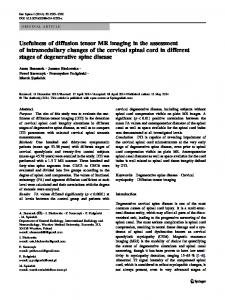
Figure 1. Measurement of tumor size on 3D-MDCT image and pathological map. Tumor size was measured in two directions, transverse size (TS) and vertical size (VS), in an A–P view of the 3D-MDCT image and on a pathological map created by pathological examination of sliced (3– 5 mm intervals) surgical specimens.
3D Multidetector-row CT image of breast cancer 121 opacity function condition and the optimal setting of the color map for the 3D-MDCT images so that these images showed good correspondence with the pathological map of tumor extension (learning set). Under these optimal conditions, 3D-MDCT tumor images were created for the next 103 patients (validation set), and the tumor size on the 3D-MDCT images was compared with pathological tumor size. Measurement of tumor size Tumor size was measured with Virtual Placee software (Medical Imaging Laboratory Inc.) on A-P views of the 3D-MDCT volume images. The size was recorded at two rectangular directions, i.e., the transverse size (TS) parallel and the vertical size (VS) perpendicular to the slice of the pathological specimen (Figure 1). When spotty or linear augmentations were observed around the main tumor, the 3D-MDCT images were observed in more detail by rotating and slicing them at various angles to determine whether these augmentations were cancer nests. The pathological tumor size was measured in the same directions (TS and VS) with the aid of a pathological map of the tumor extension. Statistical analysis Tumor size determined by means of 3D-MDCT images and of pathological maps was compared by using Pearson’s correlation coefficient test. All statistical analyses were performed with StatViewe software (SAS Institute Inc., Cary, NC).
Results Patient characteristics and representative 3D-MDCT images Patient characteristics of the learning set (n ¼ 40) and validation set (n ¼ 103) were not significantly different (Table 1). Representative 3D-MDCT images are shown in Figure 2. Figure 2A shows a case of invasive ductal
Table 1. Patients characteristics in the learning set and validation set
Age (average)
Learning set
Validation set
(n = 40)
(n = 103)
22–83 (53.7)
30–75 (51.7)
Menopausal status Pre menopausal
17
54
Post menopausal
23
49
IDC
35
88
ILC
2
5
DCIS
3
10
£2
25
53
2