Oberman, L. M., Hubbard, E. M., McCleery, J. P., Altschuler, E. L., Ramachandran, V. S., &. Pineda, J. A. (2005) EEG evidence for mirror neuron dysfunction in ...
1
Using mu rhythm perturbations to measure mirror neuron activity in infants Pär Nyström1, Therese Ljunghammar1, Kerstin Rosander1, Claes von Hofsten1 1
Department of Psychology, Uppsala University, Trädgårdsgatan 20, Box 1225, SE 75142 Uppsala, Sweden. Corresponding author: Claes von Hofsten
2
Abstract The Mirror Neuron System hypothesis stating that observed actions are projected onto the observer’s own action system assigns an important role to development, because only actions mastered by the observer can be mirrored. The purpose of the present study was to investigate whether there is evidence of a functioning mirror neuron system (MNS) in early infancy. High-density EEG was used to assess the mu rhythm perturbations in an action observation task where the infants viewed a live model. To reduce noise ICA decompositions were used. The results show a higher desynchronization of the mu rhythm when 8-month-old infants observe a goal-directed action than when they observe a spatially similar non-goal-directed movement. This provides evidence that the MNS is functioning at this age level. Importantly, the results show that mu rhythm perturbations can be used as a tool for studying MNS activity in infants.
3
Introduction It has been suggested that other people’s actions are understood by mapping them onto one’s own motor representation of that action (Flanagan & Johansson, 2003, Rizzolatti, Fadiga, Gallese, 1996). It is suggested that this mapping is accomplished by a neuronal system, the Mirror Neuron System (MNS), activated both by the execution of one’s own goal-directed actions and by the perception of someone else performing the same actions (Rizzolatti et al., 1996; Fadiga & Craighero, 2004). It has even been suggested that the MNS plays a crucial role in many social activities, like imitation learning (Buccino et al., 2004), theory of mind, empathy, and the development of language (Théoret & Pascual-Leone, 2002). Thus, the mirror neurons principle could provide a unifying framework for human social cognition (Gallese et al., 2004, Oberman et al, 2007), and has consequently attracted attention from many research fields (Berthouze & Metta, 2005). Neurophysiological evidence indicates that the MNS is a distributed system with at least three primary nodes, the ventral premotor area (Area 44 in humans), the STS, and the IPL. It was in the left ventral premotor area that the mirror neurons were first observed. Rizzolatti et al. (1996) showed that the same neuron fired when the monkey picked up a peanut and when they saw someone else do it. Fogassi et al. (2005) found that most motor IPL neurons coding a specific movement (e.g., grasping) showed markedly different activations when this act was part of different actions (e.g., for eating or for placing). Studies using fMRI have found elevated activation in STS, IPL and Area 44 in adult humans during action observation (Rizzolatti & Craighero, 2004). Using MEG, Nishitani and Hari (2002) showed that observed and imitated lip movements activated first the STS, then the IPL, and finally the Area 44. In order to understand other people’s actions in the way proposed by the MNS hypothesis, that is, in terms of one’s motor representations, the corresponding movements must be mastered by the subject. In other words, actions that are not mastered cannot be understood in this way. This makes the MNS hypothesis very interesting from a developmental point of view because it states that motor development plays a crucial role for the understanding of other people’s actions. If mastery of actions is essential for the understanding of other people’s actions, it should develop in parallel with or ahead of such understanding. The problem is that direct evidence of brain activity in young children from fMRI and MEG is generally not available below preschool age. The importance of this question, however, is reflected in several explicit requests for empirical neurophysiological data to start to resolve speculations (Lepage & Théoret, 2007; Bertenthal & Longo, 2007; Kilner & Blakemore, 2007). One indirect piece of evidence for the MNS is found in people’s proactive eye movements during action observation. When subjects perform actions, they move the eyes to the goal proactively (Land & Hayhoe, 2001). The purpose of such eye movements is to guide the hand to the goal. If the MNS hypothesis holds, the proactive gaze shifts performed during action performance should be produced during action observation as well. Because the eyes are free to move when observing such actions, the MNS hypothesis predicts that subjects should produce eye movements similar to those produced when they perform the tasks. In accordance with this, Flanagan and Johansson (2003) found that adult subjects performed proactive eye movements to the goal of a displacement action both when they performed the action themselves and when they observed someone else perform the action. Such indirect behavioral evidence for MNS activity in infancy was obtained by Falck-Ytter, Gredebäck and
4 von Hofsten (2006). They found that 12-month-old but not 6-month-old infants looked proactively at the goal when a hand transported an object there. For both age groups of infants, the eyes just tracked the object when the same object motion was shown outside the context of an action. It should be noted that 12-month-olds but not 6-month-olds displace an object they have grasped to a different location in a goal-directed way. They might move the grasped object to the mouth, however. In accordance with this, Kochukhova and Gredebäck (2009) found that observed hand movements involved in eating actions were accompanied by proactive eye movements to the mouth of the model in 6-month-old infants. Other studies of social development have found that towards the end of the first year of life, infants rapidly develop social skills requiring understanding of other people’s actions. For instance, from about 11 months of age they begin to point to objects in the surrounding that they want other individuals to attend to (Butterworth, 2003; Liszkowski, Carpenter & Tomasello, 2006; Tomasello, Carpenter, & Liszkowski, 2007). Infants have been shown to imitate other people’s actions from 6 months of age (Barr, Rovee-Collier & Campanella, 2005; von Hofsten & Siddiqui, 1993) and to perform deferred imitation from 9 months of age (Meltzoff, 1988). From around 8 months of age, infants babble in social settings (Ejiri,1998). In summary, the present results suggest that the development of the mirror neuron system either precedes the emergence of these social functions or develop in parallel with them. A neurophysiological method suitable for developmental research is EEG, due to its lightweight and few physical constraints, and that it therefore can be applied to awake subjects of all ages. MNS activity has been identified in adults using EEG by analysis of the mu rhythm (Pineda, 2005; Muthukumaraswamy et al., 2004a; related experiments has been performed by using MEG as well, e.g., Nishitani & Hari, 2000; Hari et al., 1998). There are at least three reasons to associate the mu rhythm with the MNS. First, these studies show that the mu rhythm oscillates at approximately 9-13 Hz in adults but gets suppressed both when a subject performs an action and when the subject observes someone else perform the same action. Second, an important feature of mirror neurons is that they are tuned to goal-directed actions. The mu rhythm is also modulated by the goal-directedness of actions It is suppressed more when adults either perform or observe goal-directed actions compared to non-goal-directed actions (Muthukumaraswamy & Johnson, 2004; Muthukumaraswamy, Johnson & McNair, 2004). This functional response has also been found in 4-11-year-old children (Lepage & Théoret, 2006) and has recently been observed in infants (Southgate…) . Third, the mu rhythm is a sensorimotor rhythm that appears to consist of several rhythms with different origins both in motor areas and in parietal sensory areas (Pineda, 2005). This is consistent with the locations of mirror neurons. Mu rhythm suppression can therefore be considered being an established measure of MNS activity. The infant alpha rhythm between 5-9 Hz show strong resemblances to the adult mu rhythm, and it has therefore been suggested that it is the infant mu rhythm (Stroganova et al., 1999; Marshall et al., 2002). Orekhova et al. 2006 found that the action related part of the alpha rhythm emerge at around 5-8 months and increase in frequency and amplitude over age. Other studies have also studied infant alpha rhythms (Orekhova, Stroganova & Posikera, 2001) although no study has explicitly tested the functional reactivity of the infant alpha e mu rhythms in both action execution and action observation tasks (Southgate, 2009a,2009b; . Only one study has previously shown significant mu rhythm perturbations associated with the MNS in infants (van Elk et al., 2008) but then only in an action observation paradigm. One reason why EEG studies of the mu rhythm in an action performance condition has not yet
5 been reported is that infants tend to introduce large artefacts into the EEG signal when they move. Is it then possible to measure the infant mu rhythms without a performance condition? The best option would be to investigate mu rhythm suppression during action observation at mu sources in motor and sensory areas, and determine whether the suppression is synchronized with the goal of the action. With this paradigm it might be possible to identify infant mu rhythms. This study will therefore assume that the infant mu rhythms have the same characteristics as the adult mu rhythms and investigate the mu perturbations in an action observation situation. Another reason for the sparse literature on the infant mu rhythms is probably that infant EEG is plagued by short recording sessions (due to short attention spans) and eye movement artefacts since infants do not want to sit and watch passively (Thierry, 2005). In a traditional EEG paradigm where the channels receive mixed signals from different parts of the brain as well as muscle and eye movement artefacts, the result is often a very low signal to noise ratio where the mu rhythm is masked. By using a blind source separation technique, such as the independent component analysis (ICA), many of these problems can be solved. In principle ICA can be viewed as an oblique rotation method for PCA dimensions or sphered raw data. ICA is thus similar to principal component analysis (PCA), but whereas PCA aims at finding the factors that explains the most variance, ICA aims at finding components that are most statistically independent of each other (Delorme & Makeig, 2004). Since the MNS in infants (measured by the desynchronizing mu rhythm) cannot be expected to account for much variance but still has unique statistical properties, ICA appears to be a suitable tool. Unfortunately, ICA introduces a component selection problem. Because the mu rhythm can be expected to decompose into one or a few components from each subject, only these components should be considered in the analysis. We used 3 conditions to solve the independent component selection/testing problem. In one condition, the base-line mu rhythm activation was estimated when the subject observed a non-moving human model. In the other two conditions, the model was moving. In one of the movement conditions, the model reached for and grasped an object and in the other he moved his arm in a similar way but just placed his hand on a table-top at the end. Then, the difference in mu rhythm activation between the baseline and the two movement conditions was used for the selection of EEG sources. Finally, the difference between these two conditions was analyzed. If the MNS is activated by the goal-directed reaching movement, then the mu rhythm should be more desynchronized in this condition compared to the observation of a simple arm displacement. A recent study of 6-month-old infants used a similar method to extract mu rhythm activation elicited by video presentations of a passive and an active actor (Nyström, 2008). Although no evidence of a consistent difference in the mu rhythm was obtained for these two situations, an ERP analysis of the same data showed different responses for goal-directed and non-goaldirected movements. The insignificant mu rhythm desynchronization could either be due to the young age of the infants, the suboptimal viewing conditions, or both. In the present study we optimized the experiment in two ways. First, 8-month-old instead of 6-month-old infants were studied. If MNS develops in infancy, its indicators should get more distinct with age. Secondly, live actors instead of video recorded ones were used. In earlier studies of adults (Järveläinen et al., 2001; Shimada & Hiraki, 2006) and 7-month-old infants (Shimada & Hiraki, 2006), live actors were found to elicit stronger MNS activation compared to video taped ones. In fact, although Shimada and Hiraki (2006) found that action observation gave rise to activation in the motor areas in both the live and the TV setting, only the activation in the live setting was significant. With these two improvements the present study investigated whether infant MNS activity could be assessed by mu rhythm perturbations.
6
Methods Participants In the present study 36 healthy 8-month-old infants participated. The infants were tested on a simple reaching task, and all infants reached successfully for objects presented to them. The parents were given written information upon arrival to the lab and signed a written consent form in accordance with the Helsinki Declaration. The experiment was approved by the Ethics committee at Uppsala University. Two infants were excluded before analysis due to fussing or inattention, and 2 infants were excluded due to technical problems. All parents received a gift certificate of 100 Swedish kronor (approximately € 9) at a local toy store.
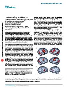
Procedure The infants were seated in front of the scene at a distance of approximately 1 meter where the events were shown and a high density 128 channels EEG net (EGI, Corp., Eugene, Oregon) of the appropriate size was applied to the infant’s head. The infants were shown from a lateral viewpoint a live male model sitting in front of a table. On the table, a short railway track with a toy train was placed (see Figure 1). First, the model was shown a baseline condition where the model sat passively (static condition). Then, two hand movement conditions were shown. In one of them (goal-directed condition), the model grasped the toy train and moved it from a starting position to a new position further away from the model. When the toy was in the new position the model performed the second condition. In this condition (non-goal-directed condition), the model moved his hand toward the first toy position (now empty) and simply placed the hand flat on the table. As the railway track was slanted toward the original starting position, the train returned there when an electronic trigger was released at the end of these events. Except for the very first condition that for practical reasons was always a goal-directed movement, these conditions were presented in randomized order by sometimes interleaving the static condition and sometimes not, or presenting the goal-directed or non-goal-directed condition twice before proceeding to any of the other conditions. The conditions were repeated until the subject was no longer interested or started to fuss.
EEG recordings and processing The 128 EEG channels were sampled at 250 Hz, with an analogue hardware band pass filter at 0.1 to 100 Hz (EGI, Corp., Eugene, Oregon). During recording, the infants observed the model either sitting still or performing the two actions described above. The EEG was timelocked by the model when the hand touched the toy or the table, or after approximately 1 second of sitting passively. Manual triggers hidden from the infants’ view were used to time lock the events. The triggers were operated by the model and triggered by the hand that performed the action (the triggers were hidden under a cloth on the table and under the models foot behind the table for the static condition). The model released the trigger with the moving hand as it arrived at the table-top or grasped the object. In the case where the object was grasped, the trigger was released by the little finger of the grasping hand situated on the far side of the object and thus not visible by the subject. The grasping action was performed by the thumb and three of the other fingers. After training the triggering over a few hundred trials during pilot testing, the temporal synchrony between the grasping and the triggering was quite high. The timing precision was determined in a separate experiment where a trigger was
7 placed on the object reached for in addition to the trigger used during the action observation. For a sequence of 182 control measures of the trained experimenter, the mean time difference was 9.7 ms and the standard deviation 27.1 ms. The experimenter observed the behaviour of the subject during the whole session, and conditions were never presented unless the infant was in an attentive and passive mode. The resulting EEG recordings consisted of between 10 – 49 trials in each condition from every subject (mean = 21.06; SD = 7.40). The data was transferred to the MATLAB v.7.2 environment and analyzed using the EEGLAB v5.03 toolbox. First, 29 of the outermost sensors were removed due to bad contact in most infants, although the net was properly positioned. The continuous EEG was then band pass filtered from 2 to 20 Hz using two-way least-squares FIR filtering to remove noise and to focus on the frequencies where most brain related signals appear. The data was segmented into trials from -1s to 1s after time-lock (touch of toy or table, or after approximately 1 second of resting). A modified artefact rejection routine for high density EEG (Junghöfer et al., 2000) removed bad trials and sensors based on their maximum values, standard deviations and range values. Next, the EEG was transformed to average reference as recommended by the EEGLAB guidelines. Modifications of the artefact rejection routine were removal of interpolation, which could violate the assumption for the ICA of independent measurements from each channel. Bad trials and channels were simply excluded from the dataset to prevent spreading of bad data when rereferencing. A natural-gradient logistic infomax independent component analysis was performed on the data (the Runica algorithm, (Delorme & Makeig, 2004), which resulted in as many independent components as remaining channels minus one for each subject (mean = 88, ranging from 78 – 98 components).
Selection of independent components Although each subject’s data was decomposed into many components only a few of them were assumed to reflect mirror neuron activity. The other components were assumed to mask the signal of interest by introducing noise and neural signals not related to the MNS. To reduce noise and to focus on components with mirror neuron properties we performed the two step component selection procedure described below. First we excluded components that reflected artefacts by the following criteria. Components with any abnormal ICA weight, >2.7 SD of all weights within a component, were considered artefactual and were excluded. The value of 2.7 SD was decided by visual inspection of all components to retain components with dipole like scalp projections and to exclude components originating from channel pops or movement artefacts. Two EEG experts knowledgeable of dipole source projections and four novices visually inspected the components that showed mu suppression. The area under the receiver operating characteristic (ROC) curve (AUC) was 0.95, and justified the use of a maximum of 2.7 was SD in any ICA weight as a rather conservative threshold. Also, the maximum absolute amplitudes of components were calculated to identify outlier values that could bias the subsequent frequency analysis. Trials with abnormal values (>3 SD) were thus excluded and also components with less than 10 trials in any condition. Secondly, we selected components related to mu rhythm desynchronization from the remaining components. Frequency spectra of the three conditions were extracted. To speed up the computations we used Welch’s method (hamming windows of 256 samples length and 128 samples overlap) instead of the EEGLAB timef function at this point. The results were converted to dB values across a 1 Ohm reference load to simplify visual presentation of the power spectra. Components with a power peak greater than 1 dB in the static condition and a decrease in the
8 power peak from this value greater than 1 dB for the two movement conditions were selected for further statistical analysis. This procedure selected components that showed mu desynchronization in the live movement conditions but without making any distinction between the goal-directed and non-goal-directed condition. The peak power was calculated as the max difference between the power spectrum and the linear interpolation of the power values at the boundaries of the 8-month-olds mu band (5-9 Hz). This definition of the peak power would account for overall differences in the power spectra between conditions. The definition of the 5-9 Hz interval was based on previous studies of infant alpha rhythms (Stroganova et al., 1999). An example of the frequency spectra used to select components is shown in Figure 2 together with the mean spectrum of all selected components. It should be noted that the highest amplitude in this frequency interval was 7 Hz in agreement with Stroganova et al. (op.cit.). In total 43 components with 10 – 49 trials (mean = 21.06; SD = 7.40) from each condition originating from 23 subjects were selected. Each subject contributed with between 1 and 5 components (mean = 1.87; SD = 1.12). If the ICA did not decompose any component with mu characteristics the subject was excluded. There could be several reasons. First, the mu rhythm is subject to individual differences. Second, the mu rhythm might not have developed in some subjects. Third, the measurements could have been contaminated by some minor artefacts that the ICA could not decompose. In either case the subject would not have any signal to contribute with, and would have to be excluded. The components that were not selected were subtracted from the raw EEG to create datasets pruned from noise, artefacts and neural activity that were not related to mu desynchronization. Thereby only the selected components were represented in the scalp channel activity of the pruned datasets. These components accounted for 1.4% of the variance in the raw data (each component ranging from 0.1% - 3.7%). To control the validity of the selection procedure, the selected components could be interchanged with random components. The resulting datasets should not show any significant effects, as randomly selected signals tend to cancel out each other.
Statistical analysis First, the statistical procedure was performed channel wise on the raw EEG datasets (n = 32). The same procedure was then repeated on the pruned datasets (n = 23) as a more elaborate analysis of the signal, and five times using randomly selected components. A time/frequency analysis using discrete wavelet transforms were performed on each channel’s (or component’s) conditions using the standard EEGLAB timef function. The window size was 64 samples (256 ms) wide, and Hamming windows was applied 200 times at an average step of 2.191 samples (8.76 ms). The EEGLAB timef function returned 20 frequency bands ranging from 2.0 Hz to 49.8 Hz and 200 time points ranging from -800ms to 800ms from time lock. However, as we were mainly interested in the 5-9 Hz frequency band, this interval was averaged and the goal-directed and non-goal-directed conditions were compared with reference to this measure at every time point. As multiple significance test inflates the risk of Type 1 errors only clusters of 10 or more adjacent significant p-values (p