438
Current Cancer Drug Targets, 2007, 7, 438-446
Using Nutrigenomics to Evaluate Apoptosis as a Preemptive Target in Cancer Prevention Keith R. Martin* Department of Nutrition, Arizona State University, Mesa, AZ 85212, USA Abstract; Apoptosis, a form of programmed cell death, is a pivotal defense against cancer and is essential in maintaining tissue homeostasis. Many diseases including cancer have been associated with aberrantly regulated apoptotic cell death, thus elucidation of events associated with both apoptosis and carcinogenesis provides the opportunity for dietary intervention with the plethora of bioactive components in the diet. Apoptosis occurs primarily through two well-recognized pathways in cells including the intrinsic, mitochondrialmediated pathway and the extrinsic, death receptor-mediated pathway. Dietary components can modulate apoptosis through effects on protein expression and function, mRNA expression, and on the human genome, either directly or indirectly, to modulate gene expression. Thus, apoptosis is an emerging target of dietary bioactive agents. However, apoptosis is a complex process, with numerous specific targets within each pathway that may or may not overlap. Furthermore, biological systems are also extremely complex and exhibit properties that extend far beyond observations associated with each independent cellular process. This is further complicated by the temporal nature of many of these effects. As a result, it is critical to evaluate the entire biological system from the nutrigenomics perspective to include critical evaluation of DNA polymorphisms or SNPs of a gene, expression of that specific gene, expression of specific processed mRNA (alternative splicing), protein production from that mRNA, post-translational modification of the resultant protein, and formation of respective metabolites. Evolution of the fields of nutrigenetics, epigenomics, transcriptomics, proteomics, and metabolomics has begun to permit this approach so that a comprehensive picture emerges from not only a single cell but tissues and whole organisms. Studies such as these can ultimately be used to study tumors to understand the molecular events that accompany carcinogenesis and perturbations that occur during cell death processes and how an individual’s response to diet can impact these processes.
Keywords: Apoptosis, nutrigenetics, epigenomics, transcriptomics, metabolomics, proteomoics, bioactive agent, DNA methylation, histone. INTRODUCTION An accumulating body of scientific evidence suggests that many cancers are preventable, especially since diet and nutrition are key factors in the modulation of cancer risk [1]. In fact, dietary habits are estimated to contribute to 35% of all human cancers and as high as 70% for some cancers [2, 3]. The capacity of diet with its thousands of inclusive bioactive molecules to independently alter gene and protein expression is generally well-accepted [4]. The recent elucidation of the 25,000 or so genes in the human genome and the greater than 100,000 functionally distinct proteins produced from these genes has suggested myriad potential influences and interactions. Moreover, the inherent variations of single genes within the population also contribute greatly to potential differences in responses to nutritional intervention. Exploring and defining the relationships between the genetic complement of an individual, the genes contained, the gene products, the metabolites formed, and the thousands of components of the diet are critical in determining not only those at risk for disease but also in determining those most likely to benefit through specific alterations in dietary habits [5]. The study of nutrigenomics encompasses such a blend of multiple contributing factors to human nutrition permitting comprehensive assessment of individual responses to diet at the molecular level. These can be subdivided into the areas of nutrigenetics, nutritional epigenetics, proteomics, and metabolomics [6]. Emerging evidence suggests that dietary components can influence numerous cellular processes within each of these disciplines that are critical to cancer evolution. For example, one key process frequently altered in carcinogenesis is apoptosis, a form of programmed cell death, and emerging data have shown dietary bioactive agents can modulate apoptosis although the means of regulation has been less clear particularly regarding epigenetic influences [4]. Indeed, proof-ofprinciple studies are needed to provide scientific confirmation that previously unproven experimental therapies actually confer therapeutic effects in animal models specifically within the context of
*Address correspondence to this author at the Department of Nutrition, Arizona State University, 7001 E. Williams Field Road, Mesa, AZ 85212, USA; Tel: 480-727-1728; E-mail:
[email protected]
1568-0096/07 $50.00+.00
apoptosis. In this manner, proof-of-principle studies provide the first measurable evidence that an experimental therapy might also work in humans by altering apoptosis or programmed cell death. Aberrantly regulated apoptotic cell death is a hallmark in the pathogenesis of cancer. Conversely, appropriately regulated apoptosis is a potent defense against cancer since this process eliminates neoplastic cells [7]. As a result, intense interest has been focused on illumination of the basic mechanisms associated with apoptosis and has lead to formulation of numerous therapeutic strategies to prevent or treat cancer. Most key molecules in cellular apoptosis regulation have been identified and can be targeted by therapeutic strategies. Aside from pharmacological agents, an additional, obvious approach is nutritional modulation of cancer through the diet with its thousands of bioactive components and their interaction with molecular targets and processes [8, 9]. Apoptosis is a complex process with numerous points of potential modulation including RNA, DNA, and protein. Thus, a logical cancer preventive approach would be dietary consumption of bioactive components with subsequent induction of apoptosis at any one or more of the myriad apoptotic pathway targets. As a result, it is critical to better understand the process of apoptosis and the molecular targets that may be modulated. DEFINITION AND BIOCHEMISTRY OF APOPTOSIS Deregulated apoptosis has been implicated in cancer and is estimated to contribute to approximately half of all medical illnesses. Apoptosis is a distinct biochemical process necessary for maintenance of tissue homeostasis and eliminates single cells that are aged, dysfunctional, or damaged by external stimuli, as well as potentially deleterious cancer cells. To distinguish this process from other forms of cell death such as autophagy, oncosis, and necrosis, apoptosis has further been defined as a sequence of events with a specific morphology that is based on cellular metabolism leading ultimately to cellular death. Apoptosis is phenotypically and morphologically characterized by chromatin condensation, nuclear fragmentation into mono- and oligonucleosomal units, cell shrinkage, and plasma membrane blebbing followed by macrophagic engulfment to prevent inflammation. The distinctive phenotypic morphology has lead to the term apoptosis, which is Greek for “falling of the leaves” to describe these observations. © 2007 Bentham Science Publishers Ltd.
Using Nutrigenomics to Evaluate Apoptosis
Current Cancer Drug Targets, 2007, Vol. 7, No. 5
439
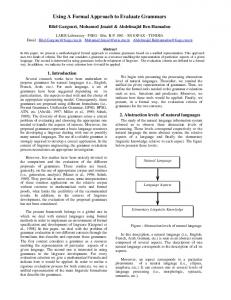
Fig (1). Cellular targets in apoptosis modulated by dietary bioactive components.
REGULATION OF APOPTOSIS Compelling evidence suggests that dietary bioactive agents may trigger apoptosis through numerous intracellular molecular targets, as well as by both intra- and extracellular stimuli including DNA damage, cell cycle disruption, hypoxia, detachment of cells from their surrounding tissue, and loss of trophic signaling [10]. Apoptosis occurs via many purported routes but primarily there are two wellrecognized pathways in cells [11, 12]. These include the intrinsic mitochondria-mediated pathway and the death receptor-mediated extrinsic pathway, which involves interaction between death receptors such as TRAIL, TNFR1, and FasL (Fig. 1). The intrinsic pathway is characterized by alterations in mitochondrial polarization and release of mitochondrial proteins including cytochrome c, endonuclease G, Smac (secondary mitochondria-derived activator of caspase)/DIABLO (direct inhibitor of apoptosis protein (IAP) binding protein with low pI), Omi/HtrA2, apoptosis-inducing factor (AIF), and its homolog AIF-homologous mitochondrion-associated inducer of death (AMID) all of which may be targeted by dietary agents [10]. Cytochrome c released to the cytoplasm during apoptosis can bind and activate Apaf-1 (apoptotic protease activating factors) promoting binding and activation of procaspase 9, an initiator caspase. Subsequently, procaspases can be cleaved to form effector caspases such as caspases 3, 6, and 7 and trigger apoptosis. Alteration of mitochondrial Bcl-2/Bax protein ratios are potential molecular targets because both are integrators of concurrent cellular pro-death and pro-survival signals. This balance influences cellular fate and whether the cell dies or not depending on the release of apoptogens from the mitochondria. Other proteins with similar Birc (baculoviral inhibitor of apoptosis repeat-containing) gene homology
domains are targets and include death suppressors including Bcl-2 and Bcl-xL, IAP family and the external death receptor-mediated pathways. Bioactive agents that bind or interfere with IAP, as well as other protein targets, may prevent the anti-apoptotic function in favor of apoptosis [13]. The extrinsic apoptosis pathway is triggered by members of the TNF (tumor necrosis factor) receptor superfamily of almost twenty cytokine receptors consisting of TNFR1, Fas, and TRAIL (TNFrelated apoptosis inducing ligand). These proteins recruit adapter proteins including FADD to cytosolic death domains with subsequent binding to pro-caspases, i.e., caspase 8. These contain a protein interaction motif (the death effector domain or DED) that binds a complementary domain in FADD. Next, protein/protein interactions involving death domains lead to intracellular recruitment of the deathinducing signaling complex (DISC) and activation of the initiator caspases 8 and 10 [14]. NUTRIGENETICS: APOPTOSIS
NUTRIENTS,
GENETICS,
AND
Nutrigenetics explores the effects of nutrients on stable changes in gene expression that do not involve DNA mutation and includes the study of individual genetic variations, which contribute greatly to individual and population-based responses to diet [15]. At this time, over 3 million SNPs, single nucleotide polymorphisms, are estimated to occur with one appearing every ~1500 base pairs primarily as a replacement of cytosine with thymine. SNPs may occur anywhere in the genome with either little influence or with marked alteration of gene expression, alternative splicing of genes, and function of
440 Current Cancer Drug Targets, 2007, Vol. 7, No. 5
Keith R. Martin
transcription factors, which can subsequently affect cancer biology and apoptosis. Polymorphism (MTHFR)
in
Methylenetetrahydrofolate
Reductase
Currently there are 50-100 genes involved either directly or indirectly in folate metabolism including receptors, binding proteins, enzymes, tissue specific gene products, and downstream factors that rely upon folate-derived metabolites [16]. Severe folate deficiency in humans is associated with an increased incidence of several forms of cancer such as colorectal cancer largely through increased genomic instability and altered programmed cell death (or apoptosis). A polymorphism in MTHFR, which causes the substitution of cytosine to thymine at nucleotide 677, is the most common SNP variant known in folate metabolism occurring in 5-20% of the population worldwide. Homozygous individuals have a 30% reduction in enzyme activity and reduced conversion of 5,10-methylene tetrahydrofolate to 5-methylene tetrahydrofolate, the form that circulates in the plasma supporting increased need for dietary folate to maintain tissues levels [17]. The MTHFR polymorphism has been associated with apoptosis. For example, in a study of the etiology of orofacial clefting, RNA interference technology was used to silence the MTHFR gene product in primary cultures of murine embryonic palatal mesenchymal cells. In these cells, gene silencing caused increased apoptosis in deficient cells when compared to control cells. Supplementation with folic acid prevented the teratogenic effect of MTHFR gene silencing [18]. In a study of postnatal brain development, mice with a homozygous knockout of the MTHFR gene displayed reduced size of the cerebellum and cerebral cortex, which was attributed, in part, to increased cell death or apoptosis. Supplementation of MTHFR(+/-) dams with an alternate methyl donor, betaine, reduced these abnormalities in the homozygous progeny suggesting a role for MTHFR in apoptosis and brain function [19]. In a human study, subjects with two genetic MTHFR polymorphisms (677C>T and 1298A>C) were compared regarding increased risk for chronic lymphocytic leukemia (CLL) and predicted disease progression. Progression-free survival was significantly longer in patients displaying the MTHFR 677CC and the MTHFR 1298A/C or CC genotypes. Moreover, spontaneous apoptosis of CLL cells in vitro was significantly increased in the group with MTHFR 677CC and MTHFR 1298AC polymorphisms. For those with the MTHFR haplotype (677CC plus 1298CC or A/C), the combination was the best independent prognostic factor for cancer prognosis. Collectively, there appears to be a potential link between specific SNPs of MTHFR, cancer, and apoptosis although more research is needed to better define this relationship. Polymorphisms of Glutathione Peroxidase Glutathione peroxidase (Gpx) is a second commonly occurring SNP and is functionally dependent, in part, on dietary selenium. A role for selenium in the prevention of cancer continues to surface in the literature. Some of the effects may relate to the incorporation of selenocysteine in antioxidant enzyme systems such as Gpx [20]. Cytosolic Gpx polymorphisms are associated with increased cancer risk and the loss of one of the copies of this same gene may contribute to malignant progression and metastasis [21]. These data along with differential expression patterns reported, in part, for the other 25 known selenoproteins in tumor versus normal tissues support a role for selenoproteins in the protection by selenium specifically in liver, prostate, colorectal and lung cancers [22]. Inadequate dietary selenium is linked to increased apoptosis. As a result, supplementation of individuals with deficient dietary intakes should exhibit enhanced antioxidant protection due to increased expression of the selenoprotein Gpx and thioredoxin reductase [23]. Moreover, higher levels of selenium supplementation can be expected to affect other functions related to tumorigenesis including carcinogen metabolism, immune function, cell cycle regulation, and
apoptosis [23]. It has been proposed in studies using genetically altered mice deficient in Gpx that selenium deficiency can trigger an inflammatory cascade and ultimately contribute to inhibition of hydroperoxide-mediated apoptosis suggesting a relationship between Gpx isoforms and apoptosis [24]. Exaggerated exposure to selenium can also induce apoptosis. For example, exposure of prostate cells to selenium as methylseleninic acid or selenite has been reported to lead to DNA fragmentation and caspase-mediated cleavage of poly(ADPribose (PARP) associated with apoptosis [25]. Cytochrome P450 and GST Polymorphisms Cytochrome P450 is a large gene family encoding a large number of enzymes that mediate the metabolism of carcinogens, endogenous substrates, and dietary components [26]. As a result, polymorphisms in the CYP genes can potentially modify individual responses to diet and ultimately cancer risk. For example, cigarette smoke places some smokers with specific CYP 1A1 polymorphisms at greater risk for cancer. In contrast, bioactive components of cruciferous vegetables can increase CYP1A1 activity altering metabolism and decreasing cancer risk. It is possible that the effect of bioactive components on pathways such as apoptosis depends on the isoform since expression of some isoforms increase cancer risk with carcinogen exposure while others decrease risk [9, 27]. Moreover, dietary isothiocyanates can induce apoptosis of pre-cancerous cells and tumor cells through activation of caspase-8 and potentiation by JNK1 and can be affected by GST SNPs [28]. In case-control studies, leukemogenesis in children was associated with DNA variants in genes associated with these pathways and the combination of genotypes seemed to be more predictive of risk than either of them independently [29]. EPIGENETICS: APOPTOSIS
NUTRIENTS,
GENOMICS,
AND
The field of epigenetics is the study of modifications of DNA and DNA binding proteins that alter the structure of chromatin without altering the sequence of DNA nucleotides and include DNA methylation, histone modifications, alteration of methyl-CpG binding proteins and modulation of chromatin remodeling proteins, which all influence interactions with transcription machinery and consequently gene expression [30]. Dietary components including both nutrients and non-nutrients play essential roles in these epigenetic events with heritable changes occurring in gene function via methylation of newly synthesized DNA by 5-cytosine DNA methyltransferase (DNMT). Folate participates in the generation of S-adenosyl methinonine (SAM), which functions as a donor in the methylation of cytosine residues in DNA and is associated with gene silencing. Covalent attachment of biotin to histones (DNA-binding proteins) also plays a role in gene silencing and in the cellular response to DNA damage, a process linked closely to apoptosis. Lastly, tryptophan and niacin are converted to NAD which is a substrate for PARP of histones and other DNA-binding proteins. PARP of these proteins participates in DNA repair and apoptosis. Clearly, epigenetics can regulate gene expression and affect apoptosis, which ultimately governs an individual’s cancer susceptibility and dictates dietary recommendations [31, 32]. Methylation Changes in DNA methylation patterns in the promoter region of genes either in the form of hypomethylation or hypermethylation have profound effects on gene expression associated with apoptosis in numerous types of cancer. In fact, cancer cells routinely display dysregulated apoptosis presumably to evade programmed cell death [33]. Hypermethylation typically down regulates gene expression associated with the cell cycle, DNA repair, growth signaling, angiogenesis, and apoptosis [34]. This is supported by observations that DNA methylation inhibitors such as azacitidine and decitabine can induce functional re-expression of aberrantly silenced genes in cancer, causing growth arrest and apoptosis in tumor cells [35].
Using Nutrigenomics to Evaluate Apoptosis
One of the most compelling examples of the link between diet and methylation is a study focusing on nutrients that donate methyl groups, i.e, folate and vitamin B12, provided in the diets of female mice during pregnancy [36]. Provision of these vitamins led to changes in chromatin methylation and gene expression where the former silenced the agouti gene. As a result, instead of giving birth to large yellow pups, which would be the normal results, the mouse dams delivered thin brown pups clearly demonstrating the in vivo impact and response to dietary bioactive agents. Moreover, this proves how subtle differences in nutrition can affect subsets of genes in the genome via methylation during embryonic development [37]. In other studies focusing on cancer, high levels of methylation at three molecular markers of cancer permitted the identification of normal samples from prostate cancer samples during early recurrence from those without recurrence. In a breast cancer study, 86% of women with low methylation levels at the transcription factor PITX2 were metastasis-free at 10 years compared to 67% of patients with high methylation levels [30]. In a human study, thirty-eight patients with premalignant gastric lesions were given folic acid three times per day [38]. Both the epithelial apoptosis rate and the p53 tumor suppressor expression in gastric mucosa significantly increased after folic acid treatment when compared to controls. It is noteworthy that loss of genomic methylation can cause p53-dependent apoptosis [39]. Hypermethylation also affects expression of DAPK and APAF-1, which are directly linked to apoptosis and the cancer process and, as a result, are promising therapeutic targets [40-42]. Clearly, bioactive nutrients involved with methylation, such as folate and B12, exerted epigenetic effects in processes linked to cancer and apoptosis. Moreover, vitamin B6, riboflavin, and zinc are coenzymes and cofactors, respectively, at different stages of the process making them also bioactive agents in methylation and resultant gene expression changes. Many other individual dietary components can influence DNA methylation including zinc, polyphenols, alcohol, coumestrol, and vitamin A as examples [6, 43]. Numerous plant-derived, non-nutritive components can also exhibit epigenetic effects. For example, epigallocatechin gallate (EGCG), the major polyphenol in green tea, can inhibit DNMT activity and reactivate methylation-silenced genes in cancer cells (Fig. 1). Treatment of human esophageal cancer cells with EGCG for as little as 12 hours caused a concentration- and time-dependent reversal of hypermethylation of the tumor suppressor p16(INK4a), retinoic acid receptor beta (RARbeta), and O(6)-methylguanine methyltransferase (MGMT) genes. This occurred in conjunction with changes in mRNA expression. Furthermore, reactivation of methylation-silenced genes by EGCG was also demonstrated in human colon cancer HT-29 cells, esophageal cancer KYSE 150 cells, and prostate cancer PC3 cells. In other studies, tea polyphenols including catechin, epicatechin, and EGCG and bioflavonoids including quercetin, fisetin, and myricetin also inhibited DNMT- and DNMT1-mediated DNA methylation in a concentration-dependent manner with EGCG exhibiting the highest potency [5]. EGCG has also been shown to protect against photocarcinogenesis in a SKH-1 hairless mouse model by significantly inhibiting UVB-induced global DNA hypomethylation [44]. The dietary polyphenol epicatechin inhibited noncompetitive methylation of DNMT by increasing the formation of S-adenosyl-L-homocysteine (SAH) but the effect of EGCG was largely due to direct inhibition of the DNMTs, which has been supported by computational modeling. Data show that the polyphenol monomer gallic acid, a building block of EGCG, inhibited DNMT by its high affinity interaction with the enzymatic catalytic site [5]. Dietary bioactive agents that exert epigenetic effects, i.e., alter DNA methylation, have been shown to induce apoptosis. For instance, EGCG inhibits cell growth and induces apoptosis in a variety of cancer cell lines including the human prostate DU145 (androgen insensitive) and LNCaP (androgen sensitive) cell lines [4547]. Experimental studies reveal that other dietary components
Current Cancer Drug Targets, 2007, Vol. 7, No. 5
441
including isoflavones, indole-3-carbinol (I3C), 3,3'-diindolylmethane (DIM), curcumin, EGCG, and apigenin modulate cell signaling pathways, i.e., NF-kB, Akt, MAPK, and p53, activate apoptosis signals, and induce programmed cell death in precancerous or cancer cells, resulting in the inhibition of carcinogenesis [48]. Several isothiocyanates, including phenethyl isothiocyanate (PEITC), found naturally in cruciferous vegetables, induce growth arrest and apoptosis in prostate cancer cells in culture and when tested as orthotopic xenografts. PEITC also inhibited the activity and level of histone deacetylases (HDACs), and induced histone acetylation and methylation necessary for chromatin unfolding in prostate cells. PEITC also demethylated the promoter region of genes and restored the unmethylated, silenced glutathione-S-transferase (GSTP1) in both androgen-dependent and androgen-independent LNCaP cancer cells. The capacity of a dietary bioactive agent such as PEITC to affect both DNA and chromatin coupled with the capacity to induce apoptosis suggests extensive connectivity among many different epigenetic mechanisms offering novel strategies in cancer chemoprevention. The results with EGCG, PEITC, and other plant-derived polyphenols discussed above demonstrate for the first time that DNA methylation can be modulated by commonly consumed dietary constituents. Furthermore, these same dietary components activate apoptosis in cancer cells. Collectively, these data support the potential of dietary bioactive agents to influence apoptosis through epigenetic mechanisms with a resultant impact on an individual’s risk for cancer. Histone Modifications Several modifications of histones have been reported to have epigenetic effects associated with apoptosis, and all are likely to depend on diet (Fig. 1). For example, biotin modulates the expression of more than 2000 genes in human cells and biotinylation of histones contributes to gene silencing, cell proliferation, apoptosis, and plays a role in the cellular response to DNA damage [49, 51, 52]. It has been demonstrated that biotin contributes to post-translational modification of histones and that biotinidase has the requisite catalytic activity to mediate attachment of biotin covalently to histones [50]. Others have specifically shown that biotinylated species of histone H4 may be involved in the formation of heterochromatin structures and in gene silencing, which can likely affect apoptosis-associated genes and ultimately protein products [49]. The amino acid tryptophan, the vitamin niacin, and PARP contribute to modification of histones and are critical events in gene expression. PARP of histones and DNAbinding proteins play a key role in DNA repair, recombination, genomic stability, and apoptosis [51]. Tryptophan and niacin are converted to NAD, which is a substrate for PARP in the modification of histones and other DNA-binding proteins. As a result, it is not surprising that PARP of proteins correlates with the dietary niacin supply in mammals and that provision of nutrients reasonably above the minimal daily requirement, but not excessive, can exert additional protective effects. Histone deacetylation is now thought to be a pre-requisite for DNA methylation and is associated with gene silencing in human cancer genes, i.e., p21 and Bax. In fact, genes silenced through DNA promoter methylation often display low histone acetylation suggesting co-regulatory mechanisms in gene silencing [52]. Moreover, apoptosis is associated with global DNA hypomethylation and histone deacetylation events in leukemia linking the processes. Several HDAC inhibitors, which can restore apoptosis, are in clinical trials with significant activity against a spectrum of both hematological and solid tumors at doses that are well tolerated by patients [53]. For example, valproic acid, a chemical compound in clinical use, inhibits the enzyme that adds acetyl groups to histone proteins and increases the effectiveness of radiation therapy, i.e., induction of apoptosis, for certain brain tumors. The dietary agent sodium butyrate, a potent histone deacetylase inhibitor, has been shown to induce gene expression and apoptosis in MCF-7 cells, a breast cancer cell line that lacks caspase-3 activity. The results also
442 Current Cancer Drug Targets, 2007, Vol. 7, No. 5
showed decreased glutathione levels after butyrate suggesting the pro-apoptotic effects are associated with oxidative stress and HDAC inhibition [54]. In a second study, co-administration of sodium butyrate, suberoylanilide hydroxamic acid (SAHA), or trichostatin with perifosine synergistically increased oxidative stress and induced apoptosis in U937 human monocytic cancer cells, as well as HL-60 and Jurkat leukemia cells suggesting an efficacious combinatorial anti-leukemic strategy [55]. Regardless of mechanism, HDAC inhibitors appear capable of reactivating genes that have become inappropriately silenced whose normal function is to suppress tumor growth. Dietary factors may beneficially correct in cancer cells the imbalance between histone acetyltransferase (HAT) and HDAC activities because cancer cells appear to be more sensitive than nontransformed cells to HDAC inhibitors [56]. Three dietary agents have been shown to exhibit HDAC inhibitory activity using in vitro and in vivo models and include butyrate, diallyl disulfide (DADS), and sulforaphane (SFN). DADS is found in garlic and sulforaphane is an isothiocyanate (ITC) found in cruciferous vegetables such as broccoli and broccoli sprouts. SFN induces cell cycle arrest and apoptosis in cancer cells, inhibits tubulin polymerization, activates checkpoint 2 kinase, and inhibits HDAC activity [57]. ITCs have also been shown to induce apoptotic cell death via both p53-dependent and independent pathways [58]. The latter findings suggest that SFN may be effective during the post-initiation stages of carcinogenesis, as a suppressing agent [56, 59]. Hyperacetylation of histone proteins can also occur with altered transcription patterns that ultimately induce apoptosis. This suggests chromatin modification can occur in the absence of DNA damage although key DNA damage signaling proteins are activated and apoptosis triggered [60]. More research is needed to further elucidate the process of excessive acetylation. APOPTOSIS AND NUTRITIONAL TRANSCRIPTOMICS The transcriptome is the set of all mRNA molecules, or transcripts, in a single cell or in a population of cells and can vary greatly with external environmental conditions such as dietary exposure. In essence, the transcriptome provides a snapshot of the genes that are actively expressed at any given time and can be targeted to specific processes. For instance, more than 100 genes have been identified as being differentially expressed during apoptosis [61]. Dietary phenolic compounds including ellagic acid and resveratrol have been shown to modulate more than 550 genes involved in cellular proliferation and apoptosis in hormone-dependent human prostate cells after incubation for 48 hours [62]. Apoptosis and cancer pathway-specific mini-microarrays have been used to screen for differentially expressed genes in four human colon adenocarcinoma cell lines in response to folate deficiency. The expression of the seven most notably and consistently affected genes were confirmed by real time RT-PCR supporting that folate deficiency affects the expression of key genes related to apoptosis. The idea that diet can affect gene expression is generally wellaccepted, but the diverse tissue and organ-specific effects of bioactive dietary agents on gene expression patterns in the context of diet, genomics, and health is less clear [63]. When one considers the temporal aspect of gene expression, the complexity increases significantly [64]. There is evidence that dietary consumption of fruits and vegetables protects against cancer. In studies with mice, animals were fed vegetables in their diets and the effects on gene expression in the colonic mucosa were measured. Results indicated that 39 genes exhibited significant, dose-related modulation. For 15 genes, this could explain reduced cancer risk from vegetable consumption [65]. Gene expression changes in the lungs of mice fed different vegetables showed that individual vegetables had a higher potential for modulating anti-carcinogenic genes associated with apoptosis in favor of lung cancer prevention [65]. After energy restriction, rats prone to mammary carcinogenesis differentially expressed Bcl-2,
Keith R. Martin
CARD, and IAP functional gene groupings, as revealed by microarray analysis, directly involved in apoptosis [66]. Moreover, the activities of caspases and mitochondrial proteins associated with inhibition of apoptosis were down regulated. In a study of prostate cancer, dietary soy was provided in a SCID-human model and gene expression analyzed. In animals fed soy, gene expression of multiple genes involved in apoptosis were altered both in vitro and in vivo [67]. In a compelling human study, men that were either native Japanese or Japanese American were divided into low fat-high soy or high fat-low soy groupings. After radical prostatectomies, tissues from normal and neoplastic tissue were analyzed by microarray analysis to determine expression of numerous tissue biomarkers, i.e., Bcl-2 and caspase-3, for apoptosis. Normal prostate tissue could be differentiated from cancerous tissue and genes associated with caspases and 5-lipoxygenases were expressed more in native Japanese than Japanese-Americans [68]. Similar results have been found in other studies of hepatocellular carcinomas in rats. Rats were fed choline-deficient diets and gene expression was profiled. A total of 146 genes were differentially expressed including those in apoptosis [69]. I3C and its metabolite DIM from cruciferous vegetables also induce programmed cell death and regulate many genes associated with apoptosis. Moreover, the compounds inhibited the binding of transcription factors that inhibit apoptosis suggesting combinatorial control on cis regulatory elements are critical in modulating co-expression of genes [70, 71]. Transcription factor expression may be altered by dietary bioactive components and the process of transcription prevented. For example, activation of NF-kB promotes survival and cellular proliferation and down regulation sensitizes cells to apoptosis. Sulforaphane, PEITC, curcumin, green tea, 6-gingerol, resveratrol and soy can strongly inhibit NF-kB activation and, as a result, enhance pro-apoptotic signals in numerous models of cancer including prostate cells [19, 72]. Experimental data indicate that genistein, I3C, curcumin, EGCG, and apigenin also inhibit NF-kB activation and DNA binding in different cell lines derived from cancer tissues [67]. Genistein inhibits NF-kB activation via phosphorylation of IkB, and prevents the translocation of NF-kB subunits to the nucleus in epithelial and myeloid cells possibly through inhibition of MEKK1 kinase activity [67]. Curcumin and resveratrol have been shown to inhibit IKK, alter AP-1 activity, inhibit nuclear translocation, and suppress constitutive and inducible NF-kB activation. EGCG, apigenin, and curcumin significantly inhibit, in a time and dose-dependent manner, activation and translocation of NF-kB to the nucleus by suppressing the degradation or phosphorylation of IkB [73]. EGCG can concurrently stabilize p53 and negatively alter the ratio of Bax/Bcl-2 proteins inducing apoptosis. p53 is a sequence specific transcription factor that mediates apoptosis and is the most frequently mutated in human cancer [76, 77]. p53 has also been demonstrated to exhibit a direct apoptogenic role at the mitochondria where it translocates and interacts with BclxL and Bcl-2 proteins to induce mitochondrial permeabilization [74]. EGCG dose-dependently increases p53 expression in prostate cells with wild type p53 but not DU145 prostate cells with mutant p53 [75]. Resveratrol, a polyphenol in grapes, also stabilizes and activates p53 [78]. The AP-1 transcription factor is oncogenic and antagonizes apoptosis in neoplasia. Numerous dietary components such as polyphenols inhibit AP-1 activation, as well as other anti-apoptotic transcription factors. Dietary components such as resveratrol, curcumin, and green tea can inhibit AP-1 and NF-kB activation, as well as other anti-apoptotic transcription factors [79-81]. APOPTOSIS AND PROTEOMICS Compelling evidence suggests that dietary bioactive agents may trigger apoptosis through numerous molecular protein targets (Fig. 1). For example, dietary bioactive agents can alter mitochondrial membrane function, disrupt the mitochondrial transition pore (MTP), and/or dissipate the mitochondrial membrane potential (MMP) through enhanced release and activation of an array of proteins,
Using Nutrigenomics to Evaluate Apoptosis
Current Cancer Drug Targets, 2007, Vol. 7, No. 5
443
which induce apoptosis. For example, curcumin found in turmeric and capsaicin found in chili peppers can open the MTP, induce mitochondrial swelling, and collapse the MMP leading to induction of apoptosis [52, 73, 82]. Flavonoids such as baicilin and botanical agents such as nordihydroguaiaretic acid induce apoptosis in various cancer cells through increased cytochrome c release and disruption of MMP [73, 82, 83]. EGCG, resveratrol, and carotenoids depolarize mitochondria in numerous human cell lines including prostate, melanoma, and lung cells, induce cytochrome c release and induce apoptosis. In metastatic mouse mammary carcinoma cells, these components can alter Bcl-2/Bax protein ratios, increase Apaf1 formation, and cleave caspase and PARP proteins [82, 84-87]. Flavonoid-rich grape seed extract can also induce apoptosis in human prostate cells by increasing cytochrome c release and PARP cleavage [88]. Collectively, numerous diverse dietary bioactive agents can induce apoptosis by altering mitochondrial physiology.
diterpenoids induce apoptosis in human prostate cells by upregulation of Fas ligand [95]. The polyphenols curcumin, EGCG, and resveratrol downregulate protein phosphorylation, inhibit the MAPK pathway, and prevent ligand binding of growth factor receptors including EGF, FGF, and PDGF [103]. Moreover, resveratrol promotes protein interaction between p53, ERK, and p38 kinase, through phosphorylation, stabilization, and activation of p53 in epidermal cells [78]. In cells with mutated oncogenic Ha-ras, green and black tea polyphenols potently inhibited ERK phosphorylation and AP-1 activity [104]. ITCs increase activity of JNK, MAPK, ERK, and p38 kinase in HT29, HL-60 cells and PC3 cells and human head and neck squamous cell carcinoma lines [98]. When considering garlic compounds, DADS induces JNK1, S-allylmercaptocysteine induces JNK1 activation and JNK activity, and ajoene activates MAPK (JNK, p38, ERK1/2) in different cell types [93]. I3C inhibits signaling through protein kinase B (PKB) and binding of NF-kB to DNA [105].
Bioactive components can regulate intracellular location of proapoptotic proteins (Bcl-2, Bcl-xL) or anti-apoptotic proteins (Bax and Bak) from the mitochondria. In breast, prostate, and other human cell lines, I3C, DIM, curcumin, and genistein alter the ratio of the antiapoptotic proteins Bcl-2 and Bcl-xL and the pro-apoptotic protein Bax causing apoptosis [20, 63, 89, 90] Sulforaphane, an ITC, increases expression of Bax. The polyphenol resveratrol decreases Bcl-2 and Bcl-xL levels and increases Bax levels [24, 91]. Others have shown that genistein can increase phosphorylation of Bcl-2, upregulate pro-apoptotic Bax, and downregulate Bcl-2 altering programmed cell death by modulating Bax homodimerization [92]. DAS and DADS, found in garlic, increase Bax expression, DADS reduces Bcl-xL expression, and DAS, S-allyl cysteine (SAC), and ajoene can decrease Bcl-2 expression [93]. Collectively, numerous diverse dietary bioactive agents can induce apoptosis by modulating the Bcl-2 family of proteins making these critical intracellular targets.
During apoptosis, a large number of proteins involved in signal transduction are post-translationally modified as alluded to above. In classical proteomics, the protein separation by two dimensional gel electrophoresis and protein identification by mass spectrometry provides a rapid and high resolution composite enabling elucidation of >100 proteins that are altered during apoptosis [106]. Moreover, the use of standard apoptosis assays with proteomics and computer simulation has revealed differential expression of 61 apoptosis-related genes in cells treated with toxin [107]. In other cell systems, more than 383 proteins have been shown to be significantly modulated with increased expression of ~35 and reduced expression of ~30 compared to controls [19]. Applied SELDI and FT-ICR technologies have been used in proteomics to analyze tumor and non-tumor samples resected from the liver of fish. This methodology permitted identification of protein and metabolite profiles that were specific to liver tumors [108]. This technology has been extended to humans where differential protein expression in the early stages of human colon cancer has shown in tumor tissue that mitochondrial apoptosis (intrinsic pathway) was occurring [109]. The low molecular weight range of the circulatory proteome contains tremendous information and proteomics allows collection of functional data of protein degradation, modification, translocation, and synthesis all of which are important in an individual’s response to diet [77].
Bcl-2 proteins may also be inhibited by other proteins with similar Birc (baculoviral inhibitor of apoptosis repeat-containing) gene homology domains. Plant-derived naphthoquinones and diterpenes, found in citrus peel, upregulate Bid, respectively, in human kidney, brain tumor, and prostate cancer cells [94, 95]. Bcl-2 interacting protein (t-BID), links the intrinsic and extrinsic apoptotic pathways [14]. Dietary components can induce apoptosis through activation of caspase proteins. Resveratrol increases caspase activity (6, 3, and 9) in numerous models including normal and hematopoietic cells [96, 97]. ITCs significantly induce apoptosis in cultured human and animal cell lines as well as animal tissues and cancer cell xenografts through stimulation of caspase 3-like activity and degradation of PARP [78, 98]. ITC also activates caspases in multiple pathways including caspase 9 (mitochondria), caspase-8 (death receptor) and caspase 12 (endoplasmic reticulum) in association with activation of caspase 3 [98]. I3C, ajoene, and DAS upregulate Bax, induce the release of cytochrome c, and activate caspases 3 and 9 in human breast cancer cells, human colon cells, and murine melanoma cells [79-81, 90]. Furthermore, the I3Cs induce apoptosis by down regulation of Bcl-2, Bcl-xL, IAP, XIAP, and FLIP [90]. The reduction of growth factor-induced cellular signaling permits, in many cases, the initiation of apoptotic cascades employing large numbers of proteins (Fig. 1). Dietary polyphenols can down regulate phosphorylation and ligand binding of growth factor receptors, which are essential for evasion of apoptosis [99]. Resveratrol, curcumin, polyphenols, and catechins can also inhibit growth factor signaling pathways [72, 100]. In fact, the TNF family member Apo2L/TRAIL receptor has received considerable recent attention as a therapeutic target since many cancer cells appear sensitive to Apo2L/TRAIL-induced apoptosis [69, 86, 101]. EGCG, a polyphenol in tea, directly blocks EGF binding to its receptor and interrupts signaling. Resveratrol has been shown to trigger CD95 signaling-dependent apoptosis in human tumor cells [102]. Dietary
The complex interaction of dietary bioactive agents with the proteome can probably be best illustrated with I3C. Biologically, I3C is converted to an array of oligomeric products with DIM being a major component and is considered responsible for its in vivo biological effects. In cells, I3C induces apoptosis in various tumor cells including breast cancer, prostate cancer, endometrial cancer, colon cancer, and leukemic cells. Mechanistically, I3C-induced apoptosis involves downregulation of anti-apoptotic gene products including Bcl-2, Bcl-xL, survivin, IAP, X chromosome-linked IAP (XIAP), and FADD protein-like interleukin-1-beta-converting enzyme inhibitory protein (FLIP) and upregulation of the proapoptotic Bax protein. Moreover, I3C also induces release of mitochondrial cytochrome C protein and activates caspase-9 and caspase-3 proteins. It is indeed intriguing that a single dietary component can affect multiple molecular targets within the process of apoptosis. APOPTOSIS AND METABOLOMICS Metabolomics can be defined as the dose and temporal changes in cellular small molecular weight compounds in response to diet [6]. The metabolic response will depend on the quantity and timing of exposure to bioactive food components. Metabolomic analysis can also serve as a powerful tool to localize and identify critical enzymatic steps that regulate metabolic systems as a whole [110]. Although the technology is in the early stages, research is progressing in other systems with emerging application to nutrition. For example, metabolomic analysis of sulfur-containing amino acids in testes of rats exposed to cadmium revealed marked alterations in metabolism
444 Current Cancer Drug Targets, 2007, Vol. 7, No. 5
with elevated amounts of methionine and cysteine noted concurrently and a decrease in the ratio of SAH to SAM. This collectively suggests enhancement of the remethylation cycle and increased methyl formation, both of which are instrumental in epigenetic events and associate to an extent with apoptosis [110]. In a study of humans exposed to benzene, proteomics analysis show that several proteins are altered in the serum of exposed subjects, which were associated with apoptosis and immune function [61]. Metabolite levels in plasma and urine were analyzed in relation to changes in hepatic gene expression in rats that received bromobenzene, and altered levels of genes and metabolites were associated with the degree of necrosis and linked to apoptosis, glycolysis, and amino acid metabolites [111]. Colon cancer samples from humans were compared to normal tissue and metabolic pathways analyzed. The results revealed that mitochondrial apoptosis, viz., intrinsic pathway, was being induced as noted by biochemical pathway-specific markers [109]. In a study of rat glioma, apoptosis was induced with changes in pro-apoptotic Bcl2, Bak-1, caspase 9 and FAS after 0, 4, and 8 days of treatment documenting clearly metabolic perturbations in tumors [112]. Collectively, metabolomics is beginning to contribute greatly to the elucidation of perturbations in biological systems and is paving the way for the study of the effects of bioactive dietary components in whole body systems. The primary pro-apoptotic signaling events induced by dietary bioactive agents share many commonalities. However, differences have been noted regarding the temporal occurrence and duration of some events emphasizing them as important considerations. For instance, both ITCs and polyphenols induce JNK transcription factor activation and ultimately induce apoptosis as shown by altered mitochondrial potential, induced cytochrome c release, and activated caspases. PEITC induces effects up to 4 hours, whereas EGCG produces identical effects only after 12 hours suggesting a temporal difference in susceptibility to changes [19]. The sustained exposure to some dietary agents may prolong activation of transcription factors and ultimately interfere with trophic growth factor signaling, which can lead to apoptosis. Thus, the duration of a signal appears important in determining the biological outcome. The identification and tracking of specific metabolites and interaction of components of a pathway is an emerging discipline that requires more focus and research as technologies develop. It appears that numerous molecular targets exist in vivo and collectively converge on several signaling pathways. This affords the possibility of determining one’s response to diet and use of combinatorial therapy with dietary agents to affect numerous targets that lead coordinately to the induction of apoptosis. APOPTOSIS, TRANSCRIPTOMICS, PROTEOMICS, AND METABOLOMICS CAN SHED LIGHT ON MOLECULAR MECHANISMS AND DIETARY INFLUENCES Accumulating evidence clearly demonstrates that apoptosis possesses numerous molecular targets for dietary bioactive agents supporting a role in the chemoprevention of cancer. Apoptosis, however, is a complex process comprised of intrinsic and extrinsic pathways of programmed cell death, with numerous specific targets within each arm. Biological systems are extremely complex and exhibit properties that extend far beyond the observations associated with the independent systems. This is further complicated by the temporal nature of many of these effects. As a result, it is critical to evaluate the entire biological system from the nutrigenomics perspective. It is encouraging that single bioactive dietary agents such as EGCG or I3C have been shown to directly and indirectly influence most, if not all, of the myriad targets within apoptosis including genes, transcripts, proteins, and metabolites. Thus, critical evaluation is essential for DNA polymorphisms or SNPs of a gene, expression of that specific gene, expression of specific processed mRNA (alternative splicing), protein production from that mRNA, posttranslational modification of the resultant protein, and formation of
Keith R. Martin
respective metabolites. Evolution of the fields of nutrigenetics, epigenomics, transcriptomics, proteomics, and metabolomics has begun to permit this approach so that a comprehensive picture emerges from not only a cell but tissues and whole organisms [112]. Studies such as these can be used to study an individual’s response to diet and better define the effects on molecular events associated with cancer. Specifically, the modulation of apoptosis, a critical event in cancer, through diet can be beneficially and selectively induced as an efficacious, effective means of chemoprevention. REFERENCES [1]
[2] [3] [4] [5]
[6] [7] [8]
[9] [10] [11] [12] [13] [14]
[15] [16]
[17]
[18]
[19]
[20] [21] [22]
[23] [24]
[25]
[26] [27] [28]
Forman, M.; Hursting, S.; Umar, A.; Barrett, J. Nutrition and Cancer Prevention: A Multidisciplinary Perspective on Human Trials. Annu. Rev. Nutr. 2004, 24, 223-254. Willett, W. Diet, Nutrition, and Avoidable Cancer. Environ. Health Perspect. 1995, 103, 165-170. Peto, R.; Doll, R.; Buckley, J.; Sporn, M. Can Dietary Beta Carotene Materially Reduce Human Cancer Rates? Nature 1981, 290, 201-208. Davis, C.; Milner, J. Frontiers in Nutrigenomics, Proteomics, Metabolomics, and Cancer Prevention. Mutat. Res. 2004, 551, 51-64. Go, V.; Nguyen, C.; Harris, D.; Lee, W. Nutrient-Gene Interaction" Metabolic Genotype-Phenotype Relationships. J. Nutr. 2005, 135, 3016S3020S. Trujillo, E.; Davis, C.; Milner, J. Nutrigenomics, Proteomics, Metabolomics, and the Practice of Dietetics J. Am. Dietetics Assoc. 2006, 106, 403-413. Reed, J.; Pellecchia, M. Apoptosis-Based Therapies for Hematologic Malignancies. Blood 2005, 106, 408-418. Finley, J. Proposed Criteria for Assessing the Efficacy of Cancer Reduction by Plant Foods Enriched in Carotenoids, Glucosinolates, Polyphenols, and Selenocompounds. Ann. Botany 2005, 95, 1075-1096. Milner, J. Functional Foods: The US Perspective. Am. J. Clin. Nutr. 2000, 71, 1654S-1659S. Sun, S.; Hail, N.; Lotan, R. Apoptosis as a Novel Target for Cancer Chemoprevention. J. Natl. Cancer Inst. 2004, 96, 662-672. Danial, N.; Korsmeyer, S. Cell Death: Critical Control Points. Cell 2004, 116, 205-219. Lockshin, R.; Zakeri, Z. Apoptosis, Autophagy, and More. Int. J. Biochem. Cell Biol. 2004, 36, 2405-2419. Salveson, G.; Duckett, C. IAP Proteins: Blocking the Road to Death's Door. Nat. Rev. Mol. Cell Biol. 2002, 3, 401-410. Perik, P.; de Vries, E.; Gietema, J.; van der Graaf, W.; Sleijfer, D.; Suurmeijer, A.; Van Veldhuisen, D. The Dilemma of the Strive for Apoptosis in Oncology. Crit. Rev. Oncol. Hematol. 2005, 53, 101-13. Wargovich, M.; Cunningham, J. Diet, Individual Responsiveness and Cancer Prevention. J. Nutr. 2003, 133, 2400S-2403S. Molloy, A. Role of Genetic Variation in Establishing Nutritional Requirements: Folate, a Case in Point. World Rev. Nutr. Diet. 2001, 89, 6875. Davis, C.; Hord, N. Nutritional "Omics" Technologies for Elucidating the Role(s) of Bioactive Food Compoennets in Colon Cancer Prevention. J. Nutr. 2005, 135, 2694-2697. Agrelo, R.; Cheng, W. H.; Setien, F.; Ropero, S.; Espada, J.; Fraga, M. F.; Herranz, M.; Paz, M. F.; Sanchez-Cespedes, M.; Artiga, M. J.; Guerrero, D.; Castells, A.; von Kobbe, C.; Bohr, V. A.; Esteller, M. Epigenetic Inactivation of the Premature Aging Werner Syndrome Gene in Human Cancer. Proc. Natl. Acad. Sci. USA 2006, 103, 8822-8827. Chen, C.; King, A. Dietary Cancer-Chemopreventive Compounds: from Signaling and Gene Expression to Pharmacological. TRENDS Mol. Med. 2005, 26, 318-328. Aboul-Fadl, T. Selenium Derivatives as Cancer Preventive Agents. Curr. Med. Chem. Anticancer Agents 2005, 5, 637-652. Rayman, M. Selenium in Cancer Prevention: A Review of the Evidence and Mechanism of Action. Proc. Nutr. Soc. 2005, 64, 527-542. Diwadkar-Navsariwala, V.; Diamond, A. The Link between Selenium abd Chemoprevention: A Case for Selenoproteins. J. Nutr. 2004, 134, 28992902. Combs, G. Chemopreventive Mechanisms of Selenium. Med. Klin. 1999, 94, 18-24. Lee, E.; Min, H.; Park, H.; Chung, H.; Kim, S.; Han, Y.; Lee, S. G2/M Cell Cycle Arrest and Induction of Apoptosis by a Stilbenoid, 3,4,5-Trimethoxy4'-bromo-cis-stilbene, in Human Lung Cancer Cells. Life Sci. 2004, 75, 2829-2839. Jiang, C.; Wang, Z.; Lu, J. Distinct Effects of Methylseleninic Acid Versus Selenite on Apoptosis, Cell Cycle, and Protein Kinase Pathways in DU-145 human prostate cancer cells. Mol. Cancer Ther. 2002, 1, 1059-1066. Bernhardt, R. Cytochromes P450 as Versatile Biocatalysts. J. Biotechnol. 2006, 124, 128-145. Milner, J. Molecular Targets for Bioactive Food Components. J. Nutr. 2004, 134, 2492S-2498S. Thornalley, P. Isothiocyanates: Mechanism of Cancer Chemopreventive Action. Anticancer Drugs 2002, 13, 331-338.
Using Nutrigenomics to Evaluate Apoptosis [29]
[30] [31]
[32]
[33] [34]
[35] [36]
[37]
[38]
[39]
[40]
[41]
[42]
[43] [44]
[45] [46] [47]
[48] [49] [50]
[51] [52]
[53]
[54]
[55]
[56]
Sinnett, D.; Labuda, D.; Krajinovic, M. Challenges Identifying Genetic Determinants of Pediatric Cancers: The Childhood Leukemia Experience. Fam. Cancer 2006, 5, 35-47. Jang, H.; Mason, J.; Choi, S. Genetic and Epigenetic Interactions between Folate and Aging in Carcinogenesis. J. Nutr. 2005, 135, 2967S-2971S. Smith, P.; Syed, N.; Crook, T. Epigenetic Inactivation Implies a Tumor Suppressor Function in Hematologic Malignancies for Polo-Like Kinase but Not Polo-Like Kinase 3. Cell Cycle 2006, 5, 1262-1264. Galm, O.; Wilop, S.; Luders, C.; Lost, E.; Gehbauer, G.; Herman, J.; Osieka, R. Clinical Implications of Aberrant DNA Methlyation Patterns in Acute Myelogenous Leukemia. Ann. Hematol. 2005, 84, 39-46. Gopisetty, G.; Ramachandran, K.; Singal, R. DNA Methylation and Apoptosis. Mol. Immunol. 2006, 43, 1729-1740. Suzuki, H.; Toyota, M.; Sato, H.; Sonoda, T.; Sakauchi, F.; Mori. M. Roles and Causes of Abnormal DNA Methylation in Gastrointestinal Cancers. Asian Pac. J. Cancer Prev. 2006, 7, 177-185. Baylin, S.; DNA Methylation and Gene Silencing in Cancer. Nat. Clin. Pract. Oncol. 2005, 2, 4-11. Waterland, R.; Jirtle, R. Early Nutrition, Epigenetic Changes at Transposons and Imprinted Genes, and Enhanced Susceptibility to Adult Chronic Diseases. Nutrition 2004, 20, 63-68. Cooney, C.; Dave, A.; Wolff, G. Maternal Methyl Supplements in Mice Affect Epigenetic Variation and DNA Methylation of Offspring. J. Nutr. 2002, 132, 2393S-2400S. Cao, D.; Sun, W.; Ou, X.; Yu, Q.; Yu, T.; Zhang, Y.; Wu, Z.; Xue, Q.; Cheng, Y. Effects of Folic Acid on Epithelial Apoptosis and Expression of Bcl-2 and p53 in Premalignant Gastric Lesions. World J. Gastroenterol. 2005, 11, 1571-1576. Jackson-Grusby, L.; Beard, C.; Possemato, R.; Tudor, M.; Fambrough, D.; Csankovszki, G.; Dausman, J.; Lee, P.; Wislon, C.; Lander, E.; Jaenisch, R. Loss of Genomic Methylation Causes p53-Dependent Apoptosis and Epigenetic Deregulation. Nat. Genet. 2001, 27, 31-39. Kong, W. J.; Zhang, S.; Guo, C.; Wang, W.; Chen, X.; Zhang, S. L.; Zhang, D.; Liu, Z.; Kong, W. Effect of Methylation-Associated Silencing of the Death-Associated Protein Kinases Gene on Nasopharyngeal Carcinomas. Anticancer Drugs 2006, 17, 251-259. Anjum, R.; Boux, P.; Ballif, B.; Gygi, S.; Blenis, J. The Tumor Suppressor DAP Kinase is a Target of RSK-Mediated Survival Signaling. Curr. Biol. 2005, 15, 1762-1767. Bialik, S.; Kimchi, A. DAP-Kinase as a Target for Drug Design in Cancer and Diseases Assocaited with Accelerated Cell Death. Semin. Cancer Biol. 2004, 14, 283-294. Davis, C.; Uthus, E. DNA Methylation, Cancer Susceptibility, and Nutrient Interactions. Exp. Biol. Med. 2004, 229, 988-995. Mittal, A.; Piyathilake, C.; Hara, Y.; Katiyar, S. Exceptionally High Protection of Photocarcinogenesis by Topical Application of (-)Epigallocatechin-3-gallate in Hydrophilic Cream in SKH-1 Hairless Mouse Model: Relationship to Inhibition of UVB-Induced Global DNA Hypomethylation. Neoplasia 2003, 5, 555-565. Adhami, V.; Ahmad, N.; Mukhtar, H. Molecular Targets for Rgeent tea in Prostate Cancer Prevention. J. Nutr. 2003, 133, 2417S-2424S. Shimizu, M.; Weinstein, I. Modulation of Signal Transduction by Tea Catechins and Related Phytochemicals. Mutat. Res. 2005, 591,147-160. Na, H.; Surh, Y. Intracellular Signaling Network as a Prime Chemopreventive Target of (-)-Epigallocatechin Gallate. Mol. Nutr. Food Res. 2006, 50, 152-159. Sarkar, F.; Li, Y. Cell Signaling Pathways Altered by Natural Chemopreventive Agents. Mutat. Res. 2004, 555, 53-64. Hassan, Y.; Zempleni, J. Epigenetic Regulation of Chromatin Structure and Gene Function by Biotin. J. Nutr. 2006, 136, 1763-1765. Petrelli, F.; Coderoni, S.; Moretti, P.; Paparelli, M. Effect of Biotin on Phosphorylation, Acetylation, Methylation of Rat Liver Histones. Mol. Biol. Rep. 1978, 4, 87-92. Oommen, A.; Griffin, J.; Sarath, G.; Zempleni, J. Roles for Nutrients in Epigenetic Events. J. Nutr. Biochem. 2005, 16, 74-77. Mai, A.; Massa, S.; Rotili, D.; Cerbara, I.; Valente, S.; Pezzi, R.; Simeoni, S.; Ragno, R. Histone Deacetylation in Epigenetics: An Attractive Target for Anticancer Therapy. Med. Res. Rev. 2005, 25, 261-309. Park, J.; Jung, Y.; Kim, T.; Kim, S.; Jong, H.; Lee, J.; Kim, D.; Lee, J.; Kim, M.; Kim, T.; et al.: Class 1 Histone Deacetylase-Selective Novel Synthetic Inhibitors Potently Inhibit Humor Tumor Proliferation. Clin. Cancer Res. 2004, 10, 5271-5281. Louis, M.; Rosato, R.; Brault, L.; Osbild, S.; Battaglia, E.; Yang, X.; Grant, S.; Bagrel, D. The Histone Deacetylase Inhibitor Sodium Butyrate Induces Breast Cancer Cell Apoptosis Through Diverse Cytotoxic Actions Including Glutathione Depletion and Oxidative Stress. Int. J. Oncol. 2004, 25, 17011711. Rahmani, M.; Reese, E.; Dai, Y.; Bauer, C.; Payne, S.; Dent, P.; Spiegel, S.; Grant, S. Coadministration of Histone Deacetylase Inhibitors and Perifosine Synergistically Induces Apoptosis in Human Leukemia Cells through Akt and ERK1/2 Inactivation and the Generation of Ceramide and Reactive Oxygen Species. Cancer Res. 2005, 65, 2422-2432. Myzak, M.; Dashwood, R. Histone Deacetylases as Targets for Dietary Cancer Preventive Agents: Lessons Learned with Butyrate, Diallyl Disulfide, and Sulforaphane. Curr. Drug Targets 2006, 7, 443-452.
Current Cancer Drug Targets, 2007, Vol. 7, No. 5 [57] [58]
[59]
[60]
[61]
[62]
[63] [64] [65]
[66]
[67]
[68]
[69]
[70]
[71] [72] [73]
[74]
[75]
[76] [77]
[78] [79]
[80]
[81]
[82]
[83]
[84]
445
Myzak, M.; Ho, E.; Dashwood, R. Dietary Agents as Histone Deacetylase Inhibitors. Mol. Carcinog. 2006, 45, 443-446. Gamet-Payrastre, L. Signaling Pathways and Intracellular Targets of Sulforaphane Mediating Cell Cycle Arrest and Apoptosis. Curr. Cancer Drug Targets 2006, 6, 135-145. Dashwood, R.; Myzak, M.; Ho, E. Dietary HDAC Inhibitors: Time to Rethink Waek Ligands in Cancer Chemoprevention? Carcinogenesis 2006, 27, 344-349. Berardi, P.; Russell, M.; El-Ostra, A.; Riabowol, K. Functional Links between Transcription, DNA Repair, and Apoptosis. Cell Mol. Life Sci. 2004, 61, 2173-2180. Smith, M. T.; Vermeulen, R.; Li, G.; Zhang, L.; Lan, Q.; Hubbard, A. E.; Forrest, M. S.; McHale, C.; Zhao, X.; Gunn, L.; Shen, M.; Rappaport, S. M.; Yin, S.; Chanock, S.; Rothman, N. Use of "Omic" Technologies to Study Humans Exposed to Benzene. Chem. Biol. Interact. 2005, 153-154, 123-127. Narayanan, B.; Narayanan, N.; Stoner, G.; Bullock, B. Interactive Gene Expression Pattern in Prostate Cancer Cells Exposed to Phenolic Antioxidants. Life Sci. 2002, 70, 1821-1839. Afman, L.; Muller, M. Nutrigenomics: From Molecular Nutrition to Prevention of Disease. J. Am. Dietetics Assoc. 2006, 106, 569-576. Martin, K. Targeting Apoptosis with Dietary Bioactive Agents. Exp. Biol. Med. 2006, 231, 117-129. van Breda, S.; van Agen, E.; van Sanden, S.; Burzykowski, T.; Kleinjans, J.; Delft, J. Vegetables Affect the Expression of Genes Involved in Carcinogenic and Anticarcinogenic Processes in the Lungs of Female C57BL/6. J. Nutr. 2005 135, 2546-2552. Thompson, H.; Zhu, Z.; Jiang, W. Identification of the Apoptosis Activation Cascade Induced in Mammary Carcinomas by Energy Restriction. Cancer Res. 2004, 64, 1541-1545. Li, Y.; Che, M.; Bhagat, S.; Ellis, K.; Kucuk, O.; Doerge, D. R.; Abrams, J.; Cher, M.; Sarkar, F. Regulation of Gene Expression and Inhibition of Experimental Prostate Cancer Bone Metastasis by Dietary Genistein. Neoplasia 2004, 6, 354-363. Marks, L.; Kojima, M.; Demarzo, A.; Heber, D.; Bostwick, D.; Qian, J.; Dorey, F.; Veltri, R.; Mohler, J.; Partin, A. Prostate Cancer in Native Japanese and Japanese-American Men: Effects of Dietary Differences on Prostatic Tissue. Urology 2004, 64, 765-771. Uematsu, F.; Takahashi, M.; Yoshida, M.; Igarashi, M.; Watanabe, N.; Suzuki, N.; Abe, M.; Rusyn, I.; Floyd, R.; Nakae, D. Distinct Patterns of Gene Expression in Hepatocellular Carcinomas and Adjacent NonCancerous, Cirrhotic Liver Tissues in Rats Fed a Choline-Deficient, LAmino Acod-Defined Diet. Cancer Sci. 2005, 96, 414-424. Uddin, R.; Singh, S. Ethanol-Responsive Genes: Identification of Transcription Factors and their Role in Metabolomics. Pharmacogenomics J. 2007, 7(1), 38-47. Sarkar, F.; Li, Y. Indole-3-carbinol and Prostate Cancer. J. Nutr. 2004, 134, 3493S-3498S. Dorai, T.; Aggarwal, B. Role of Chemopreventive Agents in Cancer Therapy. Cancer Lett. 2004, 215, 129-140. Biswal, S.; Datta, K.; Shaw, S.; Feng, X.; Robertson, J.; Kehrer, J. Glutathione Oxidation and Mitochondrial Depolarization as Mechanisms of Nordihydroquaiaretic Acid-Induced Apoptosis in Lipoxygenase-Deficient Cells. Toxicol. Sci. 2000, 53, 77-83. Mihara, M.; Erster, S.; Zaika, A.; Petrenko, O.; Chittenden, T.; Pancoska, P.; Moll, U. p53 has a Direct Apoptogenic Role at the Mitochondria. Mol. Cell 2003, 577-590. Gupta, S.; Afaq, F.; Mukhtar, H. Involvement of nuclear Factor-kappa B, Bax, and Bcl-2 in Induction of Cell Cycle Arrest and Apoptosis by Apigenin in Human Prostate Carcinoma Cells. Oncogene 2002, 21, 3727-3738. Levine, A. p53, The Cellular Gatekeeper for Growth and Division. Cell 1997, 88, 323-331. Calvo, K.; Liotta, L.; Petricoin, E. Clinical Proteomics: from Biomarker Discovery and Cell Signaling Profiles to Individualized Personal Therapy. Biosci. Rep. 2005, 25, 107-125. Roemer, K.; Mahyar-Roemer, M. The Basis for the Chemopreventive Action of Resveratrol. Drugs Today 2002, 38, 571-580. Manna, S.; Sah, N.; Newman, R.; Cisneros, A.; Aggarwal, B. Oleandrin Suppresses the Activation of Nuclear Transcription Factor-kB, Activator Protein-1, and c-Jun N-Termianl Kinase. Cancer Res. 2000, 60, 3838-3847. Manna, S.; Mukhopadhyay, A.; Aggarwal, B. Resveratrol Suppresses TNFInduced Activation of Nuclear Transcription Factors NF-kB, Activator Protein-1, and Apoptosis. J. Immunol. 2000, 164, 6509-6519. Ashikawa, K.; Majumbar, S.; NBanerjee, S.; Bharti, A.; Shishodia, S.; Aggarwal, B. Piceatannol Inhibits TNF-Induced NF-kB Activation and NFkB Mediated Gene Expression through Suppression of IkappaBalpha Kinase and p65 Phosphorylation. J. Immunol. 2002, 169, 6490-6497. Galati, G.; O'Brien, P. Potential Toxicity of Flavonoids and other Dietary Phenolics: Significance for their Chemopreventive and Anticancer Properties. Free Radic. Biol. Med. 2004, 37, 287-303. Ueda, S.; Makamura, H.; Masutani, H.; Sasada, T.; Takabayashi, A.; Yamaoka, Y.; Yodoi, J. Baicilin Induces Apoptosis via Mitochondrial Pathway as Pro-oxidant. Mol. Pharmacol. 2001, 38, 781-791. Baliga, M.; Meleth, S.; Katiyar, S. Growth Inhibitory and Antimetastatic Effect of Green Tea Polyphenols on Metastasis-Specific Mouse Mammary
446 Current Cancer Drug Targets, 2007, Vol. 7, No. 5
[85]
[86]
[87]
[88]
[89] [90] [91]
[92]
[93]
[94]
[95]
[96]
[97]
Keith R. Martin
Carcinoma 4T1 Cells in vitro and in vivo Systems. Clin. Cancer Res. 2005, 11, 1918-1927. Palozza, P.; Serini, S.; Torsello, A.; Di Nicuolo, F.; Maggiano, N.; Ranelletti, F.; Wolf, F.; Calviello, G. Mechanism of Activation of Caspase Cascade during Beta Carotene-Induced Apoptosis in Human Tumor Cells. Nutr. Cancer 2003, 47, 76-87. Hantz, H.; Young, L.; Martin, K. Physiologically Attainable Concentrations of Lycopene Induce Mitochondrial Apoptosis in LNCaP Human Prostate Cancer Cells. Exp. Biol. Med. 2005, 230, 171-179. Young, L.; Hantz, H.; Martin, K. Resveratrol Modulates Gene Expression Associated with Apoptosis, Proliferation, and Cell Cycle in Cells with Mutated Human c-Ha-ras, but does not Alter c-Ha-ras mRNA or Protein Expression. J. Nutr. Biochem. 2005, 16(11), 663-674. Dhanalakshmi, S.; Singh, R.; Agarwal, C.; Agarwal, R. Silibinin Inhibts Constitutive and TNF Alpha-induced Activation of NF-kB and Sensitizes Human Prostate Carcinoma DU145 Cells to TNF Alpha-induced Apoptosis. Oncogene 2002, 21, 1759-1767. Manson, M. Cancer Prevention-the Potential for Diet to Modulate Molecular Signalling. TRENDS Mol. Med. 2003, 9, 11-18. Aggarwal, B.; Ichikawa, H. Molecular Targets and Anticancer Potential of Indole-3-carbinol and its Derivatives. Cell Cycle 2005, 4(9), 1201-1215. Billard, C.; Izard, J.; Roman, V.; Kern, C.; Mathiot, C.; Meritz, F.; Kolb, J. Comparative Antiproliferative and Apoptotic Effects of Resveratrol, Epsiolnviniferin, and Vine-shots Derived Polyphenols (Vineatrols) on Chronic B Lymphocytic Leukemia Cells and Normal Human Lymphocytes. Leuk. Lymphoma 2002, 43, 1991-2002. Li, Y.; Upadhyay, S.; Bhuiyan, M.; Sarkar, F. Induction of Apoptosis in Breast Cancer Cells MDA-MB-231 by Genistein. Oncogene 1999, 18, 31663172. Wu, X.; Kassie, F.; Mersch-Sundermann, V. Induction of Apoptosis in Tumor Cells by Naturally Occurring Sulfur-Containing Compounds. Mutat. Res. 2005, 589, 81-102. Ding, Y.; Chen, Z.; Liu, S.; Che, D.; Vetter, M.; Chang, C. Inhibition of Nox-4 Activity by Plumbagin, a Plant-derived Bioactive Naphthoquinone. J. Pharm. Pharmacol. 2005, 57, 111-116. Huang, D.; Shen, Y.; Wu, C.; Huang, Y.; Kung, F.; Teng, C.; Guh, J. Investigation of Extrinsic and Intrinsic Apoptosis Pathways of New Clerodane Diterpenoids in Human Prostate Cancer in PC-3 Cells. Eur. J. Pharmacol. 2004, 503, 17-24. Ferry-Dumazet, H.; Garnier, O.; Mamani-Matsuda, M.; Vercauteren, J.; Belloc, F.; Billiard, C.; Dupouy, M.; Thiolat, D.; Kolb, J. P.; Marit, G.; Reiffers, J.; Mossalayi, M. D. Resveratrol Inhibits the Growth and Induces the Apoptosis of both Normal and Leukemic Hematopoietic Cells. Carcinogenesis 2002, 23, 1327-1333. Ulrich, S.; Wolter, F.; Stine, J. Molecular Mechanisms of the Chemopreventive Effects of Resveratrol and its Analogs in Carcinogenesis. Mol. Nutr. Food Res. 2005, 49, 452-461.
Received: September 01, 2006
[98] [99] [100]
[101] [102]
[103] [104]
[105]
[106] [107]
[108]
[109]
[110]
[111]
[112]
Zhang, Y. Cancer-Preventive Isothiocyanates: Measurement if Human Exposure Aned Mechanism of Action. Mutat. Res. 2004, 555, 173-190. de Graauw, M.; Hensbergen, P.; van de Water, B. Phospho-Proteomic Analysis of Cellular Signaling. Electrophoresis 2006, 27, 2676-2686. Cummings, J.; Ward, T.; Ranson, M.; Dive, C. Apoptosis Pathway-Targeted Drugs-from the Bench to the Clinic. Biochim. Biophys. Acta 2004, 1705, 5366. Bouralexis, S.; Findlay, D.; Evdokiou, A. Death to the Bad Guys: Targeting Cancer via Apo2L/TRAIL. Apoptosis 2005, 10, 35-51. Clement, M.; Hirpara, J.; Chwadhury, S.; Pervaiz, S. Chemopreventive Agent Resveratrol, a Natural Product Derived from Grapes, Triggers CD95 Signaling-Dependent Apoptosis in Human Tumor Cells. Blood 1998, 92, 996-1002. Manson, M.; Farmer, P.; Gescher, A.; Steward, W. Innovative Agents in Cancer Prevention. Recent Results Cancer Res. 2005, 166, 257-275. Chung, J.; Huang, C.; Meng, X.; Dong, Z.; Yang, C. Inhibition of Activator Protein 1 Activity and Cell Growth by Purified Green Tea and Black Tea Polyphenols in H-Ras-Transformed Cells: Structure-Activity Relationship and Mechanisms Involved. Cancer Res. 1999, 59, 4610-4617. Howells, L.; Gallacher-Horley, B.; Houghton, C. Indole-3-carbinol Inhibtis Akt/PKB and Induces Apoptosis in the Human Breast Tumor Cell line MDA MB468, but not in the Nontumorigenic HBL100 Line. Mol. Cancer Ther. 2002, 1, 1161-1172. Thiede, B.; Rudel, T. Proteome Analysis of Apoptotic Cells. Mass Spectrom. Rev. 2004, 23, 333-349. Chen, T.; Wang, Q.; Cui, J.; Yang, W.; Shi, Q.; Hua, Z.; Ji, J.; Shen, P. Induction of Apoptosis in Mouse Liver by Microcystin-LR: a Combined Transcriptomic, Proteomic, and Simulation Strategy. Mol. Cell Proteomics 2005, 4, 958-974. Stentiford, G.; Viant, M.; Ward, D.; Johnson, P.; Martin, A.; Wenbin, W.; Cooper, H.; Lyons, B.; Feist, S. Liver Tumors in Wild Flatfish: A Histopathological, Proteomic, and Metabolomic Study. OMICS 2005, 9, 281299. Mazzanti, R.; Solazzo, M.; Fantappie, O.; Elfering, S.; Pantaleo, P.; Bechi, P.; Cianchi, F.; Ettl, A.; Giulivi, C. Differential Expression Proteomics of Human Colon Cancer. Am. J. Physiol. Gastrointest. Liver Physiol. 2006, 290, G1329-G1338. Sugiura, Y.; Kashiba, M.; Maruyama, K.; Hoshikawa, K.; Sasaki, R.; Saito, K.; Kimura, H.; Goda, N.; Suematsu, M. Cadmium Exposure Alters Metabolomics of Sulfur-Containing Amino Acids in Rat Testes. Antioxid. Redox Signal. 2005, 7, 781-787. Heijine, W.; Lamers, R.; van Bladeren, P.; Groten, J.; van Nesselrooij, J.; Van Ommen, B. Profiles of Metabolites and Gene Expression in Rats with Chemically Induced Hepatic Necrosis. Toxicol. Pathol. 2005, 33, 425-433. Griffin, J.; Blenkiron, C.; Valonen, P.; Caldas, C.; Kauppinen, R. HighResolution Magic Angle Spinning 1H NMR Spectroscopy and Reverse Transcription-PCR Analysis of Apoptosis in a Rat Glioma. Anal. Chem. 2006, 78, 1546-1552.
Revised: October 20, 2006
Accepted: March 15, 2007