Physical Therapy Department, Methodist Sugar Land Hospital, Sugar Land, Texas, USA. ** School of Physical Therapy, Texas Woman's University, Houston, ...
Int. J. Morphol., 28(1):7-12, 2010.
Using Standing Postures as a Practical Alternative to Gait Analysis for Assessing Normal Neuromotor Activity Variation of the Ankle Muscle Antagonists: A Soleus H-Reflex and EMG Activity Study Uso de Posturas Erguidas como Alternativa al Análisis de Marcha para Evaluar la Variación en la Actividad Neuromotora de los Músculos Antagonistas del Tobillo: Un Estudio Utilizando el Reflejo H del Músculo Sóleo y EMG
*
E. E. Pineda; **M. A. Sabbahi & ***B. R. Etnyre
PINEDA, E. E.; SABBAHI, M. A. & ETNYRE, B. R. Using standing postures as a practical alternative to gait analysis for assessing normal neuromotor activity variation of the ankle muscle antagonists: A soleus H-reflex and EMG activity study. Int. J. Morphol., 28(1):7-12, 2010. SUMMARY: Soleus and tibialis anterior muscle EMG and soleus H-reflex are widely used to study ankle motor control during gait. Normally, the soleus H-reflex amplitude and EMG activity varies greatly through the course of walking. The examining of these events during walking requires space and resources that are generally found only in research oriented facilities, making difficult a more practical use. Earlier reports have suggested that normal variation of the soleus H-reflex and EMG could be determined from standing postures. Therefore, the main purpose of this study was to examine and determine which standing postures would reproduce the normal neuromotor variation of the ankle muscle antagonists. A total of five postures were investigated. The results of this study demonstrated that the normal variation in the soleus H-reflex amplitude and the associated EMG activity can be comparably reproduced from two selected standing postures (PO and SW). The described method presents a practical and functional alternative to gait analysis when the goal is determining normal ankle neuromotor control. KEY WORDS: Motor control; Ankle; H-reflex; EMG; Posture.
INTRODUCTION
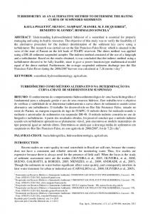
The Hoffmann reflex (H-reflex) has been long used as a means of assessing spinal reflex excitability, especially that related to the distal leg (Magladery & McDougal, 1950; Schiepatti, 1987). At the ankle level, several studies have established how the soleus muscle H-reflex amplitude varies throughout the phases of gait in healthy subjects (Crenna & Frigo, 1987; Kasai et al., 1998; Capaday & Stein, 1986; Morin et al., 1982; Petersen et al., 1999; Llewelyn et al., 1990). In general, these studies are in agreement in that the soleus H-reflex amplitude varies greatly during the course of the gait cycle. It is maximally enhanced during stance and push off and almost completely depressed or absent
during swing (Fig. 1). Functionally, the inhibition of the soleus motoneuron pool during the swing phase of gait is critical for an effective and efficient ankle dorsiflexion of the advancing foot. Although the activity from the tibialis anterior (TA) contributes to the inhibitory drive during swing through reciprocal inhibition, inhibitory effects on soleus motoneuron are already in place even before any EMG activity from the TA is detected or before any noticeably movement takes place in any segment of the lower extremity (Schneider et al., 2000). This has demonstrated the dependency of the inhibitory drive from descending, powerful central pre-synaptic mechanisms (Morin et al.; Yang & Whelan, 1993) that are movement independent.
*
Physical Therapy Department, Methodist Sugar Land Hospital, Sugar Land, Texas, USA. School of Physical Therapy, Texas Woman's University, Houston, Texas, USA. *** Department of Kinesiology, Rice University, Houston, Texas, USA. **
7
PINEDA, E. E.; SABBAHI, M. A. & ETNYRE, B. R. Using standing postures as a practical alternative to gait analysis for assessing normal neuromotor activity variation of the ankle muscle antagonists: A soleus H-reflex and EMG activity study. Int. J. Morphol., 28(1):7-12, 2009.
Therefore, the main purpose of this study was to investigate and determine the extent of the variation in the soleus H-reflex amplitude in five standing postures. A secondary goal was to describe the associated EMG activity of the soleus and TA muscles. Determining which posture achieves the most enhanced and the most inhibited soleus H-reflex amplitude might result in a more practical method to evaluate normal ankle neuromotor control.
MATERIAL AND METHOD
Subjects.Twenty-one healthy subjects (9 women, 12 men), age range 22 to 69 years (36.7 + 12.6) participated in this study. Of them, 14 consented for soleus H-reflex measures and 13 consented for EMG recording. Participating subjects had no previous history of CVA, cancer, diabetes, low back pain, or neurologic or orthopedic pathologies.
Fig 1. Normal soleus and tibialis anterior EMG & soleus H-reflex, during the gait cycle. HC= heel contact; PO= push off; SW= swing. (Stein & Capaday, 1986, 1987)
H-reflex stimulation and EMG recording. H-reflex stimulation and EMG recording were similar to previous published studies (Ali & Sabbahi, 2000, 2001; Alrowayeh et al., 2005). Briefly described, the Cadwell 5200A electromyography unit (Cadwell Laboratories, Inc., USA) was used to elicit and record the soleus H-reflex (Fig. 2). The tibial nerve was electrically stimulated at the popliteal fossa using 0-5 ms, 0.2 pps pulses at intensity equivalent to H-max. The peak-to-peak amplitude and onset latencies of four separate traces were recorded for each posture.
Early on, Crenna & Frigo suggested that a similar variation of the soleus H-reflex amplitude, compared to that from gait activity, could be obtained from standing postures (e.g., soleus H-reflex inhibition as a result of holding the foot off the ground as in the swing phase). In addition, soleus H-reflex amplitudes obtained in the upright posture in healthy subjects was reported to be more consistent (i.e., stable) compared to less functional postures such as prone lying (Ali & Sabbahi, 2000) and with high (>.80) inter and intrasession reliabilities (Hopkins et al., 2000; Ali & Sabbahi, 2001; Handcock et al., 2001). Although evoking and recording soleus H-reflex and EMG responses through gait activity are the ideal method in the research laboratory, they are too burdensome and impractical for the busy clinic, where space, time and adequate equipment are in most cases limited.
8
Fig. 2. H-reflex measures setup.
EMG from soleus and TA muscles were recorded and analyzed based in a protocol utilized in a related study (Alrowayeh et al.). EMG was obtained and recorded with
PINEDA, E. E.; SABBAHI, M. A. & ETNYRE, B. R. Using standing postures as a practical alternative to gait analysis for assessing normal neuromotor activity variation of the ankle muscle antagonists: A soleus H-reflex and EMG activity study. Int. J. Morphol., 28(1):7-12, 2009.
the Myosystem 1200 (Noraxon, Inc., USA) linked to a laptop computer (Fujitsu, Inc., Japan). A pair of round surface electrodes was placed side-by-side, along the belly of the tibialis anterior muscle and over the soleus, 1 cm below the bifurcation of the gastrocnemii and on line with the Achilles tendon. A single ground electrode was placed on the skin covering the head of the fibula. The electrodes were of the ready-to-use type (Blue Sensor M-00-S, Medicotest, Inc., Denmark), requiring no electroconductive cream (Fig. 3).
Fig. 4. PO posture.
5. Swing (SW). The subject stands on one leg while the examined leg is kept clear from the ground, then posture is held (Fig. 5).
Fig. 3 Soleus and TA EMG setup.
Experimental procedure. After the subjects read and signed the consent form, soleus H-reflex and EMG recordings of the soleus and tibialis anterior muscles were obtained in the following five postures: 1. Quiet standing (QS). The subject stands upright, relaxed. 2. Heel contact (HC). The subject stands on both heels until the ball of the feet clears the ground, then posture is held.
Fig. 5. SW posture.
The QS posture was considered as a reference posture. Measurements were carried out, while all participants rested their hands onto a walker to minimize balance changes and while using no footwear. H-reflex recording started after the subjects were stable at each of the experimental postures. Half of the participants were measured on their right leg and half on their left leg. Signal and statistical analysis.
3. Standing on single leg (SL). The subject stands on examined leg while the opposite foot is kept cleared from the ground, then posture is held. 4. Push off (PO). The subject stands on both forefeet until the heels clear the ground, and then posture is held (Fig. 4).
H-reflex. Four H-reflex amplitudes were recorded and averaged for each posture. Three parameters were measured: the H-reflex peak-to-peak amplitude, the maximum peakto-peak M-wave, and the onset latency to the first deflection. H/M ratios were subsequently calculated.
9
PINEDA, E. E.; SABBAHI, M. A. & ETNYRE, B. R. Using standing postures as a practical alternative to gait analysis for assessing normal neuromotor activity variation of the ankle muscle antagonists: A soleus H-reflex and EMG activity study. Int. J. Morphol., 28(1):7-12, 2009.
Electromyography. Two 3-sec rectified soleus and tibialis anterior EMG signals were recorded, processed and averaged for each posture. Raw EMG signal was sampled at 1000 Hz rectified and smoothed with 30 ms averaging window for online monitoring and storing. Stored data was quantified using an automatic root mean square (RMS) algorithm with a 50 ms window.
motoneuron pool (H/M) when compared to QS (100%) were 150% for PO, 25% for HC, 50% for SW, and 100% for SL.
Statistical analysis. A univariate one-way repeated measures analysis of variance (ANOVA) was used to assess soleus Hreflex amplitude (dependent variable) differences between postures (independent variable).
The averaged tibialis anterior EMG activity (Fig. 8) was reciprocal to that of the soleus H-reflex (i.e., higher tibialis anterior EMG - lower H-reflex amplitude). Tibialis anterior EMG activity was higher during HC followed by SW and PO; the lowest was during QS and SL (Table III).
Soleus and tibialis anterior EMG (RMS - mV). The averaged soleus EMG activity (Fig. 7) paralleled that of the soleus H-reflex (Fig. 6). The highest soleus EMG activity was in PO and the lowest during SW (Table II).
RESULTS
Soleus H-reflex amplitude (mV). Soleus H-reflex amplitude (mean + SD and SEM) for each posture are shown in Table I. There was a statistically significant difference in soleus H-reflex amplitude between postures. The adjusted F (Greenhouse-Geisser Epsilon method) was: F(2.61, 33.95) = 41.69, p