Sep 20, 2008 - Krässig,1 Emily R. Peterson,1 Nina Rohringer,1, â Robin Santra,1, 2 and Stephen H. ..... [24] S. E. Harris, J. E. Field, and A. Imamoglu, Phys.
Using strong electromagnetic fields to control x-ray processes Linda Young,1 Christian Buth,1, ∗ Robert W. Dunford,1 Phay J. Ho,1 Elliot P. Kanter,1 Bertold Kr¨assig,1 Emily R. Peterson,1 Nina Rohringer,1, † Robin Santra,1, 2 and Stephen H. Southworth1
arXiv:0809.3537v1 [physics.atom-ph] 20 Sep 2008
2
1 Argonne National Laboratory, Argonne, Illinois 60439, USA Department of Physics, University of Chicago, Chicago, Illinois 60637, USA (Dated: September 20, 2008)
Exploration of a new ultrafast-ultrasmall frontier in atomic and molecular physics has begun. Not only is is possible to control outer-shell electron dynamics with intense ultrafast optical lasers, but now control of inner-shell processes has become possible by combining intense infrared/optical lasers with tunable sources of x-ray radiation. This marriage of strong-field laser and x-ray physics has led to the discovery of methods to control reversibly resonant x-ray absorption in atoms and molecules on ultrafast timescales. Using a strong optical dressing field, resonant x-ray absorption in atoms can be markedly suppressed, yielding an example of electromagnetically induced transparency for x rays. Resonant x-ray absorption can also be controlled in molecules using strong non-resonant, polarized laser fields to align the framework of a molecule, and therefore its unoccupied molecular orbitals to which resonant absorption occurs. At higher laser intensities, ultrafast field ionization produces an irreversible change in x-ray absorption. Finally, the advent of x-ray free electron lasers enables first exploration of non-linear x-ray processes.
INTRODUCTION
Control of x-ray processes using intense optical lasers represents an emerging scientific frontier—one which combines x-ray physics with strong-field laser control [1]. While the past decade has produced many examples where intense lasers at optical wavelengths are used to control molecular motions [2, 3, 4, 5], extension to the control of intraatomic inner-shell processes is quite new [1, 6, 7, 8]. At first glance, it is an unusual concept to control x-ray processes using an optical or infrared radiation field since x-rays interact predominantly with inner-shell electrons, whereas longer wavelength radiation interacts with outer shell electrons. However, the inner and outer shells of atoms are coupled through resonant x-ray absorption, e.g., promotion of a K-shell electron to an empty outer shell orbital, as shown in Fig. 1. Because outer shell electronic structure can be perturbed (dressed) by an optical radiation field, one can exert control over resonant x-ray absorption using optical lasers. Reversible control is possible when the applied dressing field is gentle enough to significantly perturb outer-shell electronic structure, but is not intense enough to destroy (ionize) the atom. Let us consider the optical field amplitude necessary to achieve this control, i.e., to induce outer-shell transitions at a rate comparable to inner-shell processes. If we take the simplest case, shown in Fig. 1, where absorption of an x-ray photon leads to the ejection of a K-shell electron, a 1s−1 hole is created. The resulting atom, containing a 1s−1 hole, is unstable and decays via both radiative and non-radiative (Auger) channels [9]. These inner-shell decay rates increase with atomic number; at Z = 10 the lifetime of the 1s−1 hole state is 2.4 fs (corresponding to a 0.27 eV level width via the uncertainty principle). In order to compete with the rapid inner-shell
decay, electromagnetic transitions in the outer shell must be induced at a comparable rate. The transition rate in a driven two-level system is given by the Rabi flopping frequency, Ω12 = µ12 E/~ where µ12 = h1|ez|2i is the transition dipole matrix element between levels 1 and 2, E is the electric field amplitude. In practical units, E is related to the laser intensity via I[W/cm2 ]= (1/2Z0 )E 2 √ [V/cm], where Z0 = µ0 ǫ0 = 377 V/A is the vacuum impedance. To estimate the laser intensity required to drive Rabi oscillations at a rate comparable to inner shell decay we use atomic units which are related to the hydrogen atom: charge, e = 1; length = Bohr radius = a0 ; velocity = Bohr velocity v0 = αc; electric field = field at the Bohr radius e/a20 ∼ 51 V/˚ A; electric dipole moment = ea0 , time t0 = a0 /v0 ∼ 0.024 fs. For the hydrogen 1s → 2p1/2 transition, µ12 = 1.05ea0 . Combining this with an electric field amplitude E = 1 atomic unit, the Rabi flopping frequency will be Ω12 = 1/t0 = 1/0.024 fs. The laser-induced rate is 16× that of the decay rate of the 1s hole state in neon! (Ω12 are given in angular frequency units) Therefore, if the electric field amplitude of the optical laser can be 1 atomic unit, or equivalently a laser intensity of 3.5 × 10¹⁶ W/cm2 , outer-shell transition rates can exceed inner-shell decay rates. This intensity can now be achieved with routinely available modern lasers [10, 11]. The key development of chirped pulse amplification [12] permits increased output energies from ultrashort lasers without damage to amplifying media. Focusing a standard amplified Ti:sapphire laser (800 nm, 3 mJ, 40 fs, 1 kHz) to 15 µm produces I ∼ 3.5 × 10¹⁶ W/cm2 . Ti:sapphire lasers are very versatile; the wide bandwidth allows one to stretch the pulsewidth from tens of femtoseconds to hundreds of picoseconds, and permitting the study of atoms and molecules exposed to a wide range of electromagnetic field strengths. After the strong optical laser field, the next ingredient
2
FIG. 1: (Color online) X-ray absorption promotes a 1s electron to an empty outer shell orbital. Optical radiation induces transitions between outer shell orbitals. Inner and outer shells are coupled through resonant x-ray absorption.
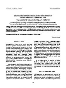
needed to study control of ultrafast inner-shell processes is a tunable x-ray source. Synchrotrons provide a convenient source of pulsed, tunable, polarized radiation from 10 eV to 100, 000 eV. This range covers inner shell edges of all elements. In Fig. 2 the three dominant photoprocesses, photoabsorption, elastic (Rayleigh) scattering and inelastic (Compton) scattering are shown for the bromine atom. Photoabsorption cross sections greatly exceed scattering cross sections over energy ranges from below to far above the respective K edges of each atom. In order to probe atoms and molecules subjected to strong pulsed optical fields, a short x-ray pulse is helpful. A typical x-ray pulse length is 100 ps at the Argonne Advanced Photon Source. For a 100 ps x-ray pulse duration, one can probe atoms and molecules subjected to 1012 W/cm2 with millijoule laser pulse energies focused to tens of microns. Shorter pulse lengths, ∼ 100 fs, at synchrotron sources are currently available using laser slicing techniques conceived by Zholents and Zolotorev [13] and put into practice at the Advanced Light Source [14], BESSY [15] and the Swiss Light Source [16]. We combine the ultrafast, ultrasmall optical and xray pulses using the x-ray microprobe method [17]. Basically, focused laser pulses (30 µm) are overlapped in time and space with microfocused x-ray pulses (10 µm). Gas-phase systems are particularly suitable for illustrating the basic principles underlying laser control of ultrafast x-ray processes. We discuss two different scenarios for modifying resonant x-ray absorption near an inner-shell edge: (1) modification of electronic structure of inner-shell-excited systems by laser dressing at 1012 – 1013 W/cm2 ; (2) control of resonant x-ray absorption by molecules through laser-induced spatial alignment at 1011 –1012 W/cm2 . We also discuss modifications to
FIG. 2: (Color online) X-ray photoprocesses for bromine. Photoabsorption (solid line) is the dominant process; the K and L edges are visible. Rayleigh scattering (dashed line). Compton scattering (dotted line). Synchrotron radiation covers the range from 10 eV to 100, 000 eV.
elastic scattering patterns from an ensemble of laseraligned molecules. Another method to modify resonant x-ray absorption is through strong-field ionization of the target particles at laser intensities in the range 1014 – 1015 W/cm2 [17, 18, 19]. Fig. 3 illustrates the microprobe and these applications. Beyond optical laser control of x-ray processes, it is also possible for strong electromagnetic fields at xray wavelengths to modify characteristic x-ray processes. The high x-ray intensities required for these modifications will soon be available with x-ray free electron lasers [20, 21, 22].
CONTROL OF X-RAY PROCESSES WITH STRONG OPTICAL FIELDS Electromagnetically induced transparency for x rays: atoms
In this subsection we describe laser-induced modifications to x-ray absorption spectra in various rare gases. Theoretical considerations are described in detail elsewhere [6, 7, 23]. Here we focus on the analogy to electromagnetically induced transparency in the optical regime and the application to imprinting ultrashort optical pulse sequences and shapes onto longer x-ray pulses. Electromagnetically induced transparency in the optical regime has been widely studied [24, 25, 26]. In a Λ-
3
FIG. 3: (Color online) X-ray microprobe and various scenarios for laser control of x-ray absorption at field strengths ranging from 1012 –1015 W/cm2 .
type medium characterized by atomic levels |1i, |2i, and |3i with energies E1 < E2 < E3 , resonant absorption on the |1i → |3i transition can be strongly suppressed by simultaneously irradiating the medium with an intense laser that couples the levels |2i and |3i. This phenomena, shown in Fig. 4a, is known as electromagnetically induced transparency, EIT. In the x-ray regime, EIT is considerably more complex. In the optical regime, levels |1i and |2i are stable to electronic decay. However, in the x-ray regime the coreexcited states, |2i and |3i are metastable. For the threelevel system in neon, depicted in Fig. 4b, the lifetime for these core-excited states is 2.4 fs (0.27 eV). Thus, at an optical field strength sufficient to compete with innershell decay, multiphoton transitions to the continuum can also play a role. Inner-shell decay rates are denoted by Γ2 and Γ3 and multiphoton transitions to the continuum are denoted by red block arrows. For the model system in neon, the calculated x-ray photoabsorption cross section for 800 nm laser dressing of the 1s → 3p transition in neon at 1013 W/cm2 with parallel and perpendicular laser/x-ray polarizations along with a fit to a three-level EIT model [7] is reproduced here for convenience in Fig. 5. At this intensity the
1s → 3p excitation at 867 eV is suppressed by a factor of 13 for the configuration in which the laser and x-ray polarizations are parallel. As can be seen in Fig. 5, the three level model reproduces most features of the calculated laser-dressed x-ray photoabsorption spectrum. In the three-level model effective linewidths of Γ3 = 0.68 eV and Γ2 = 0.54 eV account for the laser-ionization broadening. These parameters effectively reproduce both the lineshapes for the parallel and perpendicular configurations in the vicinity of the 1s → 3p resonance. The ability to control x-ray absorption in Ne at the 1s → 3p resonance allows one to imprint pulses shapes of the optical dressing laser onto long x-ray pulses [7]. This idea is illustrated in Fig. 6. With a 2 mm-long gas cell with one atmosphere of neon, the transmission of an x-ray pulse resonant with the 1s → 3p transition will be only 0.07%. A typical x-ray pulse from a synchrotron source has a duration of 100 ps. Such an x-ray pulse may be overlapped in time and space with one or several, ultrashort intense laser pulses. Those portions of the x-ray pulse that overlap with the laser are transmitted through the gas cell. In the case shown in Fig. 6, where the two dressing laser pulses have a peak intensity of 1013 W/cm2 , the intensity of the two transmitted
4
FIG. 4: (Color online) (a) Optical EIT. (b) X-ray EIT in neon.
x-ray pulses is roughly 60% of the incoming pulse. The time delay between the two x-ray pulses can be controlled by changing the time delay between the two laser pulses, opening a route to ultrafast all x-ray pump-probe experiments. With an analogous strategy, controlled shaping of short-wavelength pulses might become a reality. A disadvantage of this method is that it is applicable only at certain x-ray energies; the Ne case operates at 867 eV. It would be interesting to be able to extend these concepts to the hard x-ray regime. Calculations on the laserdressed spectra near the K edge in krypton [6] and argon [27] have been done. In argon and krypton, K edges are at 3.2 keV and 14.3 keV, respectively. The difficulty is that the inner shell decays are significantly more rapid; for Ne, Ar, Kr the decay widths of core-excited 1s−1 np levels are 0.27, 0.6 and 2.7 eV. The increased widths have two consequences. First, the 1s → np transitions are not fully resolved as the level width increases, making it difficult to isolate a three-level system. For example, the 1s−1 5p, 1s−1 6p, transitions near the krypton K edge are blended such that it is smooth edge. Second, the laser intensity needed to perturb the x-ray absorption cross section roughly scales as the square of the level width. Thus, when a dressing field of 1013 W/cm2 is applied, the Kr absorption cross section changes by less than 20%. One possible way to overcome these issues is to use rare gas ions as the EIT medium. The singly ionized krypton atom has a prominent, isolated resonance due to the 4p hole orbital [17, 19, 28]. X-ray absorption spectra for Kr, Kr1+ and Kr2+ are shown in Fig. 7. In addition to the isolated resonance, ions can withstand higher
dressing intensities without ionization. The critical field strength, Ecrit that one can apply to an atom without ionizing it can be estimated by combining the laser and 2 Coulomb potential, Ecrit = EIP /(4Ze3 ), where EIP is the ionization potential, Z is the charge of the residual ion, e is the electron charge [29]. For Kr, Kr1+ and Kr2+ the ionization potentials are 14.0, 27.9 and 41.8 eV, respectively. Thus, one should be able to apply a laser intensity 9× higher for Kr2+ than neutral krypton to generate a sizable change in absorption cross section. Of course, generating a sufficient density of ions to realize a practical hard x-ray pulse shaper would be a challenge.
X-ray absorption and scattering using laser-aligned molecules
In the presence of a strong non-resonant linearly polarized laser field, molecules align due to the interaction of the laser electric field vector with the anisotropic molecular polarizability [5, 30]. The alignment process is of intrinsic interest and of interest in applications to spectroscopy and photophysics, quantum control of molecular dynamics, high-harmonic generation, chemical reactivity, liquids and solvation and structural determinations by x-ray or electron diffraction. For asymmetric molecules having three distinct moments of inertia, 3D alignment has been achieved both in the presence of a laser-aligning field (adiabatic alignment) [31] and under field-free conditions (impulsive alignment) [32]. Xray probes of laser-aligned molecules are of considerable
5
FIG. 5: (Color online) Photoabsorption near the 1s → 3p resonance in neon. Results from ab initio calculations and a three-level model are shown. From [7].
FIG. 7: (Color online) X-ray absorption spectra for Kr, Kr1+ and Kr2+ .
FIG. 6: (Color online) Generation of ultrafast x-ray pulses using laser dressing of Ne. See the text for details.
current interest [8] because of proposals to determine structure of non-periodic specimens, such as individual biomolecules, by x-ray scattering [33, 34] using x-ray free electron lasers. Both x-ray absorption and scattering from a randomly oriented ensemble of gas phase molecules are significantly modified if the molecules are aligned. In x-ray absorption, both the near-edge and extended structure [extended x-ray absorption fine structure (EXAFS)] can be significantly altered. The near-edge structure is altered when one uses resonant polarized x-ray absorption to detect the alignment of the molecular axis relative to the lab frame [8]. Excitation of a 1s electron to the lowest unoccupied molecular orbital, LUMO, often results in an isolated resonance near the K-edge of a molecule. The LUMO is fixed relative to the molecular frame and the
transition strength from a highly localized K shell orbital to the LUMO defines the alignment of the molecule relative to the x-ray polarization axis [35]. For the molecule that we studied, CF3 Br, the LUMO is an antibonding σ ∗ orbital with substantial Br 4pz character, where z refers to the C–Br axis. The molecular symmetry dictates that x-ray absorption on the Br 1s → σ ∗ resonance occurs only when the x-ray polarization vector has a nonvanishing projection on the C–Br axis. Since the laser polarization axis defines the molecular alignment axis, one achieves control of x-ray absorption by simply rotating a waveplate. After aligning in space, time and x-ray energy, i.e., tuning to the 1s → σ ∗ resonance at 13.476 keV, the control of resonant x-ray absorption in the near-edge region can be readily achieved [8, 36], as shown in Fig. 8. In the EXAFS region, modifications to the polarized x-ray absorption will also be induced by molecular alignment. Pictorially, the effect for Br2 is shown in Fig. 9. When the Br2 molecules are aligned parallel to the x-ray polarization axis, the modulation of the absorption cross section due to Br 1s → ǫ p photoelectron backscattering from the adjacent atom is maximal, and in a perpendicular configuration minimal. Thus, the magnitude of the EXAFS modulation is controlled by the angle of the molecular axis relative to the x-ray polarization axis. The equation describing the modulation around an atomic background is given by χ(k) = −
X 3 cos2 ϑi 2 Fi (k) e−2σi k e−2Ri /λi (k) 2 k R i i
× sin(2kRi + 2δl + βi ) ,
(1)
6
FIG. 8: The LUMO of CF3 Br is an antibonding σ ∗ orbital with substantial Br 4pz character, where z refers to the C– Br axis. X-ray absorption on the 1s → σ ∗ resonance as a function of the angle between the polarization axis of the aligning laser and that of the x-ray probe.
where k is the photoelectron wavevector, ϑi is the angle between the x-ray polarization and the ith atom, Ri is the distance between the central and ith atom, σi is the Debye-Waller factor, Λi is the range parameter for photoelectron scattering and δl and βi describe the outgoing photoelectron phase shift [35]. Polarized EXAFS measurements on laser-aligned molecules will permit one to determine changes in structure due to e.g. laser aligning fields, though the utility of EXAFS for absolute bond distances may be limited. Laser control of the angle between the molecular and x-ray polarization axes will permit control of the EXAFS modulation amplitude, determination of the atomic background and, for more complex molecules, the determination of bond angles. Elastic scattering will also be modified since the contributions from the aligned molecules can be summed linearly and a scattering pattern reflective of the molecular structure will emerge. A simulation of the x-ray scattering pattern from an ensemble of molecules aligned by a laser pulse of arbitrary shape was made. First the molecular response to the laser pulse was calculated [36] and then the x-ray scattering [37] from this ensemble was simulated. In Fig. 10 elastic scattering patterns for an isotropic sample, for clamped nuclei and for an alignment of hcos2 θi(t) = 0.80 are shown. As the standard method for absolute molecular structure determination, x-ray diffraction from laser-aligned molecules will then allow us to investigate changes in molecular structure in the presence of strong laser aligning fields with the eventual goal of a predictive understanding using an ab initio electronic structure code such as dalton to calculate molecular hyperpolarizabilities.
FIG. 9: Schematic of the polarization dependence of the EXAFS modulations in Br2 .
MODIFYING CHARACTERISTIC X-RAY PROCESSES WITH STRONG X-RAY FIELDS
In the previous section we have seen how it is possible to control resonant x-ray absorption with optical fields. With focused x-ray free electron lasers it will be possible to create intensities such that characteristic x-ray processes are altered. A simple estimate may be made for the 1s → 3p transition in neon. Here the transition dipole matrix element is 0.01ea0 [38] rather than the 1.05ea0 for the hydrogen 1s → 2p1/2 transition. Equating the Rabi frequency with the core-excited decay rate (2.4 fs)−1 suggests that an intensity of 1018 W/cm2 is required to saturate this transition. Focusing the LCLS output at 800 eV (1013 photons / 233 fs) to a spot size of ∼ 1 µm will yield this intensity. At high intensity, the ejection of a second electron from the 1s shell can be more rapid than Auger decay to create hollow neon, Ne[KK] [39]. The intensity dependence is shown in Fig. 11. As one can see, hollow neon is formed preferentially at higher intensity; indeed Ne[KK] can be the dominant species, compared to the usual 1% fraction in the weak field limit. Because the LCLS output originates from SASE (self-amplified spontaneous emission), the output radiation is chaotic, consisting of a random number of intensity spikes (coherent regions) of random amplitude. Therefore, one might expect an enhancement of two-photon processes (such as Ne[KK] formation). In the lower part of Fig. 11, the Auger yield from an ensemble average of chaotic pulses is compared with the yield from an average pulse shape. As one can see, the chaotic enhancement for Ne[KK] production is at most a factor of 1.3 in the unsaturated regime. This is in contrast to visible radiation where one expects
7
FIG. 10: Elastic scattering from isotropic, fixed-in-space and aligned Br2 with hcos2 θi > (t) = 0.80.
a factor of 2. The critical quantity is the ratio of the FEL coherence time to the Auger lifetime. The larger the ratio, the closer one will be to the maximal enhancement of 2. Finally, we note that fully stripped neon can be produced in the 1 µm focus of the LCLS beam by sequential single photon processes, providing the photon energy is greater than the binding energy of Ne9+ [39].
OUTLOOK
The era in which characteristic x-ray processes can be considered invariant is at an end. We have demonstrated that placing atoms and molecules in strong optical fields can significantly affect resonant absorption and elastic scattering. Currently, we can control x-ray absorption with application of a strong-optical field to a gaseous medium; the control mechanism is EIT in atoms and laser-constrained rotation in molecules. One may be able to create an x-ray amplitude pulse shaper using these tools. X-ray scattering from aligned molecules is not far off; simple estimates show that a mere 108 x-rays/pulse at 1 kHz will suffice as obtainable with a pink beam at the Advanced Photon Source [8]. In addition, we look forward to being able to experimentally observe multiphoton processes in the hard x-ray regime when LCLS is first operational in 2009. Control of molecular alignment combined with subsequent ion/electron imaging techniques will provide a means to disentangling x-ray damage mechanisms in complex molecules. We thank Dohn A. Arms, Eric M. Dufresne, Eric C. Landahl, and D. Walko for valuable assistance during experiments at the Sector 7 beamline at the Advanced Photon Source. C.B. was partly supported by a Feodor Lynen Research Fellowship from the Alexander von Humboldt Foundation. This work and the Advanced Photon Source were supported by the Chemical Sciences, Geosciences, and Biosciences Division of the Office of Basic Energy Sciences,Office of Science, U.S. Department of Energy, under Contract No. DE-AC02-06CH11357.
FIG. 11: (Color online) Top: Yield of Ne[K] and Ne[KK] as a function of focused LCLS intensity at a photon energy of 1050 eV. Bottom: Ratio of Auger yields from an ensemble average of chaotic SASE pulses to that from an averaged pulse. From [39].
†
[1]
[2] [3] [4] [5] [6] [7] [8]
[9] [10] [11] [12] [13] [14]
∗
Present address: Department of Physics and Astronomy, Louisiana State University, Baton Rouge,
[15]
Louisiana 70803, USA Present address: Lawrence Livermore National Laboratory, Livermore, California 94551, USA R. Santra, R. W. Dunford, E. P. Kanter, B. Kr¨ assig, L. Young, “Strong Field Control of X-ray Processes” in Advances in Atomic, Molecular and Optical Physics, Volume 56, (Elsevier, Amsterdam, Netherlands, 2008). S. A. Rice, M. Zhao, Optical Control of Molecular Dynamics, (Wiley, New York, 2000). M. Shapiro, P. Brumer, Principles of the Quantum Control of Molecular Processes, (Wiley-Interscience, New York, 2003). H. Rabitz, R. de Vivie-Riedle, M. Motzkus, K. Kompa, Science 288, 824 (2000). H. Stapelfeldt, T. Seideman, Rev. Mod. Phys. 75, 543 (2003). C. Buth, R. Santra, Phys. Rev. A 75, 033412 (2007). C. Buth, R. Santra, L. Young, Phys. Rev. Lett. 98, 253001 (2007). E. R. Peterson, C. Buth, D. A. Arms, R. W. Dunford, E. P. Kanter, B. Kr¨ assig, E. C. Landahl, S. T. Pratt, R. Santra, S. H. Southworth, L. Young, Appl. Phys. Lett. 92, 094106 (2008). M. O. Krause, J. Phys. Chem. Ref. Data 8, 307 (1979). T. Brabec, F. Krausz, Rev. Mod. Phys. 72, 545 (2000). G. A. Mourou, T. Tajima, S. V. Bulanov, Rev. Mod. Phys. 78, 309 (2006). D. Strickland, G. Mourou, Opt. Commun. 56, 219 (1985). A. A. Zholents and M. S. Zolotorev, Phys. Rev. Lett. 76, 912 (1995). R. W. Schoenlein, S. Chattopadhyay, H. H. W. Chong, T. E. Glover, P. A. Heimann, C. V. Shank, A. A. Zholents, M. S. Zolotorev, Science 287, 2237 (2000). S. Khan, K. Holldack, T. Kachel, R. Mitzner, and T. Quast, Phys. Rev. Lett. 97, 074801 (2006).
8 [16] P. Beaud, S. L. Johnson, A. Streun, R. Abela, D. Abramsohn, D. Grolimund, F. Krasniqi, T. Schmidt, V. Schlott, and G. Ingold, Phys. Rev. Lett. 99, 174801 (2007). [17] L. Young, D. A. Arms, E. M. Dufresne, R. W. Dunford, D. L. Ederer, C. H¨ ohr, E. P. Kanter, B. Kr¨ assig, E. C. Landahl, E. R. Peterson, J. Rudati, R. Santra, S. H. Southworth, Phys. Rev. Lett. 97, 083601 (2006). [18] R. Santra, R. W. Dunford, L. Young, Phys. Rev. A 74, 043403 (2006). [19] S. H. Southworth, D. A. Arms, E. M. Dufresne, R. W. Dunford, D. L. Ederer, C. H¨ ohr, E. P. Kanter, B. Kr¨ assig, E. C. Landahl, E. R. Peterson, J. Rudati, R. Santra, D. A. Walko, L. Young, Phys. Rev. A 76, 043421 (2007). [20] J. Arthur et al., Linac Coherent Light Source (LCLS) Conceptual Design Report, SLAC-R-593, 2002. [21] T. Tanaka and T. Shintake, SCSS X-FEL Conceptual Design Report, RIKEN Harima Institute/SPring-8, 2005. [22] M. Altarelli et al., The Technical Design Report of the European XFEL, DESY 2006-097, 2006. [23] C. Buth, R. Santra, and L. Young, in this proceedings. [24] S. E. Harris, J. E. Field, and A. Imamoˇ glu, Phys. Rev. Lett. 64, 1107 (1990). [25] K. J. Boller, A. Imamoglu, and S. E. Harris, Phys. Rev. Lett. 66, 2593 (1991).
[26] S. E. Harris, Phys. Today 50, No. 7, 36 (1997). [27] C. Buth and R. Santra, to be published. [28] L. Pan, D. R. Beck, S. M. O’Malley, J. Phys. B. 38, 3721 (2005). [29] S. Augst, D. Strickland, D. D. Meyerhofer, S. L. Chin, J. H. Eberly, Phys. Rev. Lett. 63, 2212 (1989). [30] B. Friedrich and D. Herschbach, Phys. Rev. Lett. 74, 4623 (1995). [31] J. J. Larsen, K. Held, N. Bjerre, H. Stapelfeldt, and T. Seideman, Phys. Rev. Lett. 85, 2470 (2000). [32] K. F. Lee, D. M. Villeneuve, P. B. Corkum, A. Stolow, and J. G. Underwood, Phys. Rev. Lett. 97, 173001 (2006). [33] R. Neutze, R. Wouts, D. van der Spoel, E. Weckert, and J. Hajdu, Nature 406, 752 (2000). [34] J. C. H. Spence and R. B. Doak, Phys. Rev. Lett. 92, 198102 (2004). [35] J. St¨ ohr, NEXAFS Spectroscopy (Springer, New York, 1996). [36] C. Buth and R. Santra, Phys. Rev. A 77, 013413 (2008). [37] P. Ho and R. Santra, to be published. [38] N. Rohringer and R. Santra, Phys. Rev. A (in press). [39] N. Rohringer and R. Santra, Phys. Rev. A 76, 033416 (2007).