Sep 5, 2018 - vere cracking of enzyme crystals in their presence. For the mutants, the difference spectra actuated by the substrate are the same in lineshape ...
VOl. 268, No.25. Issue of September 5, pp. 18481-184&1,1993 Printed in U.S.A.
THEJOURNAL OF BIOLOGICAL CHEMISTRY
Q 1993 by The American Soeiety for Bioehemistry nnd Molecular Biolow, Inc.
Utilization of Conformational Flexibility inEnzyme Action-Linkage between Binding, Isomerization, and Catalysis* (Received for publication, January 27, 1993)
Joshua 0.Goldsmith#Qand Lawrence C. KuollII From the $Department of Chemistry, Stanford University, Stanford, California 94305 and the TDepartment of 3Wrogical Chemistry, Merck Research La6oratories, West Point, Pennsylvania 19486
to distort the structuresof substrates to those of the products (2), and Pauling postulated in1946 that enzymes are complementary in structure to the activated complexes of the reactions theycatalyze (3,26). Togetherwith Koshland's inducedfit hypothesis that substrates play a role in optimizing the catalytic conformation of enzymes (4), these ideas have forged today's tenet that enzymes utilize structural flexibility, driven by binding energy, to maximize reaction rate. An extensive discussion of this subject has been given by Jencks (5). Experimentally, conformational changes have been seen to take place in a large number of enzymes. Isomerization of proteins and enzymes, mediated by substrates, has been documented by spectroscopic and physical techniques, most lucidly with use of x-ray crystallographic studies from which the function of structural changes may be extrapolated, e.g. hexokinase (6) and triosephosphate isomerase (7). Similarly, correlation of binding and catalysis has also been shown, notably by the application of site-directed mutagenesis to probe ionic, steric, and hydrophobic effects of binding on reaction rate for tyrosyl-tRNA synthetase and for subtilisin (8, 9, 27). These results all point to an intimate relationship between binding, protein conformational changes, and catalysis. For the Escherichia coli enzyme ornithine transcarbamoylase (EC 2.1.3.3), which catalyzes the carbamoylation of Lornithine in theurea cycle, binding of substrates and release of products are bothordered (10,ll). In the forward reaction, which is thermodynamically favorable ( K , lo6; 11-13), carbamoyl phosphate is the first substrate bound and inorganic phosphate is the second product released (see Reaction 1).In earlier studies employing absorbance difference spectroscopy (14,15), analytical ultracentrifugation ( X ) , and classical single-crystal cracking experiments (16), carbamoyl phosphate was shown to induce protein conformational changes upon binding. Kinetic and thermodynamic studies further revealed that the substrate-induced isomerization activated the enzyme for catalysis by lowering the energy of Emil Fischer's early lock-and-key metaphor has had a pro- activation for transcarbamoylation by -6 kcal/mol (17, 28). found influence on the study of enzyme mechanisms (1). Together, these observations suggested to us a possible corHaldane suggested in 1930 that binding energy may be used relation between binding, isomerization, and catalysis. Here, we report findings that substrate-product conversion cata* This work was supported by Grant DK38089 from the National lyzed by E. coli ornithine transcarbamoylase is linked quanInstitute of Diabetes and Digestive and Kidney Diseases and Grant titatively to theinduced-fit protein isomerization whose magDIR 8820594 from the National Science Foundation. The costs of nitude is in turntied to theaffinity of the substratecarbamoyl publication of this article were defrayed in part by the payment of page charges. This article must therefore be hereby marked "aduer- phosphate for the enzyme.
An intimate relationship between protein conformational changes and catalysis has often been suggested. The present study employs ligand-induced ultraviolet difference spectra and kinetic parameters determined for Escherichia coli ornithine transcarbamoylase and its site-specific mutants toevaluate the linkage between binding, isomerization, and reaction rate. For the wild-type enzyme, the lead substrate carbamoyl phosphate introduces a large difference ab1,800 sorbance in the enzyme upon binding (Atmu M" cm"; Miller, A. W.,and Kuo, L. C. (1990) J. Biol. Chem. 265,16023-15027). The spectrum is the same in lineshape as that produced bythe bisubstrate analog N-(phosphonacety1)-L-ornithineand is 80% as intense. Both substrate and analog cause gross protein conformational rearrangements as evident by swift and severe crackingof enzyme crystals in their presence. For the mutants, the difference spectra actuated by the substrate are the same in lineshape as that of the wild type but vary in intensity. A wide range of substrate affinity and steady-state kinetic constants are also observed for the mutants. When the binding energy of carbamoyl phosphate and the activation energy for transcarbamoylation are calculated for the wild-type and mutant enzymes, they are found to be inversely correlated to the intensity of protein difference absorbance elicited by the lead substrate. Together with analyses of steady-state kinetic parameters derived for various plausible reaction schemes, the experimental data suggest that carbamoyl phosphate induces the committed isomerization in ornithine transcarbamoylase for transition state binding. Our results provide a unique demonstration that aninduced-fit isomerization, triggered by binding, either controls or contributes significantly to the rate of an enzyme-catalyzed reaction.
-
tisement" in accordance with 18U.S.C. Section 1734 solelyto indicate this fact. 8 Recipient of a National Science Foundation Research Experience for Undergraduates Award CHE8712942. 11 Recipient of a Pew Scholars Award 87-0629B HE from the Pew Charitable Trustsand a Research Career Development Award DK01721 from the National Institutes of Health during the tenure of this investigation. To whom correspondence should be addressed.
-
EXPERIMENTALPROCEDURES
Site-directed mutagenesis was accomplished with the procedure of Zoller and Smith (18) as modified by Norris et al. (19). The point mutants employed in this study are a collection of all site-specific mutants prepared in our laboratory for various projects. Excluded in this study are tryptophan-deleted mutants which display extinction coefficients different from that of the wild type and theArglOgmutant
18481
18482
Conformational Flexibility in Enzyme Action 0
I1
L-ornithine
-
NH~CPOS- + NH~(CH~)~CH(COT)NH,+ carbamoyl-P
0
II
NH~CNH(CH~)~CH(CO;)NH; + HPO:t-citrulline phosphate
REACTION1 (20) which is allosteric in substrate binding. The double mutants are prepared specifically for this study. The criteria applied to ensure that only the designated site is modified have been described previously together with details of the mutagenic experiments and the expression systems (17,281. Enzyme purification, steady-state kineticassays, and dataanalysis were performed as documented in earlier studies (11, 17, 28). Preparation of enzyme solutions, collection of ultraviolet absorption spectra, andconstruction of difference spectra have been reported (14).
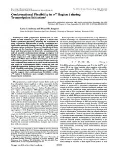
absorbance subsequent to binding. The broad band of the spectrum spanning from 245 to 320 nm may be attributed to a blue-shifted 'La transition of tryptophan while the narrow peaks at 281,287, and 294 nm are characteristic of red-shifted 'Lbands of tryptophans and/or tyrosines (22, 23). Nonspecific salt effects have been ruled out as a contributing factor to theultraviolet absorption of the enzyme. Physical evidence of gross protein conformational alterations correlating with the difference ultraviolet absorption of the enzyme has been RESULTS AND DISCUSSION provided by single crystal soaking experiments (16). Only A series of site-specific mutants of ornithine transcarbam- carbamoyl phosphate, phosphonacetamide, andthe bisuboylase are employed in this study. We have shown previously strate analog, phosphonacetyl-L-ornithine(PALO),'cause with spectroscopic measurements and kinetic arguments that single crystals of the enzyme to crack, indicating that subremoval of the charge and/or bulk of an amino-acid residue stantially large protein conformational rearrangements occurs of E. coli ornithine transcarbamoylase does not alter unduly in the crystal to generate destruction of its packing lattice.' the secondary and tertiary structures of the enzyme (17, 20, Eisenstein and Hensley (15) have found a difference spectrum 21,28). However, substitution of many of the residues of the for PALO interaction with yeast ornithine transcarbamoylase enzyme does affect the extent of the carbamoyl phosphate- nearly identical to that shown in Fig. 1. These authors have elicited protein isomerization as well as the values of the also observed a 2.8%increase in thesedimentation coefficient steady-state kinetic parameters K,, kcat, and KM for the for- of the enzyme, upon carbamoyl phosphate binding, demonward reaction catalyzed by the enzyme. strating alarge global change in protein shape and/or volume. Fig. 1 shows the ultraviolet difference spectra of E. coli The combined results indicate that the characteristic differornithine transcarbamoylase caused by a saturating concen- ence absorbance of the enzyme upon carbamoyl phosphate tration of carbamoyl phosphate. The difference spectrum of binding is the result of a ligand-induced isomerization and one of the site-specific mutants caused by a saturating con- may be exploited to gauge the extent of the specific protein centration of carbamoyl phosphate is also shown in thisfigure. conformational changes. Other than a diminution in thepeak-to-trough amplitude, the To evaluate the linkage between binding, conformational mutant difference spectra are essentially identical to that of changes, and the rate of substrate-product conversion of orthe wild-type enzyme. On the other hand, for both the wild nithine transcarbamoylase, we have examined the relationtype and mutantenzymes, L-ornithine and otherL-amino acid ligands of ornithine transcarbamoylase, such as L-norvaline ships between the extent of the induced-fit protein isomerior L-citrulline, produce no change in the ultraviolet absorb- zation, the binding energy of carbamoyl phosphate (AG),and ance of the enzyme. A detailed analysis (14) of the ligand- the activation energy for transcarbamoylation (A&*). The elicited spectral changes of ornithine transcarbamoylase has extent of the carbamoyl phosphate-induced isomerization of been presented earlier. Briefly, computer simulations have the enzyme is monitored with the peak-to-trough amplitude shown that thecarbamoyl phosphate-induced difference spec- (AAE"~-''') between 245 and 270 nm in the difference spectrum may beaccounted for by spectral perturbations resulting trum of the binary enzyme-carbamoyl phosphate complex. from -0.5-0.7 nm blue and red shifts of the enzyme ultraviolet The binding energy AG,, is determined with the relationship A(& = -RT.ln(l/&,)
(Eq. 1)
where R is the gas constant, T is the absolute temperature, and K , is apparent dissociation constant of carbamoyl phosphate from the reactive binary enzyme-carbamoyl phosphate complex. For an ordered Bi Bi reaction E A + B $ EA + B e EAE, K, represents the apparent equilibrium constant governing the equilibrium between the free E, A, and the binary complex EA (24). The activation energy of enzymecatalyzed transcarbamoylation, ACT*, is calculated from the rate constant of the binary enzyme complex reacting with L-
+
FIG.1. Carbamoyl phosphate-induced limitingdifference and its spectra of E. coli ornithine transcarbamoylase (-)
site-directed LysW4 Ala mutant (- - -) at pH 8.5. The enzyme concentration was 9.05 p~ giving an absorbance of 0.96 OD at 280 nm. The carbamoyl phosphate concentration was 2 mM and was mole of saturating (14). The A6 is the change inextinctionper holoenzyme. Nonspecific salt effects have been ruled out as thecause for the difference spectra (14). The lineshape of the difference spectrum recorded for the enzyme-carbamoyl phosphate complex of all mutant enzymes is essentially identical to thatof the wild type.
The abbreviations are: PALO, N-phosphonacetyl-L-ornithine; extent of induced-fit protein isomerization monitored as the peak-to-trough amplitude in the ultraviolet difference spectrum between 245 and 270 nm; A(&, binding energy of carbamoyl phosphate; A&*, activation energy for transcarbamoylation. Single crystals of deoxyhemoglobinlend the classic demonstration of large scale protein conformational changes. Upon oxygenation, deoxyhemoglobincrystals crack and shatter. This phenomenon is not observed with crystals of myoglobin and hemoglobin H (a tetramer containing only 0 subunits); both are known not to undergo the isomerization which gives rise to allosteric transitions (29). AAF"'O,
Conformational Flexibility Action Enzymein
18483
the data in Fig. 3 show that when induced-fit isomerization becomeslarger the free energy of activation for substrate turnover becomes smaller. We note that the line drawn in A&* = -RT.ln(kJKMom) + R T . l n ( b / h ) (Eq.2) each figure isnot intended to imply that a linear relationship necessarily exists between AAc and AG or AAc and A&'. where kB isthe Boltzman constantand h isthe Planck More importantly, it is clear that there are no outliers in constant. Thektis thecatalytic turnover rate of the enzymic either plot. In otherwords, no mutant binds carbamoyl phosreaction and K M "is~ theMichaelis constant for the substrate phate tightly while exhibiting little or no induced-fit isomerL-ornithine. Both K, and kat/KMom may be obtained readily ization, or vice versa (Fig. 2), and no mutant displays a large from steady-state kinetic assays. induced-fit isomerization while functioning as a poor catalyst, Plotted in Figs. 2 and 3 are, respectively, the values of AG or vice versa (Fig. 3). These results suggest that binding, and A&* against the intensities of carbamoyl phosphate- isomerization, and catalysis are quantitatively coupled. induced absorbance difference for ornithine transcarbamoylTo explain the results in Figs. 2 and 3, four plausible kinetic ase and itssite-specificmutants. It is seen that A& and A&' schemes are employed. In Scheme 1, the spectroscopically are both inversely correlated to AAc245-270.The data in Fig. 2 observed protein conformational changes are neglected. In show that thetighter the binding of carbamoyl phosphate the Scheme 2, the free enzyme isomerizes between two forms(E greater the extentof the substrate-elicited isomerization, and and E ' ) but onlyone is catalytically essential and binds carbamoyl phosphate. Scheme 3 shows an isomerizing binary complex but only the unisomerized form is productive and l*O1 Oils2 1 binds L-ornithine (orn). In Scheme 4, the binary complex isomerizes but it is the isomerized binary complex (E'-cp) which binds ornithine to react. Qualitatively, it can be seen that: 1) Scheme 1 does not include an induced protein isomerization step andis incompatiblewith experimental findings; 2) the isomerization in Scheme 2 is kinetically isolated from transcarbamoylation and cannotaffect Lt/KMom;3 ) the isomerization in Scheme 3 is inhibitory. Only Scheme 4 offers a mechanism in which the induced-fit isomerization is catalyt-MOL I I i I 1 -6.0 4.0 -2.0 ically competent and also kinetically significant? To further assess these four schemes, the steady-state kiAGb, kcal/mol netic parameters for Schemes 1-4 have been derived (Table FIG. 2. Correlation of thebindingenergyofcarbamoyl I). An inspection of Table I reveals that thekinetic parameters phosphate, A(&, with the intensity of the carbamoyl phmderived for Scheme 1 are not functions of the isomerization phate-induced difference protein absorbance, A A C " ~ ' ~ for ~, E. coli ornithine transcarbamoylase andits site-directed mu- equilibrium, Kim.For Scheme 2, the kat/KM0" term is indetants. The binding energies are calculated with the apparent disso- pendent of isomerization and is incompatible with the results ciation constants, K,, obtained from steady-state kinetic assays for in Fig. 3. For Scheme 3, numerical estimates (see Table I) each enzyme at pH 8.5 and 25 "C. The AAtZM2" values represent the using the Kk term yield a line with a negative slope ina plot peak-to-trough amplitudes of the difference spectra of the enzymes of AAc uersw A&, in agreement with results in Fig. 2; but between 245-270 nm (see Fig. 1).Standard single-letter amino acid gives a line with a codes are used in the diagram. W T denotes the wild-type enzyme. simulation with use of theterm Ala, positive slope in AAc uersus At&* plot which is incompatible The point mutations are: Arg7 + Gly, A r c + His, Cysa LyesB+ Ala, H is'" +Phe, Cys'" + Ala, Trplg2+ Ile, Cys"' Ala, with the results in Fig. 3. Only the analytical forms of the & and Cys2" "+ Ala. The double mutations are: (Cys" + Ala, His'3S+ and kat/KMorn terms derived for Scheme 4 are compatiblewith Phe), (Cys" + Ala, CYS"~"+ Ala), (LyssB Ala, His'= -P Phe), and the observed qualitative trend of the results in Figs. 2 and 3, (Lys" + Ala, CysZ7' Ala). respectively. Consequently, this experimental results support ornithine to give products, LJKM"", as related by the relationship (25),
8
"+
"+
"+
I6Oo1
7
1
Oilso
9
II
13
IS
AGT*, kcal/mol FIG. 3. Relationship between the activation energy, ACT:, of the enzyme-catalyzed transcarbamoylation and the intensity of the carbamoyl phosphate-induced difference protein absorbance, AAtnrb*70, for ornithine transcarbamoylase and its site-specific mutants. The activation energies are calculated with the pseudo second-order rate constant Lt/KMm obtained from kinetic assays conducted under steady-state turnover conditions a t pH 8.5 and 25 'C. The AAc-~~O values are the peak-to-trough amplitudes of the difference spectra of the enzymes between 245-270 nm. See Fig. 2 legend for other details.
E"Cp
SCHEMES 1-4 3For simplicity, E' is employed in the schemes to denote the isomerized forms of the enzyme inboth its binary and ternary complexes although they are unlikely to be identical. See Ref. 14 for discussion.
Conformational Flexibility
18484
in Enzyme Action
TABLEI Steady-state kinetic parameters for Schemes 1-4 For all schemes, K , = k-,/k, where k, denotes the rate constant of isomerization for E + E' and k--, denotes that of the reverse process. The experimentally observed variable, AAc, is inversely proportional to Kim.Using a wide range of values for K-, (O.OOl-~OOO), the qualitative trend of the change in AAc uersus A 4 and A&* can be readily evaluated using the analytical forms of the kinetic parameters given here for each of the schemes. The relationships between K, and A 4 and thatbetween kat/K~'"and A&* are given in the text. Scheme 1
Scheme 2
Scheme 3
Scheme 4
- (1 + K-,) k-1
kl
k b m
KM""
+ k-z
kz
-8
I
I
I
I
I
-7
-6
-5
-4
-3
kcal/mol and a Acfb of -5.9 kcal/mol. The contribution of isomerization toward catalysis is clearly elucidated with use of the Arg67 mutants; in the absence of ultraviolet-detectable protein conformational changes, A&$ of the mutantreactions are raised by as much as 6 kcal/mol! The role of additional protein conformational changes on catalysis, mediated by the second substrate L-ornithine (14), is unknown at present. Resolution of the enthalpic and entropic contributionsto the AG and AGTs terms should also be of interest in further elucidating the role of the substrate-induced proteinisomerization in catalysis. -2
Binding energy, kcaVmol
FIG.4. Relationship between the binding energy, A G , of carbamoyl phosphate and theactivation energy, A%* for the E. coli ornithine transcarbamforward reaction catalyzed by oylase. Values of A 4 and A&* are those given in Figs. 2 and 3. the contentions that binding and isomerization for ornithine transcarbamoylase occur sequentially, the observed enzyme isomerization takes place in the binary complex, and the isomerized complex is kinetically significant. The relationships revealed in Figs. 2 and 3 imply that changes in AG and A&* for the enzymic reaction must also be coupled. This is indeed found to be the case. A linear free energy plot of Acfb versus A&* is given in Fig. 4. It can be seen that the data points aresufficiently correlated in a linear fashion to warrant theconclusion that there exists a quantitative relationship between affinity and catalysis. Together, the results in Figs. 2-4 reveal that an induced-fit protein isomerization, triggered by binding of thefirst substrate, either controls or contributes significantly to the rateof the forward reaction catalyzed by E. coli ornithine transcarbamoylase. CONCLUSIONS
With acombination of steady-state kinetic, mutagenic, and spectroscopic experiments, we have been able to link binding, isomerization, and catalytic efficiency in the mechanism of action of an enzyme. Our data show that the catalytic efficiency of ornithine transcarbamoylase is quantitatively governed by a gross protein structural isomerization, the extent of which is in turn affected by the affinity of the substrate. The wild-type reaction proceeds with an optimal A&' of 8
REFERENCES 1. Fischer, E.(1894)Ber. Dt. Chem. Ges. 27,2985-2993 2. Haldane, J. B. S. (1965)Enzymes, pp. 179-193,M. 1. T.Press, Cambridge, MA
3. Paiiiig, L. (1946)Chem. Eng. News 24,1375-1377 4. Koshland, D. E., Jr. (1958)Proc. Natl. A d . Sci. U.S. A. 44,98-104 5. Jencks, W. P. (1975)Adu. Enzymol. Reht. Areas Mol. BioL 43,219-410 6. Anderson, C. M., Zucker, F. H., and Steitz, T.(1979)Science 204, 375380 7. Jose h, D., Petako, G. A., and Karplus, M. (1990)Science 249,1425-1428 8. Fer&, A. R., Knill-Jones, J. W.,Bedouelle, H., and Winter, G. (1988) Biochemistry 27,1581-1587 9. Estell, D. A., Graycar, T. P., Miller, J: V., Powers, D.B., Burnier, J. P., Ng, P. G., and Wells, J. A. (1986)Sctence 233,659-663 10. Legrain, C., and Stalon, V. (1976)Eur. J. Biochem. 63,289-301 11. Kuo, L. C., Henberg, W., and Lipscomb, W. N. (1985)Biochemistry 24, 4754-4761 12. W z p i e s , B., Legrain, C., and Stalon,V. (1978)Eur. J. Biochem. 89,203L LL
13. Reichard, P. (1957)Acta Chem. Scand. 11,523-536 14. Miller, A. W., and Kuo, L. C. (1990)J. Biol. Chem. 265,15023-15027 15. Eisenstein, E., and Hensley, P. (1986)J. BioL Chem. 261,6192-6200 16. Kuo L. C., and Seaton, B. A. (1989)J. Biol. Chem. 264,16246-16248 17. Kuo: L. C., Miller, A. W., Lee, S., and Kozuma, C. (1988)Biochemistry 27, QQ9L38LnQR9 " Y "
18. Zoller, M. L., and Smith, M. (1982)Nucleic Acids Res. 10,6487-6500 19. Noms. K.. Noms. F.. Christiansen,. L... and Fiii, N. (1983)Nucleic Acids Res.'ll; 5103-5112 20. Kuo, L. C., Zambidis, I., and Caron, C. (1989)Science 245,522-524 21. Kuo, L. C., Caron, C., Lee, S., and Henberg, W. (1990)J. Mol. Biol. 211, 271-280 22. Andrew, L. J., and Forster, L. S. (1972)Biochemistry 11,1875-1879 23. Herskovits, T. (1972) T. Methods Enzymol. 11,748-775 24. Se 1 I H. (1975) Enzyme Kinetics: Behavior and Analysis of R id f&il&rium and Steady State Enzyme Systems, pp. 560-565,John Wyey & Sons, Inc., New York 25. Fersht, A. (1985)E m me Structure and Mechanism, 2nd Ed., p. 311, W. H. Freeman & Co., hew York 26. Paulin L (1948)Nature 161,707-709 27. Wells %.N. C., and Fersht, A. R. (1986)Biochemistry 25,1881-1886 28. Kuo, i.C., Miller, A. W., Lee, S., and Kozuma, C. (1989)Biochemistry 28, 4522 29. Dickerson, R. E., and Geis, I. (1983)Hemoglobin: Structure, Function, Evolution, and Pathology, pp. 117-164,Benjamin/Cumminga Publishing Co., Menlo Park, CA.
We note that while the Arg67 + Gly mutant reaction displays the asthe wild-type reaction (17, 2% lowest katit holds the same so the drop in turnover rate arises from the entropy term of A@.