Academic Sciences Asian Journal of Pharmaceutical and Clinical Research Vol 5, Suppl 4, 2012
ISSN - 0974-2441
Vol. 4, Issue 3, 2011 ISSN - 0974-2441
Research Article
VALIDATED SPECTROFLUORIMETRIC METHOD FOR ESTIMATION OF PIPERINE IN AN AYURVEDIC FORMULATION SEJAL PATEL, NIRAJ VYAS* Ramanbhai Patel College of Pharmacy, Charotar University of Science and Technology, CHARUSAT Campus, Changa, Gujarat, India. Email:
[email protected] Received: 28 September 2012, Revised and Accepted: 22 October 2012 ABSTRACT A simple, accurate, precise and sensitive spectrofluorimetric method was developed for the analysis of piperine in an Ayurvedic formulation. The fluorescent nature of piperine proved to be of immense value in the development of the spectrofluorimetric method. The excitation and emission wavelengths for piperine were 339 nm and 450 nm respectively and the instrument was Perkin Elmer LS 55 Fluorescence Spectrometer. The method was validated in terms of linearity, precision, accuracy, limit of detection and limit of quantification. The proposed spectrofluorimetric method provides a faster and cost effective qualitative and quantitative control for routine analysis of piperine in an Ayurvedic formulation. Keywords: Piperine, Fluorescence Spectrometer INTRODUCTION Piper nigrum Linn. and Piper longum Linn. are oldest spices in the world, commonly known as ‘Black pepper’ and ‘Long pepper’ respectively. They belong to family piperaceae. The fruits of both the plants have variety of activity including CNS depressants, antipyretic, analgesic, hepatoprotective1, bioavailability enhancer2, Antioxidant3. The phytoconstituents of P.nigrum and P. longum fruits include volatile oil, other minor alkaloids such as piplartin, piperlogumine, piperidine, starch, resin and piperine which is pungent alkaloid is the main therapeutically active constituent of this plant4-6. The methods previously reported for estimation of piperine include UV7, HPLC8, and HPTLC9. The above methods for estimation of piperine are time consuming and costly. Methanolic solution of piperine showed intense blue fluorescence when observed under UV light (366 nm); therefore, it was thought to develop a more sensitive and simple spectrofluorimetric method for the estimation of piperine. Using standard sample preparation procedures and fluorescence spectrometry detection, we established a rapid and simple assay for piperine quantitation in any herbal formulation. EXPERIMENTAL Material and Methods: Standard piperine was purchased from Sigma Aldrich, Mumbai. Analytical grade Methanol was purchased from Merck chemicals, India. The spectrofluorimetric study was carried out with a Perkin Elmer LS 55 Fluorescence Spectrometer, to determine levels of fluorescence in the Piperine. The light source used was a xenon 150 w lamp with an optical system composed of two automatic monochromators, one for excitation and the other for emission of a mesh type to enable a suitably wide selection of excitation and emission wavelengths. A quartz cell was used. The detection system comprised of a R450-01 Photomultiplier which transformed the fluorescent radiation emitted by the piperine solution in the cell into an electrical signal. Preparation of sample extract: 1 gm of powder of sample (Trikatu Churna) was accurately weighed and transferred into flask and extracted with 10 ml methanol and shaken vigorously for 10-15 min, and kept standing overnight at room temperature. The mixture was filtered through Whatmann filter paper no.41 and the filtrate was used for further analysis. Preliminary Analysis: A preliminary analysis was carried out to determine the wavelength at which maximum intensity is exhibited by pure piperine. For this purpose, 100 ng/mL sample of pure piperine was prepared in methanol. This solution was scanned spectrofluorimetrically to
obtain the excitation and emission wavelengths. The λ max shown by piperine had an excitation at 339 nm and an emission at 450 nm. Preliminary Spectrofluorimetric Screening Of The Methanolic Extract For Testing The Wavelength Range The methanolic extract of piperine was scanned in the spectrofluorimeter in concentrations of 0.1 μg/mL and 0.01 μg/mL. The scanning was performed from 200 nm to 700 nm and absorption maxima were found at 339 nm and 450 nm. This wavelength corresponded to λmax of piperine. No other absorption peaks were detected in the range screened. Scanning between 339 nm and 450 nm did not exhibit any other peak, thus the range selected for analysis was expected to screen only piperine in the test sample. METHOD VALIDATION This method was validated in terms of linearity, precision, accuracy, limit of detection, and limit of quantification. Linearity (n=6) Standard curve of piperine was prepared in methanol. First of all, stock solution containing 1000 μg/mL of piperine was prepared in methanol. Then this stock solution was used for preparing required dilutions containing 10 – 60 ng/mL of piperine. These samples were analyzed in the Spectrofluorimeter against solvent blank (methanol). The wavelength and intensity for each sample was recorded and standard curve was prepared between concentration and intensity of fluorescence. The equation of line for the standard curve for piperine was y = 1.2336x + 20.081 and correlation coefficient (r 2) value was 0.9958. Precision (n=6) The precision of the method was checked by analyzing three different concentrations of standard solutions of Piperine on the same day (intra-day precision) and on six different days (inter-day precision). The solutions were analyzed in a Spectrofluorimeter at 339 nm and 450 nm (excitation and emission wavelengths) for Piperine and the intensities were recorded and the results were expressed as %RSD. Accuracy (n=3) Accuracy of the method was studied using recovery experiments by the standard addition method. Sample of known concentration was added in equal volume (1 ml) to all the previous dilutions, and analyzed to see whether the practical concentration obtained is in correspondence with the theoretical or hypothetical concentration from the standard curve. Percentage recoveries were calculated on the basis of determination of analyte added to a sample containing a known amount of piperine.
Vyas et al.
Table-3: Intraday and Interday Precision data for Piperine Con Intraday Precision Interday Precision c. Mean %RS S.E Mean %RS S.E (ng/ Intensi D Intensit D ml) ty ± S.D y± S.D 10 30.433 1.28 0.16 29.919 1.91 0.23 ± 0.390 3 0 ± 0.571 9 3 30 57.694 1.02 0.24 57.666 1.36 0.32 ± 0.589 0 0 ± 0.786 3 1 60 91.557 0.18 0.06 91.234 0.60 0.22 ± 0.168 4 9 ± 0.604 4 5
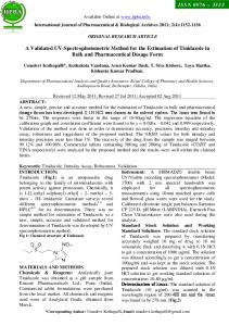
Figure 1: Structure of piperine
Table-4: Results of Accuracy study for Piperine
Table-1: Optical Characteristics and Analytical Data PARAMETERS RESULTS Excitation wavelength (nm) 339 nm Emission wavelength (nm) 450 nm Linearity (ng/ml)* 10-60 Regression equation Y=1.2336x + 20.081 Slope* 1.2336±0.01 Confidence interval for slope (At 95%) 1.223 -1.244 Intercept* 20.077±0.3857 Confidence interval for Intercept (At 19.673 -20.482 95%) Co-efficient of determination* 0.9958 LOD (ng/ml)* 1.031 LOQ (ng/ml)* 3.127 Accuracy (%) 91.928-94.365 *= Average of six determination Table-2: Data of Calibration curve Concentration Mean Fluorescence (ng/mL) Intensity ± S.D. 10 30.695 ± 0.422 20 45.658 ± 0.372 30 58.182 ± 1.008 40 69.480 ± 0.355 50 83.355 ± 0.302 60 92.168 ± 0.359
Asian J Pharm Clin Res, Vol 5, Suppl 4, 2012, 231-233
%RSD 1.374 0.816 1.733 0.511 0.363 0.390
Limit Of Detection And Limit Of Quantification (LOD AND LOQ) The limit of quantification (LOQ) of an individual analytical procedure is the lowest amount of analyte in a sample which can be quantitatively determined and the limit of detection (LOD) of an individual analytical procedure is the lowest amount of an analyte in a sample which can be detected but not necessarily quantitated as an exact value. LOQ and LOD value represent the sensitivity of the proposed analytical method. LOD and LOQ of the drugs were calculated using the following equations. LOD = 3.3 x [σ]/S LOQ = 10 x [σ]/S Where σ = the standard deviation of the response and S = the standard deviation of y intercept of regression lines
Name of the drug
Piperine
Amount of standard spiked (ng) 0 20
Average of amount Recovered (ng) 29.052 47.510
40
63.934
60
87.450
Recovery (%) ± S.D ------96.856 ± 2.87 92.587 ± 6.84 98.201 ± 3,65
RESULTS AND DISCUSSION Standard curves for piperine were prepared at excitation and emission wavelengths of 339 nm and 450 nm, respectively, using a Spectrofluorimeter. The plots of concentration versus intensity exhibited a linear relationship. The equation of the straight line for piperine was y =1.2336x + 20.081. A methanolic extract of formulation was also analyzed at the same excitation and emission wavelengths. The developed method was validated for linearity, precision, accuracy, limit of detection and limit of quantification. The linearity was found to be in the range of 10 - 60 ng/mL. The correlation coefficients (r2) were 0.9958, respectively, indicating good linearity between the fluorescence intensity and concentration. Scanning of the samples six times allowed the precision of the method to be checked. The accuracy of the method was checked by carrying out recovery studies. A sample of known concentration of standard solution of piperine was added in equal volume to the three different dilutions of the sample extract and analyzed spectrofluorimetrically to see whether the observed concentration obtained corresponded to the theoretical concentration obtained from the standard curve. The percentage recovery of piperine was found to be in the range of 91.928-94.365%. Hence, the developed spectrofluorimetric procedure is a quick and reliable method for the quantitative estimation of piperine in raw material, processed powder and in herbal preparations containing pepper. CONCLUSION The results indicate that the method proposed was simple, selective and rapid with reasonable precision and accuracy. Hence this method can be employed for the routine analysis of piperine in Pure form and in its formulations. REFERENCES 1. 2. 3. 4.
Figure 2:Calibration Curve obtained by spectrofluorimetry
A.K.Gupta, Quality Standards of Indian Medicinal Plants, ICMR, New Delhi, Vol.I, 1986, 168-173. A.M.Mujumdar, J.N. Dhule, V.K. Deshmukh, and S.R.Naik. Effect of piperine on bioactivity of oxyphenylbutazone in rats Indian Drugs. 1999, 36(2),123-126. A. Khajuria, N. Thusa, U. Zusthi and K.L. Bedi, Estimation of piperine in commercial Ayurvedic formulations. Indian Drugs. 1997 , 34(10) 557–563. C.K.Kokate, A.K.Purohit and S. B.Ghokhle. Pharmacognosy. Nirali Prakashan, (1994), 315-317.
232
Vyas et al.
5. 6. 7. 8.
9.
Asian J Pharm Clin Res, Vol 5, Suppl 4, 2012, 231-233
Evans WC. Pharmacognosy. Saunders Publication. 15th Ed. (2007) 353-355. C.P.Khare, Encyclopedia of Indian medicinal plants. Springer publication, (2006), 367-370. V. Gupta, U.K.Jain, Quantitative analysis of piperine in ayurvedic formulation by UV Spectrophotometry. Int. J. of Pharm. Sci and Res. 2011, 2(2) 58-61. R. Chauhan, J. Dwivedi and A. Siddiqui, Chemical Standardization and Quantification of Piperin from methanolic extract of Piper Nigrum by HPLC method on the basis of isolated markers. Int. J. Chem. Sci. 2008, 6(4),1726-1733. P. Hamrapurkar, K. Jadhav and S. Zine, Quantitative estimation of Piperine in Piper nigrum and Piper longum using High Performance Thin Layer Chromatography. J. of Applied Pharma. Sci. 2011, 1(3) 117-120.
233