POSTER SESSION
Validation of a Method to Replace Frozen Section During Parathyroid Exploration by Using the Rapid Parathyroid Hormone Assay on Parathyroid Aspirates Rodney K. Chan, MD; Shahrul I. Ibrahim, MD; Peter Pil, MD, PhD; Milenko Tanasijevic, MD, MBA; Francis D. Moore, Jr, MD
Hypothesis: The parathyroid hormone (PTH) content of tissue aspirates is an accurate indicator of parathyroid tissue and can replace frozen section during parathyroid surgery. Design and Setting: Prospective data collection in a tertiary care hospital with a single surgeon. Patients: One hundred sixty-seven consecutive patients completing limited parathyroid explorations. Interventions: Parathyroid adenomas removed during limited parathyroid exploration were aspirated through a 22-gauge needle into 0.5-mL isotonic sodium chloride solution and the solution held on ice in a purple-top tube. Aspirates of in situ thyroid tissue were also taken for comparison. Samples were then assessed for PTH content at the same time the serial blood samples were routinely assessed to monitor the physiologic impact of the surgery.
the identification of the tissue of origin as parathyroid in every case. Tissue aspirates from pathologically proven parathyroid tissue had a mean PTH level of at least 1691 pg/mL, with 160 of the aspirates having values exceeding the upper limit of the assay. This measure was significantly higher than values obtained from thyroid aspirates, which had a mean PTH level of 88 pg/mL (P⬍.01), reflective of blood levels at the time of aspiration. Using the 99% confidence interval of histologically confirmed parathyroid glands as the lower limit of a positive test result at 1610 pg/mL, tissue aspirate PTH assay has a sensitivity of 97% and a specificity of 100%. Conclusions: Positive identification of removed tissue as
Main Outcome Measure: The PTH content of tissue aspirates was compared with histologic identification of removed putative parathyroid tissue.
parathyroid is a necessary adjunct to limited parathyroid exploration, where decreases in false-positive blood PTH levels can be a result of operative manipulation of the neck. Analysis of tissue PTH content during the same assay that is being used for assessing blood PTH concentration is an efficient and accurate method for identifying the tissue with certainty. This measure also prevents the occasional ambiguity in frozen sections of parathyroid tissue that apparently contain thyroidlike colloid material.
Results: Elevated tissue PTH content was associated with
Arch Surg. 2005;140:371-373
I
Author Affiliations: Departments of Surgery and Pathology, Brigham and Women’s Hospital, Harvard Medical School, Boston, Mass.
N FOCUSED PARATHYROID SUR gery, accurate intraoperative recognition of parathyroid tissue is fundamental to correcting primary hyperparathyroidism. Visual identification depends heavily on the expertise of the surgeon and is more difficult where there has been prior neck surgery, irradiation, anatomical variation, or coexisting thyroid disease such as multinodular goiter or Hashimoto thyroiditis. Even in routine cases, frozen section often is used to verify the identity of the tissue. When there are other sources of doubt, as with intrathyroidal parathyroid glands or with a parathyroid gland with a microfollicular histological pattern resembling a Hürthle cell thyroid adenoma, par-
(REPRINTED) ARCH SURG/ VOL 140, APR 2005 371
affin sections are sometimes needed for definitive identification.1,2 A rapid serum PTH assay performed intraoperatively to observe a fall in parathyroid hormone (PTH) concentrations after the excision of an adenoma demonstrates the effect of removing a hyperfunctioning gland.3,4 A greater than 50% drop in the PTH level within 10 minutes is generally agreed to be indicative of cure, although the exact timing of the samples before and after excision still is under investigation. However, this technique suffers from both false negatives and positives. In the falsepositive case, unwitting manipulation of the parathyroid adenoma prior to the preremoval blood sample could result in falsely elevated PTH levels, resulting in a greater
WWW.ARCHSURG.COM
Downloaded from www.archsurg.com at Harvard University, on July 29, 2010 ©2005 American Medical Association. All rights reserved.
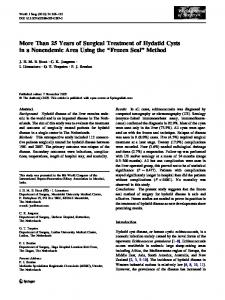
Positive Test
2000 1800
large prospective study has been performed to determine its accuracy.7 METHODS
1400 1200 1000
Negative Test
Parathyroid Hormone, pg/mL
1600
800 600 400 200 0
Lymph Node
Parathyroid
Thyroid
Figure. Graphical representation of parathyroid hormone (PTH) content in aspirates of nonparathyroid, parathyroid, and thyroid tissues. Mean PTH level of parathyroid (bar) is significantly higher than that of other tissue (P⬍.01). The large black circle at 1800 pg/mL represents values above the upper limit of the assay. The statistics assume that the greater than 1800-pg/mL value is 1800 pg/mL. Given that the real but unknown values are even higher than that, the statistical significance would be even greater and the clinical management would remain the same.
Table 1. Summary of Aspirate Data Compared With Pathologic Diagnosis*
Positive test, No. Negative test, No.
Parathyroid
Nonparathyroid
160 5
0 2
*Tissue aspirates have a sensitivity of 97% and a specificity of 100%.
than 50% fall despite the presence of additional pathologic parathyroid tissue.5 In the more common falsenegative case, manipulation of the parathyroid gland during removal but after the preremoval blood sample might cause a spike in PTH levels, resulting in an insufficient drop compared with postexcision, despite removal of all pathologic parathyroid tissue.5,6 With a false negative, further neck exploration is undertaken, with a slight increase in morbidity to the patients. However, the false-positive case is more significant because it can lead to failure of the surgery. It is possible to further compound the error by misidentifying as parathyroid the related tissues, such as ectopic thyroid, thymus, or lymph node. Thus, it continues to be important to positively associate the fall in venous PTH concentrations with accurate identification of the removed tissues as parathyroid. Measuring tissue PTH content by way of aspiration is a direct way to identify parathyroid tissue and offers an alternative to frozen section. This process theoretically eliminates some of the cost associated with pathologic processing and interpretation and might, depending on local policy, incur minimal independent additional expense. The rapid turnaround time to run this assay in parallel with the serum samples translates to no additional time in the operating room beyond that required to run the serum assay. Although some smaller studies in the literature advocate the use of this technique, no (REPRINTED) ARCH SURG/ VOL 140, APR 2005 372
From November 1997 through April 2004, we entered 167 consecutive putative parathyroid and thyroid aspirates and their corresponding resected specimens into this single-arm prospective study at Brigham and Women’s Hospital (Boston, Mass). A single surgeon performed all aspirations as well as the operations. Patients were included in the study when they were referred with signs, symptoms, and laboratory findings consistent with primary hyperparathyroidism. All patients underwent preoperative imaging with ultrasound or nuclear imaging. They were then excluded from the study if they were thought to have multiglandular disease or if limited parathyroid surgery could not be performed. Specimens were aspirated into 0.5-mL isotonic sodium chloride solution using a 22-gauge needle and then held on ice in purple-top tubes. The PTH concentration of the aspirates was determined using the dual antibody immunoassay for intact PTH (1-84), which was performed at the same time as the analysis of the serum samples. The results of the putative gland PTH values were compared with the final pathologic diagnosis; the thyroid gland PTH values served as the negative controls. Samples were analyzed by the hospital’s chemistry laboratory following the manufacturer’s instructions and using the Quick Intraoperative Biointact PTH assay kit (Nichols Institute, San Clemente, Calif ). Statistical analysis was performed using Sigmaplot (SPSS Inc, Chicago, Ill). Results are presented as means ± SE. If it was needed, we subjected comparison groups to 1-way analysis of variance and, when significance was found, applied the t test. In the statistical analysis, when the PTH measurement is greater than the limit of detection of the assay, a value of 1800 pg/mL was assumed (the highest value detected by the assay). Given that the real but unknown values are even higher than that, the statistical significance would be even greater and the clinical management would remain the same. RESULTS
Data were collected on 167 patients during the study period. Thirty-six (21%) were men, and the mean±SD age was 58.1±0.9 years. The average PTH measures before and after excision were 271.31±59 pg/mL and 43.57±4.3 pg/mL, respectively. The mean decrease in the PTH level of 227.73 pg/mL (72.58%) was consistent with removal of parathyroid adenomas. Aspirates from the putative parathyroid gland, later confirmed histologically, had a mean PTH level of 1691.83 pg/mL, which is significantly higher than thyroid aspirates with a mean PTH level of 87.90 pg/mL (P⬍.01) (Figure). One hundred sixty parathyroid aspirates, in fact, had values above the upper limit of the assay. Using the 99% confidence interval of known parathyroids as the lower limit of a positive test result (or 1610 pg/mL), tissue aspirate PTH assay has a sensitivity of 97% and a specificity of 100% (Table 1). Two thyroid aspirates exhibited high PTH values, likely from aspirating too far into adjacent structures containing parathyroid secretions. In those cases, the tests were voided and frozen sections were obtained. Five histologically confirmed parathyroid adenomas were found to have low PTH values (false negatives), likely WWW.ARCHSURG.COM
Downloaded from www.archsurg.com at Harvard University, on July 29, 2010 ©2005 American Medical Association. All rights reserved.
resulting from insufficient sampling. There were no false positives in this study. COMMENT
Correctly identifying the parathyroid gland is important in a successful parathyroid exploration, especially considering the increasing proportion of minimally invasive explorations. Even with the use of decreasing intraoperative venous PTH concentrations as an indicator for cure of hyperparathyroidism, there is still a need for positive tissue identification. A false-positive fall in PTH levels can result in the added morbidity of a failed operation and the need to surgically reexplore the neck at a later date. Identifying glands as parathyroid tissue currently relies on frozen section, which can be misleading when the parathyroid adenoma has a follicular architecture. Here we have shown that tissue PTH content can be an alternative indicator in frozen section identification. Currently, we advocate tissue aspirates as an alternative or additional indicator to confirm after removal that a putative enlarged parathyroid gland is indeed of parathyroid origin. Although such a technique might also allow for in situ identification of a parathyroid adenoma prior to resection, we have been reluctant to apply it to normal parathyroid tissue in situ. This technique samples a volume of tissue for PTH content. Although the total PTH content of an adenoma is clearly elevated, there are no generally accepted data that its PTH concentration per volume sampled differs from that of a normal gland. Second, the aspiration samples vary, depending on tissue turgor and length of needle insertion, making it difficult to sample constant volumes of tissue. Finally, the bevel of the 22-gauge needle often is larger than the thickness of a suppressed parathyroid gland, and the technique might irreversibly damage the normal glands in situ, either from barotraumas due to aspiration or from hemorrhage into the glands. For all these reasons, we did not attempt to distinguish normal parathyroid tissue from adenoma using tissue aspirates. The parallel testing of serum PTH levels and tissue aspirates eliminates the routine need for a pathologist but still provides the information necessary to perform an operation. The successful removal of a parathyroid adenoma would be reflected in a greater than 50% decrease in serum PTH levels together with a high parathyroid aspirate and a low thyroid aspirate. If the thyroid aspirate is high, the parathyroid aspirate cannot be interpreted correctly and a frozen section should be done to identify the
(REPRINTED) ARCH SURG/ VOL 140, APR 2005 373
Table 2. Scheme for Interpretation of Serum and Aspirate Parathyroid Hormone Values Venous PTH Decrease Decrease Decrease
Tissue Aspirate High Low Low
Thyroid Aspirate
Outcome
Low High Low
Success Frozen section on tissue Frozen section on tissue; consider continuing exploration No change Not applicable Not applicable Continue exploration Abbreviation: PTH, parathyroid hormone.
excised tissue. A scheme for interpreting these data is outlined in Table 2. The combination of serum PTH level, aspirate of tissue and thyroid, and clinical judgment must be used to determine whether the surgery performed was adequate. We conclude that tissue PTH content is a highly accurate measure for identifying a neck mass as parathyroid tissue. Accepted for Publication: November 29, 2004. Correspondence: Francis D. Moore, Jr, MD, Brigham and Women’s Hospital, 75 Francis St, Boston, MA 02115 (
[email protected]). Previous Presentation: This study was presented at the 85th Annual Meeting of the New England Surgical Society; October 1, 2004; Montreal, Quebec; and is published after peer review and revision. REFERENCES 1. Shidham VB, Asma Z, Rao RN, et al. Intraoperative cytology increases the diagnostic accuracy of frozen sections for the confirmation of various tissues in the parathyroid region. Am J Clin Pathol. 2002;118:895-902. 2. Schantz A. The microscopical pathology of primary parathyroid hyperplasia. Ann Clin Lab Sci. 1979;9:314-318. 3. Irvin GL III, Dembrow VD, Prudhomme DL. Clinical usefulness of an intraoperative “quick parathyroid hormone” assay. Surgery. 1993;114:1019-1023. 4. Nussbaum SR, Thompson AR, Hutcheson KA, Gaz RD, Wang CA. Intraoperative measurement of parathyroid hormone in the surgical management of hyperparathyroidism. Surgery. 1988;104:1121-1127. 5. Gauger PG, Agarwal G, England BG, et al. Intraoperative parathyroid hormone monitoring fails to detect double parathyroid adenomas: a 2-institution experience. Surgery. 2001;130:1005-1010. 6. Yang GP, Levine S, Weigel RJ. A spike in parathyroid hormone during neck exploration may cause a false-negative intraoperative assay result. Arch Surg. 2001; 136:945-949. 7. Perrier ND, Ituarte P, Kikuchi S, et al. Intraoperative parathyroid aspiration and parathyroid hormone assay as an alternative to frozen section for tissue identification. World J Surg. 2000;24:1319-1322.
WWW.ARCHSURG.COM
Downloaded from www.archsurg.com at Harvard University, on July 29, 2010 ©2005 American Medical Association. All rights reserved.