Dec 9, 2009 - the present study was to compare this new device to a validated, standardized system .... components, 1) Laptop Computer, 2) Shape-HF Data.
34
The Open Sports Medicine Journal, 2010, 4, 34-40
Open Access
Validation of a Simplified, Portable Cardiopulmonary Gas Exchange System for Submaximal Exercise Testing Andrew D. Miller1, Paul R. Woods1, Thomas P. Olson1, Minelle L. Hulsebus1, Kathy A. O’Malley1, Dean MacCarter2 and Bruce D. Johnson*,1 1
Division of Cardiovascular Diseases, Department of Internal Medicine, Mayo Clinic, Rochester, MN, USA
2
Aurora Denver Cardiology Associates, Aurora, CO, USA Abstract: Shape Medical Systems, Inc. has developed a new miniaturized, simplified system for non-invasive cardiopulmonary gas exchange quantification and has targeted their system for submaximal clinical exercise testing in order to abbreviate testing in an expanding clinical market during a climate of escalating health care costs. The focus of the present study was to compare this new device to a validated, standardized system for measures of cardiopulmonary gas exchange. Eighteen healthy adults (10 male/8 female, age 29±7 yr, BMI 23.8±2.4 kg/m2) were brought to the laboratory and instrumented with both measurement systems via in-series pneumotachs. Additionally, the Shape system included a pulse oximeter for heart rate (HR) and oxygen saturation (SaO2), while the standard system included separate 12-lead ECG and oximetry devices. The protocol included 2-min resting breathing, followed by 3-min at each of 3 workloads (50, 70, 125 watts) on a cycle ergometer. Data were collected breath-by-breath and averaged the last 30-sec of each workload. After a 15-min rest period, the pneumotach order was reversed and the study repeated. Since gas exchange data were similar (p>0.05) within a given metabolic testing system between sessions the data were pooled for comparing the Shape and Standard systems. There were no differences (p>0.05) between the systems for oxygen consumption-VO2, carbon dioxide production-VCO2, ventilation-VE, end tidal CO2-PetCO2, tidal volume-V T, respiratory rate-fb, and HR at rest or any work load. SaO2 was slightly, but significantly lower using the Shape embedded oximeter (p27), or orthopedic limitations. The study was reviewed and approved by the Mayo Clinic Institutional Review Board and all subjects reviewed and signed informed consent prior to participation.
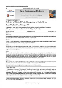
Fig. (1). Protocol for validating the Shape system for cardiopulmonary gas exchange measures. Subjects were instrumented with both systems by putting pneumotachs in series. Data were collected at rest, 50, 70 and 125 watts followed by a 15min rest period, reversing pneumotach order and repeating the testing.
Overview of the Protocol
The Shape-HF™ Cardiopulmonary Exercise Testing System (St. Paul, MN) consisted of the following significant components, 1) Laptop Computer, 2) Shape-HF Data Analyzer, 3) Patient Interface, 4) Pulse Oximeter Finger Probe (Smiths Medical PM, Inc. 3711 BCI Digital Micro Power Board with w Comfort-Clip Oximetry Finger Sensor) (see Fig. 2). The laptop computer supplied with the ShapeHF system is OEM supplied operated with Windows XP® operating software. Shape-HF system software is installed in the laptop computer.
Subjects reported to the laboratory and were simultaneously instrumented with the Shape simplified system and the “standard system”. For the Shape system they were instrumented with a pulse oximeter (for heart rate) and a pneumotach attached in series with the standard system. For the standard system, subjects were instrumented with a 12-lead electrocardiogram (for heart rate and safety monitoring), a mouthpiece in series with pneumotachographs from both systems, along with a nose clip. Both systems were started simultaneously and data were collected in the following order, 2-min rest, 3-min 50 watts, 3-min 70 watts and 3-min 125 watts on a cycle ergometer. After a brief rest period (15 minutes) the order of the pneumotachs was reversed and the test repeated (see Fig. 1). The small increment from 50 to 70 watts provided a test for sensitivity of the systems to discriminate small changes in external work. Linking the pneumotachs in series allowed simultaneous data collection, however, this could have small influences on breath timing, phase relationships and dead space and thus the data were collected with the pneumotachs under both conditions for comparison.
Shape System for Exercise Testing
Submaximal
Cardiopulmonary
Standard System for Non invasive Gas Exchange Testing The standard system included a Medical Graphics CPX/D.(Medical Graphics, St Paul, MN) integrated to a Perkin Elmer Mass Spectrometer MGA 1100 (MA Tech
Fig. (2). The Shape system consists of a plug and play analyzer, lap top and pneumotach/sensor system.
36 The Open Sports Medicine Journal, 2010, Volume 4
Additional Testing A subset of subjects (n= 4) were brought back to the laboratory on a subsequent day to repeat testing using the Shape tight fitting mask. This was to assure that the use of a mask did not alter gas exchange data quality. Data Collection and Analysis Data were collected on a breath-by-breath basis using both systems with complete datasets for each subject downloaded into Excel. Data were then averaged over the last 30-sec for each condition for each subject. Initial comparisons between exercise sessions for a given system did not reveal significant differences (p>0.05) across measures and thus data from each session were averaged for further comparisons between the Shape and standard systems. Descriptive statistics (means, standard deviations and ranges) were computed with comparisons made between systems for VT, fb, VE, VO2, VCO2, HR, SaO2, and PetCO2 values using students t-tests (�=0.05). In addition, Pearson’s correlation coefficients were calculated to determine linearity between systems and Bland-Altman plots were constructed to assess agreement between systems. Primary analyses for correlations and Bland-Altman plots focused on the measures of VO2, VCO2, VE and PetCO2 as these measures take into account the integration of the primary measures used in cardiopulmonary gas exchange. RESULTS Subject characteristics are reported in Table 1. Subjects were young men and women with normal weight and blood pressure values. The Shape system tracked closely with breath-by-breath changes measured by the standard system. Fig. (3) is an example of the breath-by-breath changes in VO2 in a subject transitioning from rest to the initial work loads (50 and 70 watts). Slight differences in phase are noted in part due to the sequence of pneumotachs and the ability to start the systems at identical time periods.
Table 1.
Subject Characteristics (n = 18)
Age (yr)
28.8 ± 7.3
Gender (M/F)
18 to 46
(10/8)
-
Ht (cm)
173.4 ± 11.4
156.2 to 203.5
Wt (kg)
72.0 ± 13.1
54.0 to 93.2
BMI (kg/m2)
23.8 ± 2.4
19.1 to 27.4
SBP (mmHg)
106.3 ± 12.0
94 to 136
DBP (mmHg)
66.6 ± 9.4
48 to 82
Ht, height; Wt, weight; BMI, body mass index; SBP, systolic blood pressure; DBP, diastolic blood pressure. 2 1.8 1.6
VO2 (L/min)
The Shape system is designed for use with a wide range of exercise testing modalities including simple stair stepping, cycle ergometry, treadmill, or other exercise modalities while the patient is connected to the Shape-HF Data Analyzer via the patient interface and pulse oximeter finger probe. The data analyzer contains discrete analyzers that measure expired oxygen and carbon dioxide, an airflow measurement subsystem that measures expired and inspired air using a fixed orifice, differential pressure pneumotach, and a pulse oximeter to measure heart rate and blood oxygen saturation. The data analyzer transmits measured data once per breath via a USB interface cable to the laptop computer. The system requires a laptop computer; however, future designs will allow standalone systems. Transmitted data includes breath count, PetCO2, VCO2, VO2, tidal volume-VT, breathing frequency-fb, heart rate-HR, barometric pressure, SaO2, dead space, peak expiratory flow, volume Inspired, resting energy, expiratory time, and inspiratory time. The patient interface consists of a fixed orifice, differential pressure pneumotach to which a respiratory gas sample line and upstream and downstream differential pressure sample lines are attached.
Miller et al.
1.4 1.2 1 0.8 Shape VO2
0.6
Standard System VO2
0.4 0.2 0 1
15
29
43
57
71
85
99 113 127 141 155 169 183 197
Time (seconds)
Fig. (3). Example of breath-by-breath plot of Shape and standard system for VO2 at rest and during the initial work intensities. Slight differences are noted in phase due to the differences in pneumotach order and slight variations in systems when attempting to start testing at identical time periods.
Reproducibility of Shape Values Relative to the Standard System (Session 1 vs 2) The coefficient of variation (CV) between sessions 1 and 2 for the Shape system ranged from 2.9 to 10.8% for VO2, 4.0 to 8.7% for VCO2, 1.9 to 3.3% for VE, 2.6 to 9.3% for HR, 0.3 to 0.4% for SaO2 and 2.9 to 4.3% for PetCO2. Typically the CV’s were higher in resting data and reduced with exercise. The exception was HR, which was highest at 125 watts. The CV’s for the standard system were similar to the Shape system during exercise, except for HR which ranged from 1.6 to 3.8%. However this might be expected when comparing a 12-lead ECG to pulse oximetry. The CV’s for VO2 and VCO2 at rest tended to be lower in the standard system at 4.1 and 5.4%, respectively. Sensitivity of Shape System for Detecting Small Changes in Cardiopulmonary Measures In order to test the ability of the Shape system to accurately measure small changes in gas exchange variables, the protocol included a relatively small 20 watt increment in workload during the exercise session (50 � 70 watts). Significant increases of similar magnitude were measured by both systems for VO2 (0.24±0.08 vs 0.23±0.07 L/min, p