RESEARCH REPORT © 2017 Human Kinetics - IJATT 22(2), pp. 29-33 https://doi.org/10.1123/ijatt.2016-0047
Validity, Reliability, and Normative Values for Clinically-Assessed Frontal Tibial Orientation as a Measure of Varus-Valgus Knee Alignment David Werner • University of Dayton; John Willson, Richard Willy • East Carolina University; Joaquin Barrios • University of Dayton
Frontal plane knee alignment can influence the development and management of various knee pathologies. Valid and reliable clinical methods for assessment are needed. The primary purposes of this study were to assess the validity and reliability of inclinometer-based frontal plane tibial orientation as a limb alignment measure, and secondarily to establish normal values in healthy individuals. Frontal tibial orientation was validated per moderately strong correlation to radiographic knee alignment. Intra- and interrater reliability were excellent. The normative mean was approximately 7°. In summary, inclinometer-based frontal tibial orientation is a valid and reliable clinical measure of frontal plane knee alignment. Key Words: inclinometry, lower limb alignment, norms
K
nee pathologies can be costly and disabling. Identifying risk factors for knee pathologies is important to help guide screening and examination procedures, as well as improve rehaKey Points bilitative treatments. Frontal tibial orientation is a valid way to Common knee patholclinically assess limb alignment. ogies include anterior cruciate ligament (ACL) Frontal tibial orientation is reliable within injury,1 patellofemoral and between raters. pain syndrome (PFPS),2 and knee osteoarthriFrontal tibial orientation is approximately tis (OA). 3 Static knee 7°. alignment is thought to contribute to ACL injury4 Frontal tibial orientation does not differ and PFPS risk,5 and is by sex. predictive of OA develINTERNATIONAL JOURNAL OF ATHLETIC THERAPY & TRAINING
opment and progression.6–8 Valid and reliable methods of clinically assessing varus-valgus alignment are needed.9 The full-limb radiograph is the most common reference standard for assessing knee alignment.10,11 This measure requires radiographically identifying the femoral and tibial mechanical axes, and measuring the angle of their projected intersection. However, there are a number of drawbacks to this approach in clinical practice, including cost, time, radiation exposure, and reliance on imaging facility. Consequently, clinically valid measures of frontal plane knee alignment are needed. In a concurrent validation study of five clinical measures of frontal plane knee alignment, the strongest correlate to radiography was frontal plane tibial orientation MARCH 2017 ❚ 29
Downloaded by Ebsco Publishing
[email protected] on 03/03/17, Volume 22, Article Number 2
(FTO) in natural stance, measured using inclinometry (r = .80).10 Another study reported on a similar assessment using a standardized foot width of 29 cm, finding comparable results (r = .83).11 Both these studies used foot maps for all testing. While an approach that is not reliant on foot maps would improve the clinical utility of FTO, it is unclear if the measure would remain valid. The reliability of assessing FTO is not well established. An intraclass correlation coefficient (ICC) of 0.94 has been reported.10 However, the analysis was poorly described and distinct intrarater and interrater results were not reported. Within-day, intrarater reliability (ICC model 3,1) was excellent in another study (ICC 0.93).12 However, without more expansive reliability analyses, it remains unclear if the measure is appropriate for between-day and between-rater use. Finally, FTO should be helpful in identifying normal and abnormal frontal plane alignment. Thus, reference norms should be generated. A normative database can then provide a standard of comparison between limbs and sexes. To date, few studies have been conducted on clinical measures of knee alignment evaluating interlimb or sex differences. Therefore, this study tested four hypotheses. First, a moderately strong correlation would be seen between radiographic frontal plane knee alignment and concurrently-assessed FTO without foot maps. Second and third, that intrarater reliability of FTO would be excellent (ICC > 0.90), and that interrater reliability would be good (ICC > 0.80). Finally, that normative data for FTO would show no interlimb differences, but would demonstrate more varus FTO values in males.
Materials and Methods Potential study participants were recruited from a university setting. Subjects were healthy, between the ages of 18–50, and claimed no history of previous or current lower limb pathology. For subjects meeting these criteria, their sex, age, and height were recorded. All subjects provided written informed consent. Twenty-two healthy subjects participated in the validation phase, undergoing concurrent radiography and inclinometry measures at a local hospital. For radiographic capture, subjects stood barefoot with knees fully extended and the feet approximated in a “feet together” position.13 Full-limb digital radiography was performed with the cathode positioned at the height of the knee with gonads shielded. Standard settings
30 ❚ MARCH 2017
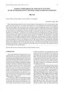
of 500 mA and 80 kV were used, allowing for adjustment as necessary by the technician. Images were assembled using three stitched images that included the entire pelvis and lower extremities. Images were exported as DICOM files for analysis using ImageJ software (National Institutes of Health, Bethesda, MD). Radiographic knee alignment was determined as the hip-knee-ankle angle, defined by the angle of the intersection between the femoral and tibial mechanical axes. The femoral mechanical axis was defined as the line from the centroid of the femoral head to the midpoint of the femoral condyles, and the tibial mechanical axis as the midpoint between the tibial spines to the midpoint of the talus. Coincident mechanical axes were described as 0° and considered neutral, with varus angles positive and valgus angles negative. Immediately after image capture, the subject remained standing still and FTO was measured bilaterally, as described in the next paragraph. To measure FTO, subjects stood in the “feet together” position with their lower limbs approximated. Subjects were instructed to bear weight evenly between both feet while maintaining full knee extension. The caliper-inclinometer device (Acuangle, Isomed, Portland, OR) was then positioned in the frontal plane of the individual. The caliper arms were equidistant in length. The end of the proximal caliper arm was positioned against the tibial tuberosity, and the end of the distal arm placed over the neck of the talus (Figure 1). The displayed angle was recorded to the nearest degree. Three measures were recorded with the mean calculated. Pearson’s product-moment correlation was used to assess the bivariate relationship between FTO measurements and digital knee alignment.10,11 Intrarater reliability between days was examined on 30 consecutive subjects. The fourth author (Examiner 1) collected data on consecutive days. To assess intrarater reliability between days, ICC (model 2,3) values were calculated.14 Data from the next 15 subjects were then collected by two graduate physical therapy students (Examiners 2 and 3), trained by Examiner 1, to assess interrater reliability. The examiner order was randomized. ICC (model 2,3) values were calculated. Finally, normative data were collected. Descriptive data including the mean, standard deviation, standard error of the measurement, and the minimum detectable difference value were calculated.15 To test the
INTERNATIONAL JOURNAL OF ATHLETIC THERAPY & TRAINING
ankle angle was slightly varus (1.0 ± 2.4°), and the average FTO measure was also varus (6.0 ± 1.5°). The correlation between hip-knee-ankle angle and FTO was moderately strong (r = .779).
Intrarater Reliability Thirty (21 female, 9 male) consecutive qualifying subjects with a mean height of 1.70 ± 0.11 m and mean age of 26.5 ± 6.1 years were measured on consecutive days by Examiner 1. Intrarater reliability was excellent (ICC [2,3] = 0.97).
Downloaded by Ebsco Publishing
[email protected] on 03/03/17, Volume 22, Article Number 2
Interrater Reliability Examiners 2 and 3 measured the next 15 (8 female, 7 male) consecutive qualifying subjects with mean height of 1.68 ± 0.10 m and mean age of 25.2 ± 2.1 years. Interrater reliability was excellent (ICC [2,3] = 0.94).
Healthy Norms with Analysis of Sex and Limb
Figure 1
Measurement of frontal tibial orientation using a caliper-inclinometer device.
hypotheses that sex differences would be observed in the absence of interlimb differences, a mixed model sex-by-limb analysis of variance was conducted with a predetermined alpha level of .05. All data were analyzed using SPSS version 21.0 (IBM Corp., Armonk, NY).
Results Validation A total of 44 (24 female, 20 male) limbs were assessed with concurrent radiographic and FTO measurements. Subjects had a mean height of 1.67 ± 0.11 m, and mean age of 27.1 ± 5.1 years. The average hip-knee-
INTERNATIONAL JOURNAL OF ATHLETIC THERAPY & TRAINING
FTO norms were then compiled using data from the 45 subjects used for reliability testing, as well as 130 additional subjects. In total, 175 (86 female, 89 male) subjects were collected (Table 1). Of note, three female subjects demonstrated enough valgus knee alignment that only their knees approximated. Age was not different between sexes, although the males were approximately 0.17 m taller (p < .001). Partly in contrast to our hypotheses, there was no sex-by-limb interaction, nor main effects for sex or limb. The final mean pooled FTO was 7.1 ± 1.9°. The standard error of the measurement was 0.21°, and the minimum detectable difference was 0.60°. The distribution skewness was 0.226, indicating there was symmetry about the mean of the normal curve. The kurtosis value was –0.217, suggesting that the data were not abnormally concentrated in the peak nor the tails of the distribution.
Discussion Knee malalignment is a risk factor for knee OA and may increase the risk of acute knee injuries.4–8 Full-limb radiography remains the reference standard for frontal plane knee alignment.16 As radiography is not always clinically appropriate or feasible, alternatives such as FTO are needed. In the current study, FTO assessment using the “feet together” position remained valid (r = .78) on a comparable level to studies using an FTO
MARCH 2017 ❚ 31
Table 1 Means and Standard Deviations for Demographic and Frontal Plane Tibial Orientation (FTO) Data with Associated p-Values for Sex Comparisons Men ( n = 89)
Women ( n = 86)
All (N = 175)
p-value
22.8 (6.6) 1.81 (0.06) 7.2 (2.0)
22.5 (4.2) 1.64 (0.07) 7.1 (2.1)
22.6 (5.5) 1.73 (.10) 7.1 (2.0)
.699 < .001 .892
Left limb FTO (°)
7.0 (1.7)
7.0 (2.0)
7.0 (1.8)
.950
Averaged limb FTO (°)
7.1 (1.8)
7.1 (2.1)
7.1 (1.9)
.942
Age (years) Height (m)
Downloaded by Ebsco Publishing
[email protected] on 03/03/17, Volume 22, Article Number 2
Right limb FTO (°)
approach with foot maps in natural stance10 (r = .80) and with feet 29 cm apart11 (r = .83). Recent efforts in radiographic knee alignment have also used the “feet together” position as it aids reproduciblity.17 We recommend the “feet together” position with limbs approximated when measuring FTO to maximize clinical utility. The reliability of measuring FTO was excellent, consistent with previous similar studies.10,12 We observed excellent intrarater (ICC 2,3 = 0.97) and interrater (ICC 2,3 = 0.94) reliability. In addition, the standard error of the measurement (0.21°) and the minimum detectable difference (0.60°) were small. Based on the current analysis, we can be 95% confident that different FTO measures on a given person should yield values within 1° of each other. Therefore, FTO represents a reliable clinical measure of knee alignment. There was no difference between limbs in the FTO measure. Interlimb symmetry has been previously reported in studies assessing static knee alignment.18 Average FTO was approximately 7° from vertical. Using a wider stance for the measure will generally decrease the FTO value, as a widened stance moves the tibia toward a more vertical (and potentially even valgus) orientation in the frontal plane, as compared with the “feet together” postition.10,11 In regard to sex, it is less clear whether lower limb alignment naturally differs. In both healthy and pathological knees, and using varying measures, there has been a greater association with valgus alignment for females.19–22 In contrast, there was no sex difference in femorotibial angle using a healthy Japanese sample.23 The current data support the notion that static alignment in healthy persons is not different between sexes. However, future studies on specific pathologies may reveal sex disparities. 32 ❚ MARCH 2017
For the “feet together” position, knees may approximate with or without feet approximating. In our study (N = 175), three female individuals presented as such (1.7%). In these three cases, the individuals exhibited FTO values of 0° or 1°. Prevalence of this occurrence may be higher in pathologic groups associated with valgus knee alignment. Measuring FTO requires the examiner to successfully identify two superficial landmarks. Proximally, the landmark is the tibial tuberosity. The tibial tuberosity is usually an easily located superficial landmark about the proximal anterior tibia, appearing fairly midline relative to the long axis of the bone. The talar dome serves as the distal tibia landmark for FTO. In most individuals, the talar dome is palpable just lateral to the descending tendon of the tibialis anterior at the level of the talocrural joint. Two anatomic confounds should be considered in the measurement of FTO. First, a laterally deflected tibial tuberosity is common.24 In such instances, the apparent inclination of the tibia relative to vertical may be increased, overestimating the inclination in the varus direction. A second anatomic factor that may confound the measure is tibial bowing, which can occur in either direction.25,26 Medial tibial concavity may increase the FTO value artificially. Inclinometer-based measurement of FTO, as described in this study, provides a clinical estimate of frontal plane lower limb alignment. The measure is simple, inexpensive, quickly performed, and easily interpreted. Foot maps are not needed. There is no radiation exposure, nor reliance on imaging facilities. However, caution must be exercised when utilizing the findings of FTO. Surgical planning for procedures such as tibial osteotomies and total knee arthroplasties requires more precise imaging modalities. However, INTERNATIONAL JOURNAL OF ATHLETIC THERAPY & TRAINING
FTO may help clinicians and clinical researchers screen or classify individuals by limb alignment, and help derive appropriate interventions.27 Further work is needed to explore whether FTO can help identify responders to interventions including foot orthoses, bracing, footwear, weight loss, and locomotor modification.
Downloaded by Ebsco Publishing
[email protected] on 03/03/17, Volume 22, Article Number 2
Conclusion In summary, the described approach for measuring FTO is a valid and reliable method to assess lower limb alignment in the absence of foot maps. In healthy individuals, the normative mean for FTO is approximately 7°, and does not differ between limbs or by sex. The utility of this measure of alignment in clinical settings should be further explored. ❚
References 1. Muneta T, Sekiya I, Yagishita K, Ogiuchi T, Yamamoto H, Shinomiya K. Two-bundle reconstruction of the anterior cruciate ligament using semitendinosus tendon with endobuttons: Operative technique and preliminary results. Arthroscopy. 1999;15(6):618–624. PubMed doi:10.1053/ar.1999.v15.0150611 2. Chesworth BM, Culham E, Tata GE, Peat M. Validation of outcome measures in patients with patellofemoral syndrome. J Orthop Sports Phys Ther. 1989;10(8):302–308. PubMed doi:10.2519/jospt.1989.10.8.302 3. Murphy L, Schwartz TA, Helmick CG, et al. Lifetime risk of symptomatic knee osteoarthritis. Arthritis Rheum. 2008;59:1207–1213. PubMed doi:10.1002/art.24021 4. Loudon JK, Jenkins W, Loudon KL. The relationship between static posture and ACL injury in female athletes. J Orthop Sports Phys Ther. 1996;24(2):91–97. PubMed doi:10.2519/jospt.1996.24.2.91 5. Boling MC, Padua DA, Marshall SW, Guskiewicz K, Pyne S, Beutler A. A prospective investigation of biomechanical risk factors for patellofemoral pain syndrome: the Joint Undertaking to Monitor and Prevent ACL Injury (JUMP-ACL) cohort. Am J Sports Med. 2009;37(11):2108–2116. PubMed doi:10.1177/0363546509337934 6. Brouwer GM, van Tol AW, Bergink AP, et al. Association between valgus and varus alignment and the development and progression of radiographic osteoarthritis of the knee. Arthritis Rheum. 2007;56:1204– 1211. PubMed doi:10.1002/art.22515 7. Sharma L, Song J, Felson DT, Cahue S, Shamiyeh E, Dunlop DD. The role of knee alignment in disease progression and functional decline in knee osteoarthritis. JAMA. 2001;286:188–195. PubMed doi:10.1001/ jama.286.2.188 8. Sharma L, Song J, Dunlop D, et al. Varus and valgus alignment and incident and progressive knee osteoarthritis. Ann Rheum Dis. 2010;69:1940–1945. PubMed doi:10.1136/ard.2010.129742 9. Nguyen A-D, Boling MC, Slye CA, Hartley EM, Parisi GL. Various methods for assessing static lower extremity alignment: implications for prospective risk-factor screenings. J Athl Train. 2013;48(2):248–257 doi:10.4085/1062-6050-48.2.08. PubMed 10. Hinman RS, May RL, Crossley KM. Is there an alternative to the fullleg radiograph for determining knee joint alignment in osteoarthritis? Arthritis Rheum. 2006;55:306–313. PubMed doi:10.1002/art.21836 11. Vanwanseele B, Parker D, Coolican M. Frontal knee alignment: three-dimensional marker positions and clinical assessment. Clin Orthop Relat Res. 2009;467:504–509. PubMed doi:10.1007/s11999-008-0545-4
INTERNATIONAL JOURNAL OF ATHLETIC THERAPY & TRAINING
12. Barrios JA, Higginson JS, Royer TD, Davis IS. Static and dynamic correlates of the knee adduction moment in healthy knees ranging from normal to varus-aligned. Clin Biomech (Bristol, Avon). 2009;24:850– 854. PubMed doi:10.1016/j.clinbiomech.2009.07.016 13. Bellemans J, Colyn W, Vandenneucker H, Victor J. Is neutral mechanical alignment normal for all patients? The concept of constitutional varus. Clin Orthop Relat Res. 2012;470:45–53. PubMed doi:10.1007/ s11999-011-1936-5 14. Shrout PE, Fleiss JL. Intraclass correlations: uses in assessing rater reliability. Psychol Bull. 1979;86:420–428. PubMed doi:10.1037/00332909.86.2.420 15. Stratford PW, Goldsmith CH. Use of the standard error as a reliability index of interest: an applied example using elbow flexor strength data. Phys Ther. 1997;77:745–750. PubMed 16. Cooke TD, Sled EA, Scudamore RA. Frontal plane knee alignment: a call for standardized measurement. J Rheumatol. 2007;34:1796–1801. PubMed 17. Song MH, Yoo S, Kang S, Kim Y, Park G, Pyeun Y. Coronal alignment of the lower limb and incidence of constitutional varus knee in Korean females. Knee Surg Relat Res. 2015;27:49–55. PubMed doi:10.5792/ ksrr.2015.27.1.49 18. Hsu RW, Himeno S, Coventry MB, Chao EY. Normal axial alignment of the lower extremity and load-bearing distribution at the knee. Clin Orthop Relat Res. 1990; (255):215–227. PubMed 19. Barrett JP, Rashkoff E, Sirna EC, Wilson A. Correlation of roentgenographic patterns and clinical manifestations of symptomatic idiopathic osteoarthritis of the knee. Clin Orthop Relat Res. 1990; (253):179–183. PubMed 20. Harvey WF, Niu J, Zhang Y, et al. Knee alignment differences between Chinese and Caucasian subjects without osteoarthritis. Ann Rheum Dis. 2008;67:1524–1528. PubMed doi:10.1136/ard.2007.074294 21. Tamari K, Tinley P, Briffa K, Aoyagi K. Ethnic-, gender-, and age-related differences in femorotibial angle femoral antetorsion, and tibiofibular torsion: cross-sectional study among healthy Japanese and Australian Caucasians. Clin Anat. 2006;19:59–67. PubMed doi:10.1002/ca.20170 22. Nguyen AD, Shultz SJ. Sex differences in clinical measures of lower extremity alignment. J Orthop Sports Phys Ther. 2007;37(7):389–398. PubMed doi:10.2519/jospt.2007.2487 23. Tamari K, Tinley P, Aoyagi K. Gender and age-related differences in axial alignment of the lower limb among healthy Japanese volunteers: comparative and correlation study. J Jpn Phys Ther Assoc. 2003;6:25–34. PubMed doi:10.1298/jjpta.6.25 24. Yoshioka Y, Siu DW, Scudamore RA, Cooke TD. Tibial anatomy and functional axes. J Orthop Res. 1989;7:132–137. PubMed doi:10.1002/ jor.1100070118 25. Howell SM, Kuznik K, Hull ML, Siston RA. Longitudinal shapes of the tibia and femur are unrelated and variable. Clin Orthop Relat Res. 2010;468:1142–1148. PubMed doi:10.1007/s11999-009-0984-6 26. Yau WP, Chiu KY, Tang WM, Ng TP. Coronal bowing of the femur and tibia in Chinese: its incidence and effects on total knee arthroplasty planning. J Orthop Surg (Hong Kong). 2007;15:32–36. PubMed doi:10.1177/230949900701500108 27. Barrios JA, Davis IS, Higginson JS, Royer TD. Lower extremity walking mechanics of young individuals with asymptomatic varus knee alignment. J Orthop Res. 2009;27:1414–1419. PubMed doi:10.1002/ jor.20904
David Werner and Joaquin Barrios are with the Department of Physical Therapy, University of Dayton, Dayton, OH. John Willson and Richard Willy are with the Department of Physical Therapy, East Carolina University, Greenville, NC. Megan N. Houston, PhD, ATC, Keller Army Community Hospital, is the report editor for this article.
MARCH 2017 ❚ 33
Copyright of International Journal of Athletic Therapy & Training is the property of Human Kinetics Publishers, Inc. and its content may not be copied or emailed to multiple sites or posted to a listserv without the copyright holder's express written permission. However, users may print, download, or email articles for individual use.