1344
Variable Procedural Strategies Adapted to Anatomical Characteristics in Catheter Ablation of the Cavotricuspid Isthmus Using a Preoperative Multidetector Computed Tomography Analysis KENTA KAJIHARA, M.D.,∗ YUKIKO NAKANO, M.D., Ph.D.,∗ YUKOH HIRAI, M.D., Ph.D.,∗ HIROSHI OGI, M.D., Ph.D.,∗ NOBORU ODA, M.D., Ph.D.,∗ KAZUYOSHI SUENARI, M.D., Ph.D.,∗ YUKO MAKITA, M.D.,∗ AKINORI SAIRAKU, M.D.,∗ TAKEHITO TOKUYAMA, M.D.,∗ CHIKAAKI MOTODA, M.D.,∗ MAI FUJIWARA, M.D.,∗ YOSHIKAZU WATANABE, M.D.,∗ MASAO KIGUCHI, R.T.,† and YASUKI KIHARA, M.D., Ph.D.∗ From the ∗ Department of Cardiovascular Medicine, Graduate School of Biomedical and Health Sciences; and †Department of Radiology, Hiroshima University Hospital, Hiroshima, Japan
Variable Strategies for CTI Ablation.
Objectives: This study aimed to investigate the anatomical characteristics complicating cavotricuspid isthmus (CTI) ablation and the effectiveness of various procedural strategies. Methods and Results: This study included 446 consecutive patients (362 males; mean age 60.5 ± 10.4 years) in whom CTI ablation was performed. A total of 80 consecutive patients were evaluated in a preliminary study. The anatomy of the CTI was evaluated by multidetector row-computed tomography (MDCT) prior to the procedure. A multivariate logistic regression analysis revealed that the angle and mean wall thickness of the CTI, a concave CTI morphology, and a prominent Eustachian ridge, were associated with a difficult CTI ablation (P < 0.01). In the main study, 366 consecutive patients were divided into 2 groups: a modulation group (catheter inversion technique for a concave aspect, prominent Eustachian ridge, and steep angle of the CTI or increased output for a thicker CTI) and nonmodulation group (conventional strategy). The duration and total amount of radiofrequency energy delivered were significantly shorter and smaller in the modulation group than those in the nonmodulation group (162.2 ± 153.5 vs 222.7 ± 191.9 seconds, P < 0.01, and 16,962.4 ± 11,545.6 vs 24,908.5 ± 22,804.2 J, P < 0.01, respectively). The recurrence rate of type 1 atrial flutter after the CTI ablation in the nonmodulation group was significantly higher than that in the modulation group (6.3 vs 1.7%, P = 0.02). Conclusion: Changing the procedural strategies by adaptating them to the anatomical characteristics improved the outcomes of the CTI ablation. (J Cardiovasc Electrophysiol, Vol. 24, pp. 1344-1351, December 2013) atrial flutter, catheter ablation, cavotricuspid isthmus, eustachian ridge, multidetector row-computed tomography Introduction The cavotricuspid isthmus (CTI) is defined as the area between the tricuspid valve (TV) and inferior vena cava (IVC), and is contiguous in anatomy to the triangle of Koch. The CTI is a critical component of the reentry circuit for CTI-dependent atrial flutter (AFL).1-5 Radiofrequency (RF) catheter ablation targeting the CTI is the optimal treatment for CTI-dependent AFL.6,7 Despite high overall success rates, No disclosures.
the ablation procedure is occasionally difficult due to variations in the anatomical characteristics of the CTI.8,9 Multidetector row-computed tomography (MDCT) has become a widely used substitute in cardiac angiography studies.10 In a preliminary study, the CTI anatomy was evaluated using 64-row MDCT, and the anatomical characteristics of the CTI that complicate the ablation procedure were identified. Subsequently, adaptations to the CTI ablation strategies according to the anatomical information provided by the preprocedural MDCT were prospectively examined in a main study. The goal was to improve and minimize the difficulty of the ablation procedure.
Address for correspondence: Kenta Kajihara, M.D., Department of Cardiovascular Medicine, Hiroshima University Graduate School of Biomedical and Health Sciences, 1-2-3 Kasumi, Minami-ku, Hiroshima 734-8551, Japan. Fax: +81-82-257-5169; E-mail:
[email protected] Manuscript received 20 April 2013; Revised manuscript received 14 June 2013; Accepted for publication 26 June 2013. doi: 10.1111/jce.12231 This is an open access article under the terms of the Creative Commons Attribution-NonCommercial-NoDerivs License, which permits use and distribution in any medium, provided the original work is properly cited, the use is non-commercial and no modifications or adaptations are made.
Methods Study Subjects The institutional review board approved the study, and written informed consent was obtained from all patients. Patients with a serum creatinine level of 1.2 mg/dL or more were excluded from the study. Patients in which adequate anatomical information necessary to perform the measurements was not obtained during the MDCT scan were
Kajihara et al.
excluded. Fortunately, the scans in all subjects provided the necessary anatomical information and it was not necessary to exclude any subjects due to an uninterpretable scan quality. Preliminary Study In total, 80 patients (63 males [79.7%], aged 59.8 ± 10.4 years) in whom CTI ablation was successfully performed using an 8 mm tip ablation catheter were included from the preliminary study. MDCT was performed within 24 hours before the ablation procedure in all patients. Typical AFL was clinically documented in 23 patients (AFL only: n = 7; both atrial fibrillation (AF) and AFL: n = 16). The remaining 57 patients with persistent or paroxysmal AF underwent a combined AF and CTI ablation with no evidence of AFL. Of the 73 patients with AF in whom circumferential pulmonary vein isolation (CPVI) was performed, paroxysmal AF was evident in 63 patients and persistent AF in 10. Main Study Three hundred and sixteen consecutive patients (males: n = 255, 80.7%, aged 60.5 ± 10.2 years) scheduled for a CTI ablation were prospectively enrolled in the main study. Typical AFL was clinically documented in 22 patients. In the remaining 294 patients (including 56 patients with AFL) with persistent or paroxysmal AF, a combined AF and CTI ablation was performed with no evidence of AFL. Multidetector Computed Tomography Protocol and Image Reconstruction MDCT data sets were acquired using a 64-slice CT scanner (Lightspeed VCT; GE Healthcare, Waukesha, WI, USA) with retrospective ECG-gated scans using a dual-shot-type injector (Nemoto-Kyorindo, Tokyo). To satisfy these conflicting issues, we used a multiphasic contrast material injection protocol. In the routine protocol, the first phase of the multistepwise protocol, 144 mgI/kg were administered during 7 seconds. In the second phase, the initial contrast concentration was 370 mgI/mL; it was gradually decreased by dilution with saline during 15 seconds. The total iodine dose in the second phase was 155 mg/kg. The scan protocol methods used in this study have been described previously.11 Image analysis software (Virtual Place Advance; AZE, Tokyo, Japan) allowed 3-dimensional (3D) viewing of multiplanar reconstruction images reformatted as cross-sectional images. The optimal image was selected during the end-diastole phase of the right atrium, which was defined as the image immediately before the opening of the tricuspid valve. The images were also reconstructed using electrocardiography (ECG) edited at the level of the anomalies of the ECG signal, which were caused by premature beats, AF, and mistriggering. Multiplanar reconstructions of the axial images were obtained by operating a cursor manually on selected cross-sectional source images orthogonal to the source images during the latest right atrial diastolic phase of the cardiac cycle [30–40% of the interbeat (R–R) interval]. Measurements of the Anatomical Parameters of the Cavotricuspid Isthmus The quantative measurements and morphologies of the CTI were analyzed in all patients. The values for the length (Fig. 1A) and angle (Fig. 1B) were obtained in long-axis 2-
Variable Strategies for CTI Ablation
1345
chamber right anterior oblique views parallel to the septum. The length of the CTI was defined as the segment beginning from the base of the end of the Eustachian valve to the base of the tricuspid valve. The angle was constructed by the intersection of the line passing through the center of the IVC and the line extending to the orifice of the IVC from the tricuspid annulus. The presence of a prominent Eustachian ridge was confirmed in the same view. The myocardial thickness of the CTI was measured at the center of the 3 parallel isthmic levels (paraseptal, central, and inferolateral) in the short-axis left anterior oblique views (Fig. 1C,D). The mean myocardial thickness of the CTI was calculated from the values of those 3 sites. The angle of the CTI was classified into 2 types (steep: 3 to ≤5 mm), and pouchlike (>5 mm), according to the depth of the CTI.10 Also, the analysis software of this study could measure only to the first decimal place. Two blinded cardiologists experienced in reading MDCT images independently analyzed all the data sets. Quantitative measurements were performed in triplicate. Electrophysiological Study and Catheter Ablation Continuous monitoring was performed using endocardial electrocardiography (bandpass filtered 30–500 Hz) and surface electrocardiography. Data were stored on a digital amplifier and recorder system (Bard Electrophysiology, Lowell, MA, USA). A 6F multipolar (15-pole) catheter (Irvine Biomedical, Irvine, CA, USA) was utilized with the distal poles (poles 1−10) placed within the coronary sinus (CS) and the proximal electrodes (poles 11−15) located from the superior vena cava to the superior right atrium via the right internal jugular vein. A decapolar electrode catheter was positioned on the lateral wall of the right atrium (Irvine Biomedical) inserted from the right femoral vein. A decapolar electrode catheter (Biosense Webster, Diamond Bar, CA, USA) was introduced via the femoral vein into the region of the bundle of His. After intravenous administration of heparin (bolus injection of 3000 IU followed by an hourly injection of 1000 IU), a 7F quadripolar deflectable catheter with an 8 mm tip electrode (Navister; Biosense Webster) was positioned on the isthmus using a preshaped long sheath with a 180◦ curve (RAMP curve, St. Jude Medical, Daig Division, Inc., Minnetonka, MN, USA). In the beginning, the ablation catheter was positioned at the ventricular aspect of the CTI at the 6 o’clock position in the LAO 45◦ projection. Ablation was carried out with the temperature set at 55 ◦ C and energy output at 40−50 W using a dragging catheter technique under anatomic and electrographic guidance.4,12,13 RF was applied with voltage outputs modulated by impedance monitoring to avoid an increase due to excessive heating.14 Ablation of the CTI was performed during either sinus rhythm (pacing from the CS) or ongoing typical AFL with the procedural endpoint of complete bidirectional isthmus block.15 Reversal of the right atrial depolarization sequence was established by a complete CTI map using a decapolar mapping catheter straddling the line of block and by recording widely separated (>120 milliseconds) local double potentials along the ablation line during atrial pacing.16 Differential pacing was performed to distinguish complete from incomplete bidirectional isthmus block. When complete conduction block of
1346
Journal of Cardiovascular Electrophysiology
Vol. 24, No. 12, December 2013
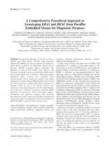
Figure 1. Quantitative measurements and morphologies of the CTI. Values for the length and angle were obtained in the long-axis 2-chamber right anterior oblique views parallel to the septum. Panel A: The length of the CTI (red double-headed arrow) was defined as the segment beginning from the base of the end of the Eustachian valve to the base of the tricuspid valve. Panel B: The angle (red curve line) was constructed by the intersection of the line passing through the center of the IVC and a line extended to the orifice of the IVC from the tricuspid annulus. Panels C and D: The myocardial thickness of the CTI was measured at the center of 3 parallel isthmic levels in the short-axis left anterior oblique view (paraseptal; blue circle, central; light blue, and inferolateral; light green). The wall thickness, manually measured on an image, took into account the visually detectable wall edges (spread light blue circle). Panel D shows the virtual endoscopic view of the right atrium around the CTI.
the CTI was not achieved after first-line ablation, the conduction gaps were mapped and ablated searching for single or narrow-split potentials with the ablation catheter moving along the original ablation line. If conduction block was not still achieved, the catheter was repositioned on the ventricular side of the CTI at the 7 or 5 o’clock position. If bidirectional block status was maintained over the 30-minute period following the achievement of the bidirectional block, the procedure was considered complete. In this study, the procedure time of the CTI ablation was defined as the duration from the beginning of the first RF application to confirmation of the bidirectional block of the CTI, excluding the observation time. The CTI was divided into a proximal part (from the coronary sinus ostium to the Eustachian valve) and a distal part (from the tricuspid annulus to the coronary sinus ostium) according to a catheter placed within the CS. The region in which a complete bidirectional conduction block was successfully achieved was recorded. For patients in whom a combined AF and CTI ablation was performed, CPVI was performed at the beginning of the procedure. The patients were mildly sedated without intubation during the procedure using short acting intravenous drugs. Ablation Protocol of the Main Study The patients were prospectively randomized and assigned to either a modulation or nonmodulation group. In the modulation group (n = 183), the ablation strategies were modulated according to a flow chart that was based on the results of the preliminary study (Fig. 2). In brief, if the thickness of the CTI was ≥2.7 mm, the CTI ablation was initiated with a high output (60 W) and high-temperature (60 °C) setting from the
beginning. If the morphology of the CTI was concave, angle of the CTI was ≤86.4◦ , or a prominent Eustachian ridge was found, the catheter was inverted until the outer curve was in contact with the isthmus at the proximal part of the CTI (Fig. 3). Thus, the catheter came in contact with the outer surface of the CTI in these cases. In the absence of any of these anatomical features, the CTI ablation was performed by the conventional method. In the nonmodulation group (n = 183), the ablation lines were created conventionally, similar to the method in the preliminary study. Verification of the bidirectional block in the CTI was also performed as in the preliminary study. Patient Follow-Up The patients were examined in our outpatient clinic 2 weeks after performance of the ablation procedure. Followup continued every 1−2 months thereafter. Patients were examined using 12-lead ECG and Holter monitoring. Interviews were conducted regarding clinical symptoms and residual palpitations. Statistical Analysis The data were tested for normal (Gaussian) distribution using the Kolmogorov−Smirnov test. Normally distributed continuous variables are presented as means ± SD. In the case of a non-Gaussian distribution, median values and quartiles are given. Comparisons between groups were performed using Student’s t-test. In cases of continuous data with a nonGaussian distribution, the Mann−Whitney U test was used. Categorical variables were compared using the Fisher’s exact test. A univariate analysis of the patient characteristics was conducted for a comparison between the difficult and
Kajihara et al.
Variable Strategies for CTI Ablation
1347
Figure 2. Flow chart of the ablation strategies in the modulation group. The ablation strategies were modulated according to this flow chart in the modulation group.
Figure 3. Catheter inversion technique. Radiographs in the right anterior oblique (RAO; A) and left anterior oblique (LAO; B) projections show the inverted ablation catheter (ABL) within the right atrium targeting the region of the Eustachian ridge. ABL = an 8 mm tipped ablation catheter; CS = a decapolar catheter positioned in the proximal coronary sinus; HB = a decapolar catheter in the region of the His-bundle; LRA = a decapolar circular catheter in the lateral right atrium.
straightforward cases, and a logistic regression analysis was performed to detect any independent significant predictors by adjusting for multiple variables. The multiple pairwise comparisons among the patient groups were tested using a Tukey–Kramer test for continuous variables. Statistical significance was defined as a P < 0.05 using a 2-sided comparison. Statistical analyses were performed using JMP 9 software (SAS Software, Cary, NC, USA).
Results Preliminary Study The baseline characteristics of the 80 subjects and ablation results of the preliminary study are summarized in Table 1. For the myocardial thickness of the CTI, there was no significant difference between the 3 parallel isthmic levels
1348
Journal of Cardiovascular Electrophysiology
Vol. 24, No. 12, December 2013
TABLE 1 Patient Characteristics and Ablation Results of the Preliminary Study Parameters Patient characteristics Gender male, n (%) Age, year BMI, kg/m2 Left ventricular ejection fraction, % Hypertrophic cardiomyopathy, n (%) Dilated cardiomyopathy, n (%) Coronary artery disease, n (%) Valvular heart disease, n (%) Anatomical data collected from MDCT Length of the CTI, mm Angle of the CTI, degree Angle type of the CTI Steep type, n (%) Gentle type, n (%) Mean thickness of the CTI, mm Prominent Eustachian ridge, n (%) Morphology of the CTI Straight aspect, n (%) Concave aspect, n (%) Pouch-like aspect, n (%) Ablation results Ablation during AFL, n (%) Procedure time, minutes Total RF application duration, seconds Total RF energy, J Fluoroscpic time, minutes
Results (n = 80) 63 (79.7) 59.8 ± 10.4 23.9 ± 2.9 64.0 ± 9.9 2 (2.5) 1 (1.2) 4 (5.0) 4 (5.0) 29.9 ± 7.6 97.6 ± 26.7 16 (20.0) 64 (80.0) 2.5 ± 0.6 11 (13.8) 46 (57.5) 20 (25.0) 14 (17.5) 3 (3.8) 21.8 ± 20.9 346.4 ± 321.4 26,482.5 ± 21,403.4 13.1 ± 11.8
AFL = atrial flutter; BMI = body mass index; CTI = cavotricuspid isthmus; MDCT = multidetecter computed tomography; RF = radiofrequency.
Efficacy of Varying the Procedural Strategies According to the Cavotricuspid Isthmus Anatomy In this prospective, randomized study, no significant difference was evident in the baseline and anatomical characteristics between the nonmodulation and modulation groups (Table 3). In the results of the CTI ablation in both groups (Table 3), the total CTI ablation procedure time was significantly shorter in the modulation group than that in the nonmodulation group. Further, the total RF duration was significantly shorter and total amount of RF energy required to establish bidirectional conduction block at the CTI was significantly smaller in the modulation group than that in the nonmodulation group. In addition, the fluoroscopic time was significantly shorter in the modulation group than that in the nonmodulation group. The results of the comparison of the procedure time between the modulation and nonmodulation groups according to the anatomical characteristics complicating the CTI ablation are shown in Fig. 4. Tukey–Kramer tests revealed that the procedure time in the patients with a concave aspect, thickness of the CTI ≥2.7 mm, angle of the CTI ≤86.4◦ or prominent Eustachian ridge was significantly longer than that in those without in the nonmodulation group (30.4 ± 23.6, 31.6 ± 23.6, 41.2 ± 26.5, 52.6 ± 37.3 vs 9.1 ± 6.8 minutes, P < 0.05). However, in the modulation group, there was no significant difference in the procedure time between the patients with anatomical difficulty and those without (13.6 ± 8.5, 12.8 ± 9.2, 13.2 ± 9.4, 12.4 ± 8.6 vs 9.3 ± 8.2 minutes). Follow-Up Results
(paraseptal: 2.61 ± 0.46 vs central: 2.41 ± 0.51 vs inferolateral: 2.38 ± 0.43) by Tukey–Kramer multiple comparison tests. Bidirectional conduction block in the CTI was achieved in all patients. The subjects in the preliminary study were divided into 2 groups according to the median procedure time: the difficult cases (n = 40; procedure time ≥16 minutes) and straightforward cases (n = 40; procedure time