Genetic Resources and Crop Evolution 46: 175–182, 1999. © 1999 Kluwer Academic Publishers. Printed in the Netherlands.
175
Variation and genomic polymorphism of lectin-related proteins in Lima bean (Phaseolus lunatus L.) seeds Lucia Lioi1 , Francesca Sparvoli2 & Roberto Bollini2 1 Istituto
del Germoplasma, CNR, Via Amendola 165/A, 70126 Bari, Italy; 2 Istituto Biosintesi Vegetali, CNR, Via Bassini 15, 20133 Milano, Italy (∗ Corresponding author: E-mail:
[email protected]; Fax +390 80 5587566) Received 26 May 1998; accepted in revised form 24 August 1998
Key words: α-amylase inhibitor, arcelin, gene pools, lectin-related protein, Phaseolus, RFLPs
Abstract Variation of the lectin and the two lectin-related proteins, AIL (α-amylase inhibitor-like) and ARL (arcelin-like) was examined in wild and cultivated accessions of Lima bean (Phaseolus lunatus L.) using electrophoresis of total seed proteins, immunoblot and RFLP analysis of lectin-related genes. Results confirm that divergence of the two major Lima bean gene pools, Andean and Mesoamerican, also apply to this protein family. All three members of the family are present in both gene pools, with differences in size, abundance and composition between gene pools, giving the possibility to distinguish Andean from Mesoamerican lectin pattern types. Both patterns show some variants, such as lack of lectin or its presence as an abundant protein. The observed variation reflects, at least in part, into genomic polymorphism. The presence of arcelin- and α-amylase-related proteins in Lima bean could represent a tool to increase our knowledge in the evolution of the lectin family in Phaseolus species. Abbreviations: α-AI – α-amylase inhibitor; AIL – α-amylase inhibitor-like; ARL – arcelin-like; LBL – Lima bean lectin; PHA – phytohemagglutinin Introduction Plant lectins are carbohydrate-binding proteins that are present in different organs and tissues of many plant species, and particularly in storage organs and seeds. Although many physiological roles have been proposed for plant lectins, the most likely functions of abundant vacuolar lectins stored in bark and seeds are as nitrogen reserve and defense from predators (Chrispeels & Raikhel, 1991; Peumans & Van Damme, 1995). Several legume species contain proteins that are clearly related to lectins. A well-studied example is the common bean (Phaseolus vulgaris L.) lectin-related protein family, composed of arcelin, α-amylase inhibitor (α-AI) and phytohemagglutinin (PHA) (Ishimoto et al., 1995; Lioi, 1991; Osborne et al., 1988). Genetic evidence shows that genes for these three proteins are located at the same locus, indicating they may have arisen by duplication and divergence from a single ancestral gene (Nodari et al., 1993).
The amino acid sequences of these proteins, deduced from their cDNA, show a high degree of homology (Mirkov et al., 1994). During evolution, these proteins have acquired different protective properties against insect predators. Arcelins are proposed to be responsible for resistance against the Mexican bean weevil, Zabrotes subfasciatus (Cardona et al., 1990), whereas α-AI confers resistance to Callosobruchus maculatus, the cowpea weevil (Ishimoto et al., 1996). In common bean, PHA and arcelin are glycosylated proteins whose mature polypeptides range between 31 and 45 kDa. PHA consists of two homologous polypeptides, PHA-E (erythroagglutinating subunit) and PHA-L (leucoagglutinating subunit) not covalently bound in tetramers and presents an high genotypic variation (Brown et al., 1982; Lioi, 1991). Seven electrophoretic arcelin variants have been so far described in wild beans collected in Mexico (AcostaGallegos et al., 1998). In their native status, arcelin polypeptides are assembled into dimers (arcelin types
176 1, 2, 5 and 6) or tetramers (arcelin types 3 and 4) (Goossens et al., 1994; Hartweck et al., 1991; Sparvoli & Bollini, 1998). Contrary to arcelin and PHA, α-AI is synthesized as a glycosylated precursor of about 40 kDa, which is proteolytically processed in the protein storage vacuoles into polypeptides of 15 – 20 kDa, characteristic of the mature inhibitor (Pueyo et al., 1993; Santino et al., 1992). Immunoblot analysis and PCR amplification of genomic DNA indicated the presence of lectins and related sequences also in P. acutifolius Gray, P. coccineus L., P. maculatus Scheele, P. microcarpus Mart. and P. polyanthus Greenm. (Mirkov et al., 1994). In Lima bean seeds, the presence of a true lectin has long been known (in: Galbraith & Goldstein, 1970). Lima bean lectin (LBL) is able to agglutinate only type A red blood cells and is made up of 31 - kDa subunits covalently linked by a single disulfide bridge to form dimers of 62 kDa (Roberts et al., 1982). Recently, the presence of two abundant lectin-related proteins in Lima bean seeds has been demonstrated (Sparvoli et al., 1998). The amino acid sequences derived from the cDNA clones coding for the two proteins has indicated that one sequence (ARL) is related to arcelin and the other (AIL) to α-AI. In the mature seed, each protein is made up of two major subunits, which represent two glycoforms of the same polypeptide precursor. The subunits of ARL (43 and 46 kDa) and AIL (40 and 42 kDa) assemble into oligomers of about 125 – 130 kDa and 88 – 100 kDa, respectively. The two proteins represent together the second major component of total seed proteins, phaseolin being the most abundant (Sparvoli et al., 1996). The high similarity between the Lima bean and the common bean lectin family as well as the finding that some accessions of P. lunatus are completely resistant to Acanthoscelides obtectus (Say) (Dobie et al., 1990) suggested these proteins could be potentially toxic and might protect seeds from predation by insects. Genetic organization of Lima bean is particularly suitable for studies on the variation and evolution of lectin-related proteins. Indeed, morphological, biochemical and molecular data (Fofana et al., 1997; Gutierrez-Salgado et al., 1995; Lioi, 1994; Maquet et al., 1997) showed that Lima beans have been domesticated independently in at least two centers, and indicated the existence of two separate groups, recognized as gene pools and denominated, following the definition proposed by Gepts for P. vulgaris (1988), as Andean and Mesoamerican.
The detection and characterization of new lectinrelated polypeptides is important both as a possible source of resistance factors against insect predation, and to study the evolutionary relatedness among the two groups. We investigated the variability of these proteins in Lima bean seeds through electrophoresis (SDS/PAGE). Different variants were subjected to immunoblot analysis to identify polypeptides crossreacting with antibodies against P. vulgaris PHA. Furthermore, RFLPs at the lectin-related protein locus, were determined to provide molecular evidence of variant diversity.
Materials and methods Plant materials. One hundred twenty eight accessions of P. lunatus, belonging to both Andean and Mesoamerican gene pools as determined by phenotype and phaseolin pattern, were analyzed; 20 of them were wild forms (W), the remaining ones were landraces (L). Accessions were mainly obtained from: Centro International de Agricultura Tropical (CIAT), Cali, Colombia (G accessions), or from the Institute of Plant Genetics and Crop Plant Research (IPK), Gatersleben, Germany (PHA accessions). In Table 1, identification number, origin, botanical status, and gene pool of accessions containing variants used as references are provided. Protein extraction, SDS/PAGE and immunoblot analysis. Total seed proteins were extracted from mature cotyledons by homogenization in 10 volumes of 20 mM sodium borate buffer, pH 9.0, for 2 h at 4◦ C and recovered in the supernatant after centrifugation for 15 min at 10000 × g. Proteins were dissociated by heating to 90◦ for 2 min in the presence of denaturing buffer (20 mM Tris–HCl, pH 8.6, containing 1% SDS, 8.3% glycerol and, when necessary, 0.5% β-mercaptoethanol (β-ME). One-dimensional SDS/PAGE was performed following the procedure described by Laemmli (1970), using 15% polyacrylamide. Gels were stained with Coomassie Brilliant Blue or blotted onto a supported nitrocellulose membrane (Bio-Rad). Immunoblot analysis was performed using rabbit antibodies against P. vulgaris PHA (Ceriotti et al., 1989) at 1:2000 dilution; they were able to recognize all Lima bean lectin-related proteins (Sparvoli et al., 1998). Peroxidase-linked anti-rabbit
177 Table 1. Identification of Lima bean (Phaseolus lunatus L.) accessions showing lectin-related protein variants, used as references Identification
Origin
PHA8152 G25832 G25916 G25915 G25849 PHA8110 PHA8111 PHA8071 PHA8151 PHA754 G25109 G25355 G25226 G26359
Zambia Peru Peru Peru Peru Zambia Argentina Romania Zambia Cuba Brazil Nigeria Costa Rica Mexico
Botanical status
Gene Pool
Pattern
L L W W L L L L L L L L W W
A A A A A A M M M M M M M M
A1 A2 A3 A4 A5 A6 M1 M2 M3 M4 M5 M6 M7 M8
G, CIAT number; PHA, IPK number; M, Mesoamerican; A, Andean; L, landraces; W, wild.
IgG was used as second antibody. Blots were developed with the ECL system (Amersham) according to manufacturers instructions. Erythroagglutination test. Erythroagglutinating activity was determined on total seed extracts. Group A red blood cells were sedimented by centrifugation and resuspended in 10 volumes of PBS (saline phosphate buffer); 0.1 ml of this suspension was added to 5 µl of protein extract, and agglutination visually determined after 1 h. Southern blot analysis. Genomic DNA was extracted from young leaves of single plants using the ‘maize DNA miniprep’ method according to Dellaporta et al. (1983). Five µg of DNA from each sample were digested with HindIII. The resulting fragments were separated on a 1% agarose gel and transferred onto positively charged nylon membrane using a standard technique. Probe labeling, hybridization and detection were carried out by the nonradioactive chemiluminescent method following the manufacturers instructions (Gene Image, Amersham). The DNA probe used was the PlLec1 cDNA clone, coding for ARL in an Andean P. lunatus (Sparvoli et al., 1998).
Results Electrophoretic analysis of seed lectin-related protein variability. One hundred and twenty-eight Lima bean accessions were analyzed by one-dimensional SDS/PAGE. When polypeptides migrating in the region of the SDS/PAGE corresponding to the position of lectinrelated proteins were considered, two basic seed protein electrophoretic banding patterns were detected, each showing several variants (Figure 1 and 2). Their frequency and distribution in cultivated and wild forms is reported in Table 2. One pattern type, with six variants, was found among the large-seeded accessions (Big Lima morphogroup and its wild form) and was therefore called Andean (A), as the probable center of origin of largeseeded forms has been located in northern Peru and southern Ecuador (Table 1). Variants A1 – A5 contain both ARL and AIL (Figure 1, lanes 1 – 5). These proteins cross-reacted with polyclonal antibodies (Figure 1c) and showed slight differences in abundance and electrophoretic mobility among variants. In reducing conditions, LBL polypeptides migrated with mobility corresponding to about 30 kDa and were clearly detectable on both Coomassie- stained gel (Figure 1a) and immunoblot (Figure 1c). In variants A3 and A4, LBL was represented by two major and equally abundant polypeptides (Figure 1c). Under non- reducing
178 Table 2. Distribution of seed lectin-related protein patterns in 128, wild and cultivated, Phaseolus lunatus L. accessions Botanical status
Landraces Wild Forms
Andean patterns A A A A 1 2 3 4
A 5
A 6
Mesoamerican patterns M M M M M 1 2 3 4 5
M 6
M 7
M 8
Total
6 0
1 1
7 0
10 2
11 0
0 4
0 2
108 20
1 1
0 1
0 2
conditions, LBL polypeptides gave rise to a number of dimeric forms which showed differences in mobility and abundance among variants (Figure 1d). This protein was particularly abundant in variant A4, where its amount was comparable to that of the other two members of the lectin family (Figure 1a, lane 4). About half the large seeded Andean landraces (variant A6 in Table 2) had a pattern that more closely resembled those of the small-seeded Mesoamerican accessions (see below). In A6 variant, the crossreactive polypeptides showed a significant change in electrophoretic mobility (Figure 1c); furthermore, LBL was undetectable (Figure 1d, lane 6) and its absence was confirmed by the lack of agglutinating activity. The other pattern type (M), with eight variants, was found among the small-seeded accessions (Sieva and Potato morphogroups and their wild forms). This pattern family was called Mesoamerican, as the probable center of origin of small-seeded forms has been located in Mesoamerica. When polypeptides were challenged with the antibodies, major immunoreactive polypeptides were detected (Figure 2a and c). These polypeptides could be divided into two groups (vertical bars in panel a) and biochemical data indicate they represent the counterparts of ARL and AIL found in Andean accessions. Mesoamerican ARL was constituted by polypeptides with molecular mass comparable to the Andean one (45 – 40 kDa), whereas AIL polypeptides (37 – 33 kDa) were smaller than the Andean ones. In the M2 and M3 variants, ARL consisted of a more intense band surmounted by a very faint one. The latter band was apparently absent in variants M4 and M7. In the M8 variant (two wild accessions), the two bands showed a slightly increased mobility and similar intensities. In this variant, the two polypeptides represented the second most abundant protein component after phaseolin (Figure 2, a, c). On the contrary, in variants M1, M5 and M6, ARL was a minor component and showed different electrophoretic mobility compared to ARL in the other patterns.
47 6
16 1
5 1
4 0
The faster migrating group, the AIL-related polypeptides, was similarly composed of two bands, with the exception of M8 variant, where only one band was detectable. In all variants, the smaller AIL component partially comigrated with the high molecular weight phaseolin subunit, therefore it could not be clearly identified in the stained gel (Figure 2a). The two immunoreactive AIL components showed similar intensity and mobility in variants M3, M4 and M7. In variant M2 a similar doublet was present, although the slower migrating component, apparently as abundant as in the latter three patterns (Figure 2a), showed a reduced crossreactivity (Figure 2c). Whether the faster migrating polypeptide of variant M6 was present in reduced amounts or it had a reduced immunoreactivity is not clear. Interestingly, genotypes showing variant M1 contained reduced amounts of all crossreactive polypeptides (Figure 2a, b). In A6 variant lectin-related proteins clearly resembled the majority of M variants (compare Figure 1c and Figure 2c), nevertheless phaseolin was of Andean type. LBL was detectable in the immunoblots of the Mesoamerican material only when the SDS-PAGE was performed in the absence of the reducing reagent β-ME. In this case the interchain disulfide bond was maintained and the dimeric form of the protein was readily detectable (Figure 2d). In the presence of the reducing agent, LBL was not detectable, because it probably comigrated with one of the other components of this protein family. Two mutant variants, M1 and M6, lacking LBL were identified (Figure 2d). Indeed, these genotypes were devoid of agglutinating activity against human group A erythrocytes. In both A and M patterns, the only other polypeptides affected by β-ME, beside LBL, were the two polypeptides of legumin, which covalently bind in the absence of the reducing agent (Figure 1 and 2, open circles).
179
Figure 1. Variability of lectin-related polypeptides in Lima bean (Phaseolus lunatus L.) seeds from the Andean gene pool. Polypeptides were separated by SDS-PAGE either under reducing (+β-ME) or non-reducing conditions (–β-ME) to detect Lima bean lectin (LBL). Gels were stained with Coomassie Brilliant Blue (a,b) or subjected to Western blot analysis (c,d). Vertical bars, major lectin-related polypeptides; dots and open circles, phaseolin and legumin proteins, respectively. Variant and accession number: lane (1) A1, PHA8152; lane (2) A2, G25832; lane (3) A3, G25916; lane (4), A4, G25915; lane (5) A5, G25849; lane (6), A6, PHA8110; lane M, Mesoamerican pattern type M7, G25226.
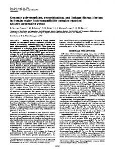
RFLP patterns. A total of 14 accessions, representing all protein pattern variants, were chosen to compare RFLP patterns (Table 1). The cDNA clone PlLec1, encoding Lima bean ARL (Sparvoli et al., 1998), was used as a probe. Washing of the blot in moderate condition of stringency (0.5× SSC, 0.1% SDS at 60◦ C) allowed recognition of sequences related to the genes of the lectin family. Hybridization patterns, obtained from genomic DNA of Mesoamerican material digested with HindIII, showed two major fragments (Figure 3). Variants M1 – M7 contained two fragments, the one with slight differences in size among samples (between 3.2 and 3.1 kb), and the other more variable of 4.1 – 6.2 kb. M8 variant pattern showed the highest variation, having the two major fragments a size of 6.8 and 4.5 kb. This high variation rate mirrors variation at the protein level (Figure 2).
Restriction pattern polymorphisms were also observed in the Andean gene pool. A6 variant had a high similarity with some Mesoamerican patterns, confirming the observation at the protein level. The remaining material yielded hybridization patterns with two major fragments in the range of 4.5 – 6.8 kb and a common one of about 2 kb.
Discussion Our data on electrophoretic variation of the components of the lectin-related protein family and related RFLP further support informations obtained by allozyme and phaseolin diversity, which indicate that Lima beans belong to two major gene pools distinguished by contrasting alleles (Gutiérrez Salgado et al., 1995; Maquet et al., 1997).
180
Figure 2. Variability of lectin-related polypeptides in Lima bean (Phaseolus lunatus L.) seeds from the Mesoamerican gene pool. Polypeptides were separated by SDS-PAGE either under reducing (+β-ME) or non-reducing conditions (–β-ME) to identify Lima bean lectin (LBL). Gels were stained with Coomassie Brilliant Blue (a,b) or subjected to immunoblot analysis (c,d). Vertical bars, major lectin-related polypeptides; dots and open circles, phaseolin and legumin proteins, respectively. Variant and accession number: lane (1) M1, PHA8111; lane (2) M2, PHA8071; lane (3) M3, PHA8151; lane (4) M4, PHA754; lane (5) M5, G25109; lane (6) M6, G25355; lane (7) M7, G25226; lane (8) M8, G26359; lane A, Andean pattern type A1, PHA8152.
All three members of the lectin-related family are present in both Lima bean gene pools. Andean ARL and AIL have higher molecular weight with respect to Mesoamerican forms. Each protein is generally organized as a double band, probably representing two glycoforms of the same polypeptide. Interestingly, arcelin-related proteins have been so far detected only in wild Mexican P. vulgaris and in a domesticated P. acutifolius (Mirkov et al., 1994). Data presented here, indicate this protein is commonly present in Lima bean instead. In both Andean and Mesoamerican gene pools, LBL is constituted of subunits which, under non reducing conditions, aggregate into dimers. Staining of the SDS-PAGE with Coomassie indicates this protein is generally a minor component in total protein extracts. Nevertheless, in some Andean material LBL
is an abundant protein. The presence of more than one LBL monomer in Andean variants was clearly detectable in variants A4 and A5 and could be due to different glycosylation of a single precursor (as for ARL and AIL) or they could be the products of different genes. Indeed, two cDNA clones coding for LBL have been isolated (Sparvoli et al., 1998). Differential oligomerization of the monomers could account for the complex patterns shown by dimeric Andean LBL. Our results indicate that variation in lectin-related protein electrophoretic banding patterns occurs in wild and cultivated forms of Lima bean. The double role proposed for these proteins, reserve proteins as well as biologically active proteins, are both limiting mutational events. As reserve proteins, they are tightly packed within protein bodies, hence any conformational modification limiting this process is probably
181
Figure 3. RFLP patterns of lectin-related genes in Andean and Mesoamerican gene pools of Phaseolus lunatus L. Genomic DNA was digested with HindIII and Southern blot was hybridized with PlLec1 cDNA clone. Plate Andean: variant and accession number: lane (1) A1, PHA8152; lane (2) A2, G25832; lane (3) A3, G25916; lane (4) A4, G25915; lane (5) A5, G25849. lane (6) A6, PHA8110. Plate Mesoamerican: variant and accession number; lane (1) M1, PHA8111; lane (2) M2, PHA8071; lane (3) M3, PHA8151; lane (4) M4, PHA754; lane (5) M5, G25109; lane (6) M6, G25355; lane (7) M7, G25226; lane (8) M8, G26359.
selected against. As biologically active proteins, any change in the amino acidic sequence or in maturation processing could inactivate their function, as demonstrated for α-AI (Nakaguchi et al., 1997). Two major groups of DNA hybridization patterns (M1–M4 and M5–M7) were found among Mesoamerican wild and cultivated forms (Figure 3). In the Mesoamerican varieties it was not possible to correlate a particular pattern, either proteic or at the DNA level, to the Sieva or Potato morphogroups. The M8 pattern exhibits DNA fragments similar in size to some Andean varieties (A1, A2, A5). Interestingly, the M8 type appears intermediate to the two morphotypes: LBL is of the Mesoamerican type, whereas the lectin-related polypeptides and the lighter phaseolin subunits show a mobility intermediate to those of the Mesoamerican and the Andean patterns. Moreover, half of Andean landraces (variant A6), with large-seeded phenotype and Andean phaseolin pattern, show lectin-related proteins resembling those of Mesoamerican type. The finding of accessions exhibiting characteristics from both gene pools have been already described in Lima bean (Maquet et al., 1997) and might represent the product of hybridizations between genotypes from both pools. Although a limited number of wild accessions have been analysed until now, the typical Andean pattern of lectin-related proteins is found in both Andean landraces and wild forms. On the contrary, A6 variant was not present among wild An-
dean accessions, supporting the hypothesis that after domestication some introgression event in Andean material from the Mesoamerican gene pool occurred.
Acknowledgements We would like to thank Dr I. Galasso for her helpful suggestions in molecular techniques, Mrs M. Attolico for technical assistance, and Dr D. Pignone for his help with Figures composition. This research work was supported by the Special Project ‘Biologia e Produzioni Agrarie per una Agricoltura Sostenibile’ of the National Research Council of Italy.
References Acosta-Gallegos, J.A., C. Quintero, J. Vargas, O. Toro, J. Tohme & C. Cardona, 1998. A new variant of arcelin in wild common bean, Phaseolus vulgaris L., from southern Mexico. Genet. Res. Crop Evol. 45: 235–242. Brown, J.W.S., T.C. Osborn, F.A. Bliss & T.C. Hall, 1982. Bean lectins. Part 1: Relationships between agglutinating activity and electrophoretic variation in the lectin-containing G2/albumin seed proteins of French bean (Phaseolus vulgaris L.). Theor. Appl. Genet. 62: 263-271. Cardona, C., J. Kornegay, C.E. Posso, F. Morales & H. Ramirez, 1990. Comparative value of four arcelin variants in the development of dry bean lines resistant to the Mexican bean weevil. Entomol. Exp. Appl. 56: 197–206.
182 Ceriotti, A., A. Vitale & R. Bollini, 1989. Lectin-like proteins accumulate as fragmentation products in bean seed protein bodies. FEBS Lett. 250: 157–160. Chrispeels, M.C. & N.V. Raikhel, 1991. Lectins, lectin genes, and their role in plant defence. Plant Cell 3: 1–9. Dellaporta, S.L., J. Wood & J.B. Hicks, 1983. A plant DNA minipreparation: version II. Pl. Mol. Biol. Reporter 1: 19–21. Dobie, P., J. Dendy, C. Sherman, J. Padgham, A. Wood & A.M.R. Gatehouse, 1990. New sources of resistance to Acanthoscelides obtectus (Say) and Zabrotes subfasciatus Boheman (Coleoptera: Bruchidae) in mature seeds of five species of Phaseolus. J. Stored Prod. Res. 26, 177–186 Fofana, B., X. Vekemans, P. du Jardin & J.P. Baudoin, 1997. Genetic diversity in Lima bean (Phaseolus lunatus L.) as revealed by RAPD markers. Euphytica 95: 157–165. Galbraith, W. & I.J. Goldstein, 1970. Phytohemagglutinin: a new class of metalloproteins. Isolation, purification and some properties of the lectin from Phaseolus lunatus. FEBS Lett. 9: 197–201. Gepts, P., 1988. Phaseolin as an evolutionary marker. In: P. Gepts (ed.), Genetic Resources of Phaseolus Beans. Kluwer Academic Publishers, Dordrecht, The Netherlands: 215–241. Goossens, A., R. Geremia, G. Bauw, M. Van Montagu & G. Angenon, 1994. Isolation and characterisation of arcelin-5 proteins and cDNAs. Eur. J. Biochem. 225: 787–795. Gutiérrez Salgado, A., P. Gepts & D.G. Debouck, 1995. Evidence for two gene pools of the Lima bean, Phaseolus lunatus L., in the Americas. Genet. Res. Crop Evol. 42: 15–28. Hartweck, L.M., R.D. Vogelzang & T.C. Osborn, 1991. Characterization and comparison of arcelin seed protein variants from common bean. Plant Physiol. 97: 204–211. Ishimoto, M., K. Suzuki, M. Iwanaga, F. Kikuchi & K. Kitamura, 1995. Variation of the α-amylase inhibitors in the common bean. Theor. appl. Genet. 90: 425–429. Ishimoto, M., T. Sato, M.J. Chrispeels & K. Kitamura, 1996. Bruchid resistance of transgenic azuki bean expressing seed αamylase inhibitor in common bean. Entomol. Exp. Appl. 79: 309–315. Laemmli, U.K., 1970. Cleavage of structure proteins during assembly of the head of bacteriophage T4. Nature 22: 680–685. Lioi, L., 1991. Electrophoretic variation and geographical distribution of the seed protein phytohemagglutinin in cultivated Phaseolus vulgaris L. J. Genet. Breed. 45: 97–102. Lioi, L., 1994. Morphotype relationships in Lima bean (Phaseolus lunatus L.) deduced from variation of the evolutionary marker phaseolin. Genet. Resour. Crop Evol. 41: 81–85.
Maquet, A., I. Zoro Bi, M. Delvaux, B. Wathelet & J.P. Baudoin, 1997. Genetic structure of a Lima bean base collection using allozyme markers. Theor. appl. Genet. 95: 980–991. Mirkov, T.E., J.M. Wahlstrom, K. Hagiwara, F. Finardi-Filho, S. Kjemtrup & M.J. Chrispeels, 1994. Evolutionary relationships among proteins in the phytohemagglutinin-arcelin-α-amylase inhibitor family of the common bean and its relatives. Plant Mol. Biol. 26: 1103–1113. Nakaguchi, T., T. Arakawa, J. S. Philo, J. Wen, M. Ishimoto & H. Yamaguchi, 1997. Structural characterization of an α-amylase inhibitor from a wild common bean (Phaseolus vulgaris): insight into the common structural features of leguminous α-amylase inhibitors. J. Biochem. 121, 350–354. Nodari, R.O., S.M. Tsai, R.L. Gilbertson & P. Gepts, 1993. Towards an integrated linkage map of common bean. 2. Development of an RFLP-based linkage map. Theor. appl. Genet. 85: 513–520. Osborn, T.C., D.C. Alexander, S.S.M. Sun, C. Cardona & F.A. Bliss, 1988. Insecticidal activity and lectin homology of arcelin seed protein. Science 240: 207–210. Peumans, W.J. & E.J.M. Van Damme, 1995. Lectins as plant defence proteins. Plant Physiol. 109: 347–352. Pueyo, J.J., D.C. Hunt & M.J. Chrispeels, 1993. Activation of bean (Phaseolus vulgaris) α-amylase inhibitor requires proteolytic processing of the proprotein. Plant Physiol. 101: 1341–1348. Roberts, D.D., M.E. Etzler & I.J. Goldstein, 1982. Subunit heterogeneity in the Lima bean lectin. J. Biol. Chem. 257: 9198–9204. Santino, A., M.G. Daminati, A. Vitale & R. Bollini, 1992. The α-amylase inhibitor of bean seed: two step proteolytic maturation in the protein storage vacuoles of the developing cotyledon. Physiol. Plant. 85: 425–432. Sparvoli, F. & R. Bollini, 1998. Arcelin in wild bean (Phaseolus vulgaris L.) seeds: sequence of variant 6 (arcelin 6) shows it is a member of the arcelin 1 and arcelin 2 subfamily. Genet. Resour. Crop Evol. 45: 383–388. Sparvoli, F., M.G. Daminati, L. Lioi & R. Bollini, 1996. In vivo endoproteolytically cleaved phaseolin is stable and accumulates in developing Phaseolus lunatus L. seeds. Biochim. Biophys. Acta 1292: 15–22. Sparvoli, F., A. Gallo, D. Marinelli, A. Santucci & R. Bollini, 1998. Novel lectin-related proteins are major components in Lima bean (Phaseolus lunatus L.) seeds. Biochim. Biophys. Acta 1382: 311–323.