Journal of
Plant Biology
, November 2008, 51(6) : 418-423
Variation in the Phenotypic Features and Transcripts of Color Mutants of Chrysanthemum (Dendranthema grandiflorum) Derived from Gamma ray Mutagenesis Geung-Joo Lee, Sung Jin Chung, In Sook Park1, Jong Suk Lee, Jin-Baek Kim, Dong Sub Kim, and Si-Yong Kang*
Advanced Radiation Technology Institute, Korea Atomic Energy Research Institute, Jeongeup 580-185, Korea 1 College of Agriculture and Life Science, Kyungpook National University, Daegu 702-701, Korea We investigated the structural genes and their transcripts for anthocyanin synthesis in Dendranthema grandiflorum ‘Argus’. Color variations in chrysanthemum mutants were obtained through gamm ray irradiation to regenerated plants from an in vitro. Normal florets were pinkish, but the mutants had white or purple ray florets and white, purple, or yellow-green disc florets. Irradiation modified both flower size and the number of ray florets. Compared with the control, levels of total anthocyanins in the mutants ranged from 4 times lower to 6 times higher for the disc florets. This disparity was even more evident, up to 14-fold greater, in the ray florets. Expression of the CHI, F3’H, F3’5’H, DFR, and LDOX genes varied among the mutants, but no dramatic changes were detected in CHS and F3H transcripts in either leaf or floret tissues. Sequence homology to known anthocyanin genes from other plant species was 61 to 84%, 62 to 74%, and 71 to 76% for CHI, F3’H, and LDOX, respectively. Our results support the proposal that such radiation-induced mutations in genes within the anthocyanin pathway are associated with variations in chrysanthemum flower color. : anthocyanin pigments, chrysanthemum, flower color, gamma ray irradiation, mutagenesis
Keywords
One method for obtaining new cultivars of ornamental plants is mutation breeding (Ahloowalia and Maluszynski, 2001; Broertjes et al., 1976; Zalewska and Jerzy, 1997). This technique is especially efficient for creating sterile, inter-specific hybrids (Miyazaki et al., 2006); native ornamentals with limited gene pools in a given species (Maluszynski et al., 1995); or plants with long juvenile periods before flowering and seed production begin (Predieri, 2001). Because the mutagens employed can cause large DNA alterations that encompass transversion as well as transition, those derived mutants have become increasingly important to functional, structural, and comparative genomics (Emmanuel and Levy, 2002; Levin et al., 2004). Thus, such an approach continues to be a viable tool in plant improvement and genetics studies (Ahloowalia and Maluszynski, 2001; Sanjay, 2007; Schum, 2003). More than 2500 mutant varieties have now been registered, with 625 representing ornamental and floral plants. Among those major flowering species are 267 varieties of chrysanthemum (http://www-mvd.iaea.org). Physical radiation, e.g., from gamma rays and X-rays, has been widely used for inducing mutations (Jain, 2005; Park et al., 2007), and several physical ion beam mutagens (i.e., heavy ions or fast neutrons) have been reported to induce a wider spectrum of mutants in flowering plants (Miyazaki et al., 2006; Okamura et al., 2003). Three major pigments (betalain, carotenoid, and anthocyanin) are responsible for visual reflection of flower color (Grotewold, 2006). Among them, anthocyanin is associated with the majority of orange, red, purple, and blue hues (Kim et al., 2007). Betalain and anthocyanin pigments rarely co-
exist in a particular plant species, and are more likely to be mutually excluded, whereas the anthocyanin and carotene pigments can occur together (Grotewold, 2006; Kimler et al., 1970). A mutation in the biosynthetic pathway of structural or regulatory genes causes a change in flower color (Nakatsuka et al., 2005). When the blockage occurs in the early steps of anthocyanin synthesis, white flowers result; a blockage in later steps leads to different flower colors because of the accumulation of a particular anthocyanin (Mato et al., 2000). Researchers have attempted to correlate the phenotypic characteristics of mutants to the particular physical, chemical, or biological mutagens used as well as to the doses that were applied by which those inductions were originally prompted. Further investigations into the molecular changes in genes of interest can facilitate our understanding, at the genomic level, of the phenotypic features when such induced mutants are available. Therefore, our objectives here were to characterize flowering features quantitatively, and to verify the difference in expression by target genes that control anthocyanin pigmentation among chrysanthemum mutants derived through gamma ray irradiation. This we combined with an in vitro culture approach. MATERIALS AND METHODS
Plant materials and morphological evaluation
We have previously described our methods for mutant induction and gamma ray irradiation treatment, with the flower-color mutants used in this study selected from that earlier mutant population (Park et al., 2007). Briefly, stem segments of Dendranthema grandiflorum (Ramat.) Kitam. ‘Argus’, with purplish flowers in a white background (Figure
*Corresponding author; fax +82-63-570-3319
e-mail
[email protected]
418
Phenotype and Transcript Variations in Chrysanthemum Color Mutants
419
Degenerate primers designed from the conserved regions of different plant species for reverse-transcription (RT) analysis. Primer set PCR conditions Gene Annealing Forward (above) and reverse (below) sequences Extension time temperature (oC) 5’-ATG GCT TCC TTA ACT GAC ATT GC-3’ CHS 56 1 min 20 s 5’-TTA TGC AAC CGA TAT AGT GGT TGG-3’ 5’-TCC THG SHG GYG CWG GKS WKA GR-3’ CHI 62 30 s 5’-GGR GAA ACD SCN TKC TYB CCR AT-3’ 5’-ATG GAC GAT AAT TCG CTG CAT G-3’ 59 1 min F3H 5’-CTA AGC CAA GAT ACT TTC AAT G-3’ 5’-TVG GAA ACY TNC CNC AYM TSG GC-3’ F3’H 62 1 min 10 s 5’-GGR TCW CGR GMW ATG GCC CAH AY-3’ 5’-GAY ATG GTK GTD GAG YTV ATG AC-3’ F3’5’H 54 1 min 5’-TYC CAR TCA AAD GMD TGM AYC AA-3’ 5’-GTG GCC ACT CCT ATG GAC TTT GA-3’ DFR 56 1 min 20 s 5’-GAA GTC GTC TAA GTG CAC GTA TT-3’ 5’-CAG CTB GAR TGG GAR GAC TAY TT-3’ LDOX 57 30 s 5’-CTC YTT NGG HGG CTC RCA RAA AAC-3’ 5’-TGG GAT GAT ATG GAG AAA ATC TGG-3’ Actin 59 50 s 5’-ATC GGC TAT GCC GGG GAA CCT AGT-3’
Table 1.
1), were used to regenerate plants on a Murashige and Skoog (MS) medium containing 1.0 mg L− NAA and 1.0 mg L− Kinetin (Park et al., 2007). Regenerated plants were irradiated with 30, 40, or 50 Gy of gamma rays. After the plants were acclimatized following four courses of sub-culturing, they were propagated vegetatively and grown under natural conditions in a plastic greenhouse. In the ensuing two-year period, no other cuttings were made for propagation. Color mutations were individually marked in the greenhouse, and five mutants that were visually distinguished as having inherited and maintained that mutated color over those two years were sampled. Their data were recorded for flower color and morphological traits, including flower diameter and number of ray florets at the full-bloom stage. We assigned the colors for ray and disc florets based on the code from the Color Chart of the Royal Horticultural Society (RHS; London). 1
1
Extraction and Quantification of Total Anthocyanin
Ray and disc florets were collected in separate tubes containing liquid nitrogen, then transferred to the laboratory where samples were immediately ground and stored at –80 C. Their anthocyanin pigment was extracted overnight at 4 C in a solution of 7% HCl (v/v) in methanol (Nissim-Levi et al., 2007). After the crude extracts were centrifuged at 15,000 rpm for 15 min, the optical density of the supernatants was measured at 530 and 657 nm with a spectrophotometer (UVIKON 923, Bio-Tek, USA) before relative anthocyanin concentrations were calculated as described by Martin et al. (2002). o
o
RNA preparation
Leaf and floret tissues were collected from the mutants
and the original ‘Argus’ control. Total RNA was isolated with Trizol reagent according to the manufacturer’s instructions (Invitrogen, Carlbad, CA, USA). All extracted RNA was treated with RNase-free DNase I at 37 C for 3 h (Promega, Madison, WI, USA) to digest contaminant DNA. The yield of total RNA was quantified with a Nanodrop (NanoDrop Technologies, USA). For RT-PCR analysis, we designed sets of primers by using information on chrysanthemum for chalcone synthase (CHS; NCBI Accession No. DQ521272), dihydroflavonol 4-reductase mRNA (DFR; NCBI Accession No. EF094936), and flavanone hydroxylase mRNA (F3H; NCBI Accession No. U86837). Because other genes involved in the anthocyanin pathway have not yet been reported for chrysanthemum, their degenerated primers were designed based on the most conserved regions known in other plant species to amplify chrysanthemum-specific cDNA fragments (Table 1). For reverse transcription, 1 μg of DNase-treated total RNA was subjected to cDNA synthesis in 5 μL of reaction mixture incubated at 42 C for 60 min. This was followed by amplification with a mixture containing RT 1X buffer, 1.25 U of AMV reverse transcriptase, 5 U of RNase inhibitor, 1 mM dNTP, 5 mM MgCl , and 0.5 pmol gene-specific primer. The resulting cDNA served as templates for subsequent PCR amplification using primers specific for the anthocyanin structural genes. PCR conditions were modified for those individual genes depending on the amplification length and base compositions (Table 1). Differential display of reversetranscribed amplifications was carried out on 1% agarose gels, fused into a T&A cloning vector (Real Biotech Corporation, USA), then sequenced to confirm amplification of the predicted genes. Multiple sequence alignments were finally made to determine homology similarity, using a CLUSTALW ®
o
in silico
o
2
420
Geung-Joo Lee et al.
Figure 1.
Table 2.
J. Plant Biol. Vol. 51, No. 6, 2008
Variations in color of ray and disc florets from wild-type ‘Argus’ control and five mutants.
Phenotypic features of selected chrysanthemum mutants and original cultivar ‘Argus’. Diameter of whole flower Diameter of disc floret (cm) (cm) a
Argus’ 4.1 ± 0.1 MT1 4.4 ± 0.2 MT2 4.5 ± 0.2 MT3 4.0 ± 0.1 MT4 4.3 ± 0.1 MT5 4.5 ± 0.1 Comprises disc (inside) and ray (outside) florets Mean (n=5) ± SE b
‘
2.1 ± 0.1 2.4 ± 0.1 2.4 ± 0.2 1.4 ± 0.1 1.5 ± 0.1 2.6 ± 0.1
Number of ray florets 21.4 ± 0.2 21.0 ± 0.5 19.4 ± 0.2 19.8 ± 0.3 26.4 ± 1.9 20.8 ± 0.2
RHS index of flower color Ray floret Disc floret 69-D 65-B 72-C 60-A 69-D 150-A 155-B 158-B 72-C 13-B 69-D 59-A
a
b
program.
RESULTS
Variations in Flower Color and Morphology
Color variations were found in flowers from mutants of Argus’ chrysanthemums (Figure 1; Table 2). After consecutive propagation events, all mutants were found to inherit and maintain a mutated flower color and shape that was distinguishable from the original. ‘Argus’ control flowers had lightpink disc and ray florets while the mutants produced white and purple ray florets and white, purple, and yellow-green disc florets (Figure 1). Except for Mutants 3 and 4, all others had wider flowers. Diameters of the disc florets from Mutants 3 and 4 were statistically smaller than the control. Mutant 4 had statistically more ray florets than all other mutants and the original. RHS indices also indicated that the ray colors from Mutants 1, 3, and 4 differed from ‘Argus’ while those of Mutants 2 and 5 were the same (Table 2). All of these mutants produced new disc colors that were unique from the original.
‘
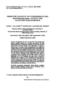
Differences in Anthocyanin Contents Total anthocyanin pigments in the disc and ray florets differed among ‘Argus’ and our color mutants (Figure 2). A quantitative comparison showed that the pinkish or purplish mutants, either in the disc or ray florets, accumulated more anthocyanin while white-flowered individuals had lower detectable amounts, as would be expected based on their colors. In the disc florets, total anthocyanin levels for the mutants ranged from 4 times lower to 6 times higher compared with ‘Argus’. In the ray florets, those relative levels were more evident, with a difference of up to 14 times more.
Sequence comparisons among ‘Argus’ and flower-color mutants
Total anthocyanin content in ray or disc florets from chrysanthemum color mutants. Figure 2.
Our visualized reverse transcriptions of mRNA indicated that the anthocyanin structural genes were expressed differently depending on tissue or mutant type (Figure 3). Degenerate primers designed from the conserved regions of other species were used to obtain chrysanthemum-specific cDNA
Phenotype and Transcript Variations in Chrysanthemum Color Mutants Sequence homologies of partial fragments from anthocyaninsynthesizing genes of chrysanthemum compared with other plant species. Partial cDNA sequence Homology Gene Common name (%) Length (bp) Accession no. Table 3.
CHI
F3’H
LDOX
Chrysanthemum Verbena Petunia Camellia Eustoma Saussurea Callistephus Lotus Soybean Chrysanthemum Osteospermum Gerbera Soybean Ipomoea Arabidopsis
500 650 654 660 651 654 654 636 645 1100 1235 1223 1229 1250 1226
Chrysanthemum Carrot Grape Eustoma Perilla Arabidopsis
500 942 948 936 948 930
AB234907 AF233637 DQ120521 AB078955 AF509335 Z67980 AJ548840 AF276302 DQ250711 DQ218417 AB061212 AY333419 NM_12088 1 AF184274 X75966 AB078959 AB003779 AK226417
67 67 72 69 84 84 61 64 74 71 62 64 63 76 74 73 74 71
fragments. The mRNA of CHS, CHI, F3H, F3’H, F3’5’H, DFR, and LDOX genes was amplified by 1.2, 0.5, 1.1, 1.1, 0.8, 1.2, and 0.5 kb, respectively, with an average of 843
Figure 3.
421
bp. Sequence analysis of those partial fragments indicated that the chrysanthemum color genes were sequentially and highly homologous to known sequences from other plants, with ranges of 61 to 84%, 62 to 74%, and 71 to 76% for CHI, F3’H, and LDOX, respectively (Table 3). Those sequences for LDOX and ANS were identically aligned, and we considered them to be the same as those explained previously (Marles et al., 2003; Turnbull et al., 2004). Expression Patterns for Anthocyanin Pathway Genes in Flower and Leaf Tissues
We compared expression patterns for anthocyanin structural genes (CHS, CHI, F3H, F3’H, F3’5’H, DFR, and LDOX) among the mutants and found that they varied depending on the gene and tissue used (Figure 3). Some, such as CHS and F3H, showed no difference in expression while CHI, F3’H, F3’5’H, DFR, and LDOX were either enhanced or repressed among the mutants. In addition, genes such as CHI, F3’H, and LDOX were not amplified (i.e., in Mutant 5, Mutant 4, and Mutants 4 and 5, respectively; Figure 3).
DISCUSSION Irradiation-induced changes in flower color and shape were diverse in this study: white, red-purple and yellowgreen disc florets; ivory to white, red-purple, and red ray florets; and variations in the lengths and widths of ray florets. When 50 Gy of gamma rays was applied as the mutagen, 17% of all individuals differed from the ‘Argus’ control in their flower color and shape (Park et al., 2007). a gamma ray irradiation has previously been proven to be useful for inducing color changes in chrysanthemum (Mandal et al., 2000).
Transcriptional pattern of genes involved in anthocyanin pathway in leaf and flower tissue of ‘Argus’ (control) and five color mutants.
422
Geung-Joo Lee et al.
Materials that were vegetatively propagated maintained their mutated colors and shapes over consecutive cuttings (Park et al., 2007). The anticipated bottleneck when using radiation, including gamma rays, ion beams, and X-rays, to induce mutants in ornamental species is a reversion of those chimeric traits to the original form when the original and mutated cells co-exist (Mandal et al., 2000; Sun et al., 2007). Interestingly, Mutants 2 and 5, where RHS indices for ray florets were the same as for ‘Argus’, had higher total anthocyanin contents than did the control. Therefore, this demonstrates that screening of color mutants that relies on either an RHS color chart or total anthocyanin content alone is not enough, but that both tools are necessary for clear evaluation. A suppression or deficiency in the LDOX gene (equivalent to ANS) is associated with a white-colored mutant from a naturally blue gentian plant (Nakatsuka et al., 2005; Turnbull et al., 2004). By comparison, we detected mRNA of LDOX in the leaves and flowers from Mutants 2 and 3, which had white ray florets and yellowish disc florets (Figure 3). However, Mutants 2, 3, and 4, in which yellowish disc florets were common, showed reduced transcripts in their leaves but enhanced mRNA in their flowers for encoding CHI, a regulation gene involved in an early step in the anthocyanin pathway (Mato et al., 2000). Floral pigments in a pink-colored Torenia mutant from the original blue plant exhibit extremely greater amounts of peonidin and cyanidin derivatives for red hues, which results from the strong expression of F3’H and DFR in the anthocyanin pathway (Miyazaki et al., 2006). Our Mutants 1 and 5, with higher total anthocyanin contents in the disc florets, had more mRNA for F3’H in the leaf tissue but not in the flower (Figure 3). Because more than one copy of the anthocyanin genes exists in many plant species, some transcripts of those copies might convey an erroneous association with flower coloration (Holton and Cornish, 1995; Jaakola et al., 2002). DFR is involved in the accumulation of all three major anthocyanins (cyanidin, pelargonidin, and delphinidin). Therefore, we presumed that the five mutants used here, which exhibited greater or lower amounts of those anthocyanins (Figure 2), had similar levels of the mRNA encoding for DFR (Figure 3) (see also Grotewold, 2006). Likewise, increased activity for F3’5’H is associated with the predominant synthesis of delphinidin derivatives for lilac to blue hues (Jeong et al., 2006; Seitz et al., 2007). Based on the limited blue-colored flowers from our mutants, such delphinidin derivatives are not likely to predominate. This is further supported by the consistent mRNA levels of F3’5’H among those color mutants (Figure 3). A red discoloration in our transgenic chrysanthemums is, however, is associated with co-expression of F3’H and F3’5’H, as found in Mutants 2 and 3 (Seo et al., 2007). Ionizing radiation, including from gamma rays, induces fragment deletions or insertions that eventually lead to changes in amino acids and a modification of leaf and stem pigmentations (Shikazono et al. 2003). In addition to the structural genes involved in the anthocyanin biosynthetic pathway, mutations in other regulatory genes or the involvement of transposable elements, e.g., MELSs (mobile ele-
J. Plant Biol. Vol. 51, No. 6, 2008
ment-like sequences) and DRs (direct repeats), might be associated with variations in flower coloration (Hoshino et al., 2001; Kim et al., 2007). For example, in yellow-flowered chrysanthemums, transcripts of a carotenoid cleavage dioxygenase (CCD) gene that accounts for white flowers are accumulated in extremely low amounts. Such a response is a case of less association by a structural gene involved in carotenoid biosynthesis (Ohmiya et al., 2006). Environmental stresses such as cold can cause pigmentation accompanied by anthocyanin accumulations in the petals or leaves of some species (Christie et al., 1994; Nakatsuka et al., 2005). However, in our study, variations in flower color are thought to have been caused by a mutation in a structural or regulatory gene involved in the anthocyanin biosynthesis pathway, rather than because of environmental factors. Our selected mutants were maintained under greenhouse conditions to avoid extremes that can cause light or temperature stresses. Therefore, all of our results demonstrate that the targeting flower color and shape of selected chrysanthemum plants are inherited from mutated origins. Among these mutants, transcript profiles indicated that various color features are associated with different mRNA levels for CHI, F3’H, F3’5’H, DFR, and LDOX -- all genes involved in anthocyanin synthesis. Therefore, we assume that a mutation in one or some pathway genes should stop or detour to the downstream pathway, eventually resulting in variations in flower color. We must still investigate the genomic structure of those genes to verify whether their expression patterns result from gain- or loss-of-function because of gamma ray mutagenesis. Although coloration can be re-directed in a few plant species by a transgenic technique that introduces some isolated genes (Shimada et al., 2001), a physical mutagen, e.g., gamma ray radiation, also is effective for acquiring a unique or complicated pigmentation of mutants with favorable morphologies. Such efforts can then meet the increasing demands for diverse color features in horticultural industries. ACKNOWLEDGEMENTS
We are grateful for research funding from the Biogreen 21 Program, Rural Development Administration (RDA), Korea (Code No. 20070301034033); and from the nuclear R&D Program, Ministry of Education, Science and Technology (MEST), Korea. Received June 30, 2008; accepted September 18, 2008.
LITERATURE CITED Ahloowalia BS, Maluszynski M (2001) Induced mutations - A new paradigm in plant breeding. Euphytica 118: 167-173 Broertjes C, Roest S, Bokelmann GS (1976) Mutation breeding of Chrysanthemum morifolium Ram. using in vivo and in vitro adventitious bud techniques. Euphytica 25: 11-19 Christie PJ, Alfenito MR, Walbot V (1994). Impact of low-temperature stress on general phenylpropanoid and anthocyanin path-
Phenotype and Transcript Variations in Chrysanthemum Color Mutants
ways: Enhancement of transcript abundance and anthocyanin pigmentation in maize seedlings. Planta 194: 541-549 Emmanuel E, Levy AA (2002) Tomato mutants as tools for functional genomics. Curr Opin Plant Biol 5: 112-117 Grotewold E (2006) The genetics and biochemistry of floral pigments. Annu Rev Plant Biol 57: 761-780 Holton TA, Cornish EC (1995) Genetics and biochemistry of anthocyanin biosynthesis. Plant Cell 7: 1071-1083 Hoshino A, Johzuka-Hisatomi Y, Iida S (2001) Gene duplication and mobile genetic elements in the morning glories. Gene 265: 1-10 Jaakola L, Maatta K, Pirttila AM, Torronen R, Karenlampi S, Hohtola A (2002) Expression of genes involved in anthocyanin biosynthesis in relation to anthocyanin, proanthocyanidin, and flavonol levels during bilberry fruit development. Plant Physiol 130: 729-739 Jain SM (2005) Major mutation-assisted plant breeding programs supported by FAO/IAEA. Plant Cell Tiss Org Cult 82: 113-123 Jeong ST, Goto-Yamamoto N, Hashizume K, Esaka M (2006) Expression of the flavonoid 3’-hydroxylase and flavonoid 3’5’hydroxylase genes and flavonoid composition in grape (Vitis vinifera). Plant Sci 170: 61-69 Kim BG, Kim JH, Min SY, Shin K-H, Kim JH, Kim HY, Ryu SN, Ahn J-H (2007) Anthocyanin content in rice is related to expression levels of anthocyanin biosynthetic genes. J Plant Biol 50: 156160 Kimler L, Mears J, Mabry TJ, Roesler H (1970) On the question of the mutual exclusiveness of betalains and anthocyanins. Taxon 19: 875-878 Levin I, Lalazar A, Bar M, Schaffer AA (2004) Non GMO fruit factories: Strategies for modulating metabolic pathways in the tomato fruit. Ind Crop Prod 20: 29-36 Maluszynski M, Ahloowalia BS, Sigurbjornsson B (1995) Application of in vivo and in vitro mutation techniques for crop improvement. Euphytica 85: 303-315 Mandal AKA, Chakrabarty D, Datta SK (2000). In vitro isolation of solid novel flower color mutants from induced chimeric ray florets of chrysanthemum. Euphytica 114: 9-12 Marles MAS, Ray H, Gruber MY (2003) New perspectives on proanthocyanidin biochemistry and molecular regulation. Phytochemistry 64: 367-383 Martin T, Oswald O, Graham IA (2002) Arabidopsis seedling growth, storage lipid mobilization, and photosynthetic gene expression are regulated by carbon: Nitrogen availability. Plant Physiol 128: 472-481 Mato M, Onozaki T, Ozeki Y, Higeta D, Itoh Y, Yoshimoto Y, Ikeda H, Yoshida H, Shibata M (2000) Flavonoid biosynthesis in white-flowered Sim carnations (Dianthus caryophyllus). Sci Hort 84: 333-347 Miyazaki K, Suzuki K, Iwaki K, Kusumi T, Abe T, Yoshida S, Fukui H (2006) Flower pigment mutations induced by heavy ion beam irradiation in an interspecific hybrid of Torenia. Plant Biotech
423
23: 163-167 Nakatsuka T, Nishihara M, Mishiba K, Yamamura S (2005) Two different mutations are involved in the formation of white-flowered gentian plants. Plant Sci 169: 949-958 Nissim-Levi A, Ovadiar R, Forer I, Oren-Shamir M (2007) Increased anthocyanin accumulation in ornamental plants due to magnesium treatment. J Hortic Sci Biotech 82: 481-487 Okamura M, Yasuno N, Ohtsuka M, Tanaka A, Shikazono N, Hase Y (2003) Wide variety of flower-color and shape mutants regenerated from leaf cultures irradiated with ion beams. Nucl Instrum Methods Phys Res Sect B 206: 574-578 Ohmiya A, Kishimoto S, Aida R, Yoshioka S, Sumitomo K (2006) Carotenoid cleavage dioxygenase (CmCCD4a) contributes to white color formation in chrysanthemum petals. Plant Physiol 142: 1193-1201 Park IS, Lee GJ, Kim DS, Chung SJ, Kim JB, Song HS, Goo DH, Kang SY (2007) Mutation breeding of a spray chrysanthemum ‘Argus’ by gamma-ray irradiation and tissue culture. Flower Res J 15: 52-57 Predieri S (2001) Mutation induction and tissue culture in improving fruits. Plant Cell Tiss Org Cult 64: 185-210 Sanjay J (2007). Mutagenesis: Generation and evaluation of induced mutations. Adv Bot Res 45: 417-434 Schum A (2003) Mutation breeding in ornamentals: An efficient breeding method? Acta Hort 612: 47-58 Seitz C, Vitten M, Steinbach P, Hartl S, Hirsche J, Rathje W, Treutter D, Forkmann G (2007) Redirection of anthocyanin synthesis in Osteospermum hybrida by a two-enzyme manipulation strategy. Phytochemistry 68: 824-833 Seo J, Kim SW, Kim J, Cha HW, Liu JR (2007) Co-expression of flavonoid 3’, 5’-hydroxylase and flavonoid 3’-hydroxylase accelerates decolorization in transgenic chrysanthemum petals. J Plant Biol 50: 626-631 Shikazono N, Yokota Y, Kitamura S, Suzuki C, Watanabe H, Tano S, Tanaka A (2003) Mutation rate and novel tt mutants of Arabidopsis thaliana induced by carbon ions. Genetics 163: 14491455 Shimada Y, Ohbayashi M, Nakano-Shimada R, Okinaka Y, Kiyokawa S, Kikuchi Y (2001) Genetic engineering of the anthocyanin biosynthetic pathway with flavonoid-3’,5’-hydroxylase: Specific switching of the pathway in petunia. Plant Cell Rep 20: 456-462 Sun M, Li P, Zhang Q-X (2007) Flower color and florescence mutants obtained using electron beam irradiation of chrysanthemum buds. Acta Hort 760: 667-672 Turnbull JJ, Nakajima J-I, Welford RWD, Yamazaki M, Saito K, Schofield CJ (2004) Mechanistic studies on three 2-oxoglutarate-dependent oxygenases of flavonoid biosynthesis. J Biol Chem 279: 1206-1216 Zalewska M, Jerzy M (1997) Mutation spectrum in Dendranthema grandiflora Tzvelev after in vivo and in vitro regeneration of plants from irradiated leaves. Acta Hort 447: 615-618