Pezeshki et al. Veterinary Research 2011, 42:15 http://www.veterinaryresearch.org/content/42/1/15
RESEARCH
VETERINARY RESEARCH Open Access
Variation of inflammatory dynamics and mediators in primiparous cows after intramammary challenge with Escherichia coli Adel Pezeshki1, Philippe Stordeur2, Hugues Wallemacq3, Frédéric Schynts2, Mieke Stevens1, Philippe Boutet3, Luc J Peelman1, Bart De Spiegeleer1, Luc Duchateau1, Fabrice Bureau3, Christian Burvenich1*
Abstract The objective of the current study was to investigate (i) the outcome of experimentally induced Escherichia coli mastitis in primiparous cows during early lactation in relation with production of eicosanoids and inflammatory indicators, and (ii) the validity of thermography to evaluate temperature changes on udder skin surface after experimentally induced E. coli mastitis. Nine primiparous Holstein Friesian cows were inoculated 24 ± 6 days (d) after parturition in both left quarters with E. coli P4 serotype O32:H37. Blood and milk samples were collected before and after challenge with E. coli. The infrared images were taken from the caudal view of the udder following challenge with E. coli. No relationship was detected between severity of mastitis and changes of thromboxane B2 (TXB2), leukotriene B4 (LTB4) and lipoxin A4 (LXA4). However, prostaglandin E2 (PGE2) was related to systemic disease severity during E. coli mastitis. Moreover, reduced somatic cell count (SCC), fewer circulating basophils, increased concentration of tumor necrosis factor-a (TNF-a) and higher milk sodium and lower milk potassium concentrations were related to systemic disease severity. The thermal camera was capable of detecting 2-3°C temperature changes on udder skin surface of cows inoculated with E. coli. Peak of udder skin temperature occurred after peak of rectal temperature and appearance of local signs of induced E. coli mastitis. Although infrared thermography was a successful method for detecting the changes in udder skin surface temperature following intramammary challenge with E. coli, it did not show to be a promising tool for early detection of mastitis. Introduction The severity of coliform mastitis is of much more concern than its incidence [1]. Pathogen, cow and environment are three interdependent factors which influence the mastitis susceptibility [1]. From the various bacterial virulence factors studied during Escherichia coli mastitis [2], only a few have been found to play an important role in the outcome of the disease. It has been accepted that the type of E. coli strain is not the main factor in classification of severity. Preventive treatments which are efficient against contagious mastitis have been shown to be inefficient in the control of E. coli mastitis [3]. The severity of bovine E. coli mastitis is mainly * Correspondence:
[email protected] 1 Department of Comparative Physiology and Biometrics, Laboratory of Genetics, Drug Quality and Registration Group, Ghent University, B-9000 Ghent, Belgium Full list of author information is available at the end of the article
determined by cow factors rather than by the pathogenecity of the invading pathogen and management [1]. It is known that the growth of E. coli in the udder cistern is specially related with the period of lactation and parity of cows. E. coli mastitis with severe clinical symptoms is more frequently observed around calving and during early lactation in dairy cows, whereas symptoms are mild to moderate during mid and late lactation. Because of hormonal, metabolic and nutritional alterations associated with pregnancy, immune system is compromised around calving (reviewed by Pezeshki et al. [4]). Cow parity is another important physiological factor that influences the severity of clinical coliform mastitis [5,6]. Clinical severe cases of coliform mastitis are mostly seen among multiparous cows rather than primiparous cows during early lactation. To our best knowledge the inflammatory status of primiparous cows
© 2011 Pezeshki et al; licensee BioMed Central Ltd. This is an Open Access article distributed under the terms of the Creative Commons Attribution License (http://creativecommons.org/licenses/by/2.0), which permits unrestricted use, distribution, and reproduction in any medium, provided the original work is properly cited.
Pezeshki et al. Veterinary Research 2011, 42:15 http://www.veterinaryresearch.org/content/42/1/15
ranking based on severity after intramammary infection of E. coli is poorly understood during early lactation. Physiological factors have been mainly studied in multiparous cows ranging from second lactation to sixth lactation [6-10]. Thromboxanes (TX), prostaglandins (PG), leukotriens (LT) and lipoxines (LX) which are the enzymatically generated products of cyclooxygenases (COX) and lipoxygenases are generated during inflammation and act as major pro-inflammatory mediators. There is evidence that TXB2 and PGE2 might have an important role in severity of E. coli mastitis in primiparous cows during early lactation [11]. Although not significantly, nonsteroidal anti-inflammatory drug treatment reduced the concentration of TXB2 and PGE2 in milk, while it had no effect on chemotactic inflammatory mediators and early innate immune molecules [11]. LTB4 is known to induce infiltration and sequestration of the neutrophils in the bovine mammary gland, whereas LXA4 serve as neutrophil “stop signals” in inflammation [12]. As excessive recruitment of neutrophils can exacerbate the inflammatory process via releasing detrimental intracellular products for tissues, persistence of LTB4 production or its ratio with LXA4 could be of particular importance in severity of the disease. We hypothesize that the magnitude of eicosanoids response plays an important role in the susceptibility to E. coli mastitis during early lactation in primiparous cows and can influence the resolution of this disease. An automated method for early detection of coliform mastitis would prevent its progression and likely reduce the costs brought about by repeated treatments. Inflammation of the udder or mastitis results in increase of the temperature. The radiated heat emitted by the skin, reflecting subcutaneous circulation and tissue metabolism could be measured using infrared thermography. Although the thermography technique has been tested for early detection of experimentally induced acute clinical mastitis using E. coli endotoxin [13] or subclinical mastitis [14], the technique has not yet been validated for evaluating the temperature changes of the udder’s skin surface after challenge with E. coli. The objectives of the current study were to investigate (i) the outcome of experimental E. coli mastitis in primiparous cows during early lactation in relation with production of eicosanoids and inflammatory indicators in blood and milk and to determine (ii) whether thermography is a valid method for evaluating the temperature changes of the affected udder.
Page 2 of 10
from 511 to 615 kg were used. Cows arrived at the experimental dairy farm (Center of Rural EconomyCER-Marloie, Belgium) 15 days before expected calving date (actual day was 22 ± 6 days (d)). The age of cows was between 23 to 34 months at calving. Cows were milked twice a day at 06:30 and 18:30 using a quarter milking device [8]. After parturition, cows were fed a daily ration according to the National Research Council system (NRC, 2001). The forages were delivered in two equal meals at 07:00 and 19:00 while the concentrate was fed in several times, according to milk production. The composition and ingredient of the lactation diet used during the experiment is reported in Table 1. Water was provided ad libitum. Experimental infections were approved by the Ethical Committee of the CER (No. META-DVA-1). After an adaptation period, animals were inoculated intramammarily with the E. coli suspension at 24 ± 6 days in milk. Bacteriological examination of the milk was carried out according to International Dairy Federation (IDF, 1981) recommendations. The cows were clinically healthy and free of major mastitis pathogens through three consecutive bacteriologically negative examinations, with a quarter foremilk somatic cell count (SCC) below 200 000 cells/mL. Daily quarter milk production (QMP) was recorded at d -7, -4, -3, -2, -1, 0, +1, +2, +3 and +6, relative to day of inoculation. On average, the concentration of preinfection b-hydroxybutyrate (BHBA), glucose and non-esterified fatty acids (NEFA) was 1.09 ± 0.42, 3.70 ± 0.42 and 0.33 ± 0.17 mmol/L, respectively. Bacteria and intramammary inoculation
The strain used in this study, to reproduce clinical signs of mastitis, was the E. coli strain P4, serotype O32:H37.
Table 1 Ingredient and nutrient compositions of the diet administered during the experimental period Ingredient (Kg/d) Grass silage
18.50
Barley
1.00
Beet pulp
2.00
Commercial concentrate 1 Nutrient composition (%DM basis)
8.00
DM (Kg/d)
17.18
NEL2 (Mcal/kg)
1.55
CP
15.03
NDF
48.52
NFC3
25.66
1
Materials and methods Experimental animals and study facilities
In total, 9 primiparous Holstein Friesian cows in early lactation ranging in weight (one week after calving)
Contained 18% CP, 3% fat, 13% cellulose, 8000 UI/kg Vitamin A, 1200 UI/kg Vitamin D3, 15 mg/kg Vitamin E, 9.6 mg/kg CU2SO4. 2 Calculated according to NRC (2001). NEL was estimated based on a cows weighing 650 kg and average 21.8 kg dry matter intake per day. 3 NFC calculated as 100 - [(NDF - NDFCP) + CP + ether extract + ash] (NRC, 2001).
Pezeshki et al. Veterinary Research 2011, 42:15 http://www.veterinaryresearch.org/content/42/1/15
The strain was maintained in a stock on lyophilization medium at -20°C. A stock of bacteria was sub-cultured in brain-heart infusion broth (CM225; Oxoid, Nepean, ON, Canada) during three consecutive days at 37°C. Subsequently, subcultures were washed 3 times with pyrogen-free PBS and resuspended in PBS [11]. Immediately before inoculation, a final concentration of 1 × 10⁴ cfu/mL bacterial suspension in pyrogen-free PBS was prepared. On d0 (the day of challenge), 30 min after morning milking, the bacterial suspension was inoculated aseptically into the left quarter’s teat cistern of all 9 cows by means of a sterile, pyrogen-free teat cannula (length, 7 cm, I.D., 2 mm; Me.Ve.Mat, Deinze, Belgium). The total volume of inoculation was 10 mL, consisting of 1 mL of bacterial suspension and 9 mL of pyrogenfree saline solution (NaCl 0.9%; Baxter N.V., Lessines, Belgium) per quarter [5,15]. Sampling procedure
Blood samples were collected in the morning on d -2, +1, +2, +3, +6 relative to the day of challenge. On the day of challenge, blood samples were collected at -0.5, +0.5, +3, +6, +9, +12, +15, +18, +21 hours (h) relative to the time of challenge. Blood samples (3 × 9 mL) were drawn aseptically from the external jugular vein by venipuncture into 3 vacutainer tubes (BD Biosciences, USA) containing Lithium-Heparin (10 IU/L), EDTA or no anticoagulant. Plasma was separated from the blood samples in tubes containing Lithium-Heparin and stored at -20°C until analysis of metabolites. Serum was harvested from blood samples in dry tubes and stored at -20°C for later analysis of haptoglobin. The blood samples in tubes containing EDTA were used for the determination of hematologic parameters. Quarter milk samples were taken on d -7, -4, -2, +1, +2, +3, +6 relative to the day of challenge. On the day of challenge, milk samples were collected at -0.5, +0.5, +3, +6, +9, +12, +15, +18, +21 h relative to the time of challenge. Equal volumes of milk samples were collected for each specific analysis within first 12 h after the challenge and at other time points mentioned above. Foremilk (5 mL) was aseptically collected for quantification of E. coli (cfu/mL), SCC and milk composition and stored at 4°C until analysis. Morning milk samples (20 mL) were collected for determination of eicosanoids and stored at -70°C prior to analysis. The collected morning milk samples (5 mL) for quantification of ions were stored at -20°C before the analysis begins. Clinical examinations
The cows were examined and scored generally for rectal temperature, skin turgor, rumen motility, general attitude [5] and specifically for udder temperature, quarter swelling, pain, teat relaxation, milk leakage and milk
Page 3 of 10
appearance [11] until six days after the intramammary inoculation of E. coli. Infrared thermographic imaging
The infrared images were taken from the caudal view of the experimental and control quarters at 0, 3, 6, 9, 10, 11, 12, 13, 14, 15 PIH. To ensure integrity of the thermal data and reduce the effect of environmental factors, some considerations were standardized in the present study: (i) all infrared images were captured within the stall and (ii) at the same distance (1.5-2 m) from the subject, when (iii) cows were in rest with no exercise. (iv) Circadian rhythm effects were controlled by scanning the animals at the same time of the day. Moreover, any extraneous debris and foreign materials like manure, straw and moisture were removed from the body surface to have clear infrared images from the skin of the udder. A hand-held portable infrared camera (ThermaCAM® E2, FLIR SYSTEMS) with thermal sensitivity of 0.12°C was used to take images of all animals. The camera was calibrated to ambient temperature and absorptive conditions on each scanning time. An emissivity value of 0.93 was set on the camera before performing the scanning. Analytical methods
The concentration of milk eicosanoids was determined with a commercially available competitive ELISA kit (Neogen, Lexington, KY, USA). Briefly, quarter milk samples were filtered using a 70-μm cell strainer (Becton Dickinson; Erembodegem, Belgium) to discard cell clusters. Milk sample was diluted with distilled water (50:50 v/v), and 1 mL of the mixture was loaded on a conditioned 100-mg C18 column (Varian, St.-Katelijne-Waver, Belgium). After washing the column subsequently with distilled water, methanol: distilled water (30:70 v/v), and hexane, the column was centrifuged at 3200 × g for 3 min to remove any trace of hexane. Finally, eicosanoids were eluted from the C18 column with 1 mL of methanol. The collected elute was dried under a stream of nitrogen. Dried samples were reconstituted in an appropriate volume of assay buffer and manufacturer’s instructions were followed for measuring the concentration of eicosanoids. The number of E. coli (cfu/mL) in milk was determined by plating out the prepared dilutions in PBS on drigalski agar (Biokar Diagnostic, Beauvois, France) and counting the colonies after 24 h of incubation at 37°C. Milk sodium, chlorine and potassium were measured using an ion-selective electrode analyzer (Ilyte; Instrumentation Laboratories, Milan, Italy). Milk SCC was determined using a fluoropto electronic method (Fossomatic 400 cell counter; Foss Electrics, HillerØ, Denmark). Milk fat, protein and lactose were determined using mid-infraredphotospectrometry (MilkoScan 4000; Foss Electrics,
Pezeshki et al. Veterinary Research 2011, 42:15 http://www.veterinaryresearch.org/content/42/1/15
HillerØ, Denmark). Total blood leukocyte count was determined using an electronic particle counter (Coulter Counter Z2; Coulter Electronics Ltd., Luton, UK). Differential blood leukocyte count was performed on blood smears using bright field microscopy as previously described [16]. Plasma tumor necrosis factor-a (TNF-a) and Interleukin-1b (IL-1b) were quantified with ELISA assays as previously described [17]. Mouse anti-recombinant bovine TNF-a and mouse anti-ovine IL-1b antibody were commercially available (Serotec Ltd., Oxford, UK). Serum was assayed for haptoglobin using the hemoglobin-binding assay method. Blood metabolites were analyzed at 37°C using a clinical auto-analyzer (ILAB 600, Instrumentation Laboratory, Lexington, MA, USA). Infrared images
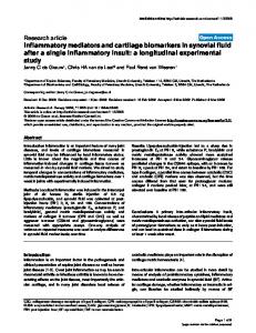
The output of the infrared camera was un-calibrated digitized images with pixel points. Pixel points were analyzed with appropriate computer software (FLIR QuickReport 1.2) to generate mean temperature of the skin surface. The mean temperature variation of four different areas between the groove of the udder and the hind leg and median suspensory ligament on caudal udder skin surface was examined. The areas were selected above the infected and control teats and centered according to the teats. The mean temperature of rectangles of 25 × 25 pixels drawn by the help of the camera software was measured (Figure 1). The two areas just above the teats showed the smallest variation in the temperature of the udder (Figure 1). An example of the
A
Page 4 of 10
infrared images at 3 and 12 PIH is shown in Figure 1A and Figure 1B, respectively. Statistical analysis
The main objective was to study the disease severity in relation with production of different inflammatory mediators/indicators and clinical parameters. Disease severity was defined as the percentage decrease of QMP in uninfected quarters at 48 PIH compared to QMP in the same quarters at -24 PIH [8]. To investigate the relationship of various parameters analyzed in milk and blood and disease severity, a mixed model was used with cow as random effect, and time, disease severity and their interaction as fixed effects. To explore the relationship between disease severity and systemic and local inflammatory responses, the Kendall’s correlation coefficient between categorical parameters and disease severity was calculated. To study the kinetics for temperature of udder skin in infected and uninfected quarters, a mixed model was used, with temperature as response variable, cow as random effect, and PIH, udder point and their interaction as categorical fixed effects. To compare the measurements at different time points with time zero, the mixed model was used, with measured parameters as response variable, cow as random effect and PIH as categorical fixed effect. For milk production, measurements at all times were compared with time -48. Significance level for multiple comparisons was adjusted by Dunnett’s procedure. A P value of