Hindawi Publishing Corporation Advances in Radiology Volume 2014, Article ID 969728, 6 pages http://dx.doi.org/10.1155/2014/969728
Research Article Variations in the Branching Pattern of the Aortic Arch Detected with Computerized Tomography Angiography Pasaoglu Lale, Ugur Toprak, Gökhan YagJz, Tunca Kaya, and SadJk Ahmet UyanJk Department of Radiology, Ankara Numune Training and Research Hospital, Ulku Mahallesi Talatpasa Bulvari No. 5, Altindag, 06100 Ankara, Turkey Correspondence should be addressed to G¨okhan Yagız;
[email protected] Received 8 March 2014; Accepted 4 June 2014; Published 29 June 2014 Academic Editor: Letterio S. Politi Copyright © 2014 Pasaoglu Lale et al. This is an open access article distributed under the Creative Commons Attribution License, which permits unrestricted use, distribution, and reproduction in any medium, provided the original work is properly cited. Objectives. The aim was to determine the prevalence of aortic arch variations in 881 patients who underwent neck CT angiography for various reasons. Material and Methods. 881 patients were included in the study who had undergone neck CT angiography between 2010 and 2013. Results. Of 881 patients, 770 (87.4%) patients with classic branching pattern of the aortic arch (AA) were observed. Variations in branching pattern were seen in 111 (12.6%) patients. The most common variation was the origination of the left common carotid artery (LCCA) from the brachiocephalic trunk (BCT). This pattern was observed in 64 (7.2%) cases. In 25 (2.8%) cases, the left vertebral artery (LVA) originated directly from the AA between the origin of the LCCA and left subclavian artery (SCA). 17 (1.9%) cases had aberrant right subclavian artery. Three (0.3%) cases showed right aortic arch. Two cases had right aortic arch with aberrant left subclavian artery. Conclusions. Variations in the branching pattern of the AA are not rare. Head and neck surgeons and interventional radiologists should be aware of aortic arch variations. CTA is a reliable imaging method for demonstrating anatomical features and variations of the AA.
1. Introduction In the classical anatomical configuration, the aortic arch (AA) is left sided and the most common branching pattern of the AA comprises of three great vessels; first the brachiocephalic trunk (BCT), then the left common carotid artery (LCCA), and finally the left subclavian artery (SCA) from right to left. The BCT branches into right SCA and right common carotid artery (RCCA). This branching pattern occurs in 64.9–94.3% of the cases and it is described as “normal” [1–6]. Variations in the branching pattern of the AA range from differences in the origins of different branches to the number of branches [2, 4]. Development of the aorta takes place during the third week of gestation [5]. Six pairs of aortic arches, the so-called branchial arch arteries, develop between the ventral and dorsal aortae. These variations of the AA can be explained by persistence of segments of the aortic arches that normally regress or disappearance of segments that normally remain, or both [1, 5, 6].
Anatomical variations in the branching pattern of AA are significant for diagnostic, surgical, and interventional procedures of the thorax and neck. The purpose of this study is to review the computed tomography angiography (CTA) appearance of anatomical variations in branching pattern and position alterations of the AA and to determine the prevalence in 881 patients.
2. Material and Methods The approval for this retrospective study was obtained from the institutional review board. 881 consecutive cases that underwent carotid CTA examination for different purposes were examined retrospectively. These CTA examinations belonged to 527 (%59.8) male and 354 (%40.2) female patients and they were performed during a 3-year period, from 2010 to 2013. The age range was 19–93 years. Mean age of all the cases was 62 (19–93), mean age of males was 59 (21–93), and mean
2
Advances in Radiology
age of females was 62 (19–91). There was not any significant age difference between male and female patients (𝑃 > 0.05). 2.1. CTA Protocol. CTA examinations were performed using a 16-detector and 64-detector scanner (Aquilion 16, Aquilion 64, Toshiba Medical Systems, Otawara, Japan). In our standard CTA protocol for brain and neck examinations, a scan area from the aortic arch to the vertex level in supine position was adopted as a field of view. During examination, an 18–20 gauge angiocath in the antecubital vein was used to inject 90–120 mL of nonionic iodinated contrast media using bolus-tracking method with an automatic injector at a rate of 5 mL/sec and 40 mL of saline solution at a rate of 4 mL/sec, respectively. The region of interest was positioned at the aortic arch, and the threshold was set to 130 HU. When the threshold was surpassed, helical scanning was automatically initiated. Scan parameters were 120 kV, 300 mA, and 420 msec rotation time with a slice thickness of 1 mm and increments of 0.5 mm, using a detector collimation of 16 × 1 mm.
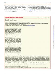
Figure 1: Three-dimensional volume rendered image shows brachiocephalic trunk (white arrow head), left common carotid artery (white arrow), and left subclavian artery (white star).
2.2. Image Analysis. The obtained axial images from CTA examinations were transferred to the work station (Vitrea 2, Vital Images Inc., Minnetonka, MN, US) for analysis. Axial source data images, multiplanar reformat images, and postprocessed (maximum intensity projection and 3D volume rendered) images were evaluated. Image analysis was performed by a radiologist experienced in CTA and all the examinations were reviewed by the same radiologist. 2.3. Statistical Analysis. Data analysis was carried out using commercially available software (statistical package for social sciences, SPSS Inc., Chicago, Illinois, US). Mean minimum and maximum values of participants’ demographic data were performed.
3. Results Eight hundred and eighty-one cases (527 males and 354 females, age range 19–93 years) were included in this study. In 770 (87.4%) cases (516 males and 354 females) the classic branching pattern of the AA was observed. This pattern which is described as “normal” in the literature gives rise to three branches: BCT, CCA, and left SCA from right to left (Figure 1). 111 (12.6%) cases (65 males, 46 females) showed variations in branching pattern. The most common variation was origination of the LCCA from the BCT (arch with two branches, BCT with LCCA, and left SCA). This pattern was observed in 64 (7.2%) cases (43 males, and 21 females) (Figure 2). In 25 (2.8%) (14 males, 11 females) cases, the left vertebral artery (LVA) originated directly from the AA between the origin of the LCCA and left SCA, providing an AA pattern with four branches (BCT, LCCA, LVA, and left SCA from right to left) (Figure 3). 17 (1.9%) (6 males, 11 females) cases had aberrant right subclavian artery (RCCA, LCCA, left SCA, and right SCA). The right SCA was the last branch of the AA coursing to the right behind the esophagus (Figure 4).
Figure 2: Three-dimensional volume rendered image demonstrates the left common carotid artery (white arrow head) originating from brachiocephalic trunk (white star) (white arrow head, left subclavian artery).
Figure 3: Three-dimensional volume rendered image shows the left vertebral artery (black arrow) originating from aortic arch between the left common carotid artery (white arrow) and left subclavian artery (black cross) (white star, brachiocephalic trunk).
Advances in Radiology
3
Figure 4: Axial maximum intensity projection image demonstrates the aberrant right subclavian artery (white arrow) originating from the last branch of the aortic arch and coursing to the right behind the esophagus (black star, right common carotid artery; black cross, left common carotid artery; white arrow head, left subclavian artery).
Figure 6: Three-dimensional volume rendered image demonstrates the right aortic arch with mirror-image type (black arrow, right subclavian artery; white arrow, right common carotid artery; white star, left brachiocephalic trunk).
Figure 5: Three-dimensional volume rendered image shows the right aortic arch with aberrant left subclavian artery (black arrow) (white star, right common carotid artery; white arrow, right subclavian artery; white arrow head, left common carotid artery).
Figure 7: Axial image of the same patient with mirror-image type right aortic arch (white star, left brachiocephalic trunk; white arrow, right common carotid artery; black cross, right subclavian artery).
3 (1 male, 2 females) cases showed right aortic arch. Two cases had right aortic arch with aberrant left subclavian artery. The vessels originated in the following order: LCCA, RCCA, right SCA, and left SCA (Figure 5). One case with right aortic arch had mirror image type (left BCT, RCCA, and right SCA) (Figures 6 and 7). In one female patient (0.1%), the right vertebral artery (RVA) originated from the AA as terminal branch. It passed behind the esophagus and trachea and entered transvers foramina at the level of the seventh cervical vertebra. BCT with LCCA was also present in the same patient. The order of the branches was BCT with LCCA, left SCA, and RVA (Figures 8 and 9). Double aortic arch was observed in one male patient (0.1%). The ascending aorta divided into two arches that passed to either side of the esophagus and trachea and reunited to form the descending aorta (Figure 10).
Figure 8: Axial maximum intensity projection image demonstrates the right vertebral artery (white arrow) originating from the aortic arch as the last branch.
4
Figure 9: Three-dimensional volume rendered image of the same patient with right vertebral artery originating from aortic arch with left common carotid artery (white arrow) originating from brachiocephalic trunk (black arrow) (black star, right subclavian artery; white star, left subclavian artery).
Figure 10: Three-dimensional volume rendered image demonstrates the double aortic arch.
4. Discussion In the present study the normal three-branch pattern was observed in 87.4% of the cases; however, in 24.9% cases, the AA showed variations from usual branching pattern. The normal three-branch pattern of the AA is found with an incidence of 64.9–94.3% according to the literature [1–6]. Our relative incidence of 87.4% lies in this wide range. The most common variation of the AA with two branches (BCT with LCCA and left SCA) is found with an incidence of 10–22% in the literature [1, 7–11]. This type is also called “bovine aortic arch” [7, 12, 13]. However, it is a misnomer because it does not in fact resemble the AA of the cattle which has only one branch that branches into right subclavian artery, a common truncus for common carotid arteries and left subclavian artery [7]. Clinical symptoms related to this variation have been reported and attributed to the widening of the mediastinum [14]. Developmentally, the two-branch pattern of the AA may be explained as follows: aortic sac normally bifurcates into right and left limbs. Left limb of the aortic sac forms the part of arch that intervenes between the
Advances in Radiology origins of BCT and LCCA. If the aortic sac fails to bifurcate, BCT and LCCA will connect to aortic sac directly resulting in bifurcated trunk or common trunk giving origin to LCCA [6, 11]. Origination of the left vertebral artery from the aortic arch is not uncommon and the reported prevalence is between 2.4 and 8% [8, 15]. The most frequent location is between the LCCA and left SCA [5]. Occasionally, the left vertebral artery is the last branch of the AA. A case with anomalous origin of both vertebral arteries as additional branches of the AA distal to the left SCA has been reported [16]. In the current study, the left vertebral artery originated directly from AA between the LCCA and left SCA was observed in 2.8% of the cases. Normally the first part of vertebral artery develops from the dorsal ramus of the seventh intersegmental artery. In cases where vertebral artery arises as a branch from AA, embryologically it is explained that this is due to the fact that vertebral artery develops from the persistent sixth cervical intersegmental artery and segment of dorsal aorta fails to disappear, so blood flows through these persisting routes [6]. It is hypothesized that anomalous origins of the vertebral arteries lead to altered hemodynamics and predispose the patient to the formation of intracranial aneurysms. Satti et al. claim that patients with such anomalies should therefore be screened for coexisting aneurysms [17]. However, within the current literature, there is no conclusive evidence to suggest that anomalous origin of the vertebral arteries predisposes an individual to cerebrovascular disorders [18]. Among our cases with left vertebral artery originating from the aortic arch, we observed only one case with aneurysm of the clinoid segment of the right internal carotid artery. Right aortic arch is an uncommon anatomical anomaly that occurs in