MBoC | ARTICLE
Dual function of the NDR-kinase Dbf2 in the regulation of the F-BAR protein Hof1 during cytokinesis Franz Meitingera, Saravanan Palania, Birgit Hubb, and Gislene Pereiraa a
Molecular Biology of Centrosomes and Cilia Unit, DKFZ-ZMBH Alliance, and bDepartment of Tumor Virology, German Cancer Research Center, 69120 Heidelberg, Germany
ABSTRACT The conserved NDR-kinase Dbf2 plays a critical role in cytokinesis in budding yeast. Among its cytokinesis-related substrates is the F-BAR protein Hof1. Hof1 colocalizes at the cell division site with the septin complex and, as mitotic exit progresses, moves to the actomyosin ring (AMR). Neither the function of Hof1 at the septin complex nor the mechanism by which Hof1 supports AMR constriction is understood. Here we establish that Dbf2 has a dual function in Hof1 regulation. First, we show that the coiled-coil region, which is adjacent to the conserved F-BAR domain, is required for the binding of Hof1 to septins. The Dbf2-dependent phosphorylation of Hof1 at a single serine residue (serine 313) in this region diminishes the recruitment of Hof1 to septins both in vitro and in vivo. Genetic and functional analysis indicates that the binding of Hof1 to septins is important for septin rearrangement and integrity during cytokinesis. Furthermore, Dbf2 phosphorylation of Hof1 at serines 533 and 563 promotes AMR constriction most likely by inhibiting the SH3-domain–dependent interactions of Hof1. Thus our data show that Dbf2 coordinates septin and AMR functions during cytokinesis through the regulation/control of Hof1.
Monitoring Editor Doug Kellogg University of California, Santa Cruz Received: Aug 20, 2012 Revised: Dec 13, 2012 Accepted: Jan 2, 2013
INTRODUCTION During cytokinesis one cell physically divides into two daughter cells. As such, cytokinesis must be tightly coordinated with cell cycle progression to ensure proper inheritance of chromosomes and cell organelles (Balasubramanian et al., 2004; Barr and Gruneberg, 2007). In yeast and animal cells, cytokinesis is driven by a contractile actomyosin ring (AMR), which drives membrane ingression, thereby closing the cell division site. In yeast, AMR contraction is accompanied by the formation of a septum, the future cell wall that covers the cell division site. AMR contraction and septum formation are interdependent and tightly coordinated
This article was published online ahead of print in MBoC in Press (http://www .molbiolcell.org/cgi/doi/10.1091/mbc.E12-08-0608) on February 27, 2013. Address correspondence to: Gislene Pereira (
[email protected]). Abbreviations used: 5-FOA, 5-fluoroorotic acid; AMR, actomyosin ring; CC, coiled coil; Cdk1, cyclin-dependent kinase 1; F-BAR, Fes/CIP4 homology Bin/Amphiphysin/ Rvsp; FCH, Fes/CIP4 homology; GST, glutathione S-transferase; HA, hemagglutinin; MEN, mitotic exit network; PP2A, protein phosphatase 2A; RLS, ring localization domain; SID, septin interaction domain; TAP, tandem affinity purification. © 2013 Meitinger et al. This article is distributed by The American Society for Cell Biology under license from the author(s). Two months after publication it is available to the public under an Attribution–Noncommercial–Share Alike 3.0 Unported Creative Commons License (http://creativecommons.org/licenses/by-nc-sa/3.0). “ASCB®,” “The American Society for Cell Biology®,” and “Molecular Biology of the Cell®” are registered trademarks of The American Society of Cell Biology.
1290 | F. Meitinger et al.
processes (Schmidt et al., 2002). Cyclin-dependent kinase 1 (Cdk1), when complexed with cyclins, is a major regulator of cell cycle progression and negatively regulates cytokinesis (Holt et al., 2009; Meitinger et al., 2012; Palani et al., 2012). In previous studies, other mitotic kinases have been reported to execute essential roles in the coordination of cytokinesis with the cell cycle progression. Among them are the conserved family of NDR-related kinases, which includes LATS1 in human cells and Dbf2 in budding yeast (Frenz et al., 2000; Vallen et al., 2000; Yang et al., 2004; Yoshida and Toh-e, 2001). Dbf2 is part of a signaling cascade, known as the mitotic exit network (MEN), that activates the mitotic Cdk1-counteracting phosphatase Cdc14 in late anaphase (Bardin and Amon, 2001; Meitinger et al., 2012). The Dbf2-dependent activation of Cdc14 drives cells out of mitosis by reverting the impact of Cdk1 phosphorylation and down-regulating its activity. Of interest, Dbf2 appears at the cell division site after Cdk1 down-regulation and is directly involved in AMR contraction and septum formation (Frenz et al., 2000). Although the mitotic exit function of Dbf2 does not seem to be conserved in higher eukaryotes, LATS1, like Dbf2, has been reported to drive AMR contraction in human cells, indicating functional conservation of the prominent role for this kinase in AMR control in yeast (Meitinger et al., 2011; Yang et al., 2004). Molecular Biology of the Cell
Dbf2 was reported to recruit the cytokinesis factors Cyk3, Inn1, and Chs2 to the cell division site (Meitinger et al., 2010). Cyk3, Hof1, and Inn1 form a complex that is most likely involved in the activation of the chitin synthase Chs2, the catalytic enzyme that generates the primary septum (Cabib et al., 1993; Nishihama et al., 2009). In addition, Dbf2 was reported to phosphorylate Chs2 (Oh et al., 2012) and the F-BAR protein Hof1 (Meitinger et al., 2011). Dbf2 function on Hof1 depends on a Cdc5 mediated pre-phosphorylation of Hof1, which creates a binding site for Mob1, the regulatory subunit of Dbf2 (Meitinger et al., 2011). Hof1 appears at the cell division site in S phase, where it colocalizes with the septin collar and is later part of the contractile AMR during the subsequent phase of cytokinesis (Lippincott and Li, 1998; Young et al., 2010). Hof1 overexpression results in an aberrant distribution of septins, which is indicative of a role in the regulation of septin structures (Lippincott and Li, 1998). However, a molecular understanding of the interaction of Hof1 with septins is lacking. Hof1 is a multifunctional protein that possesses an N-terminal F-BAR, a C-terminal SH3 domain, and a ring localization sequence (RLS) that facilitates the association of Hof1 with the AMR (Meitinger et al., 2011). The F-BAR and the RLS are essential for proper AMR contraction, whereas the SH3 domain is dispensable for it (Meitinger et al., 2011). Dbf2 phosphorylates Hof1 at several sites between the F-BAR and SH3 domains (Meitinger et al., 2011). The introduction of phosphomimetic mutations in all of the Dbf2-phosphorylation sites of Hof1 reduced the degree of colocalization of Hof1 with septins during mitosis and supported AMR constriction in the absence of Dbf2 activity (Meitinger et al., 2011). How Dbf2 regulates the septin and AMR bound pools of Hof1 remains to be established. Here we characterize the molecular function of Dbf2-dependent Hof1 phosphorylation. We find that the interaction between Hof1 and septins relies on a novel coiled-coil region next to the F-BAR domain that is phosphorylated and regulated by Dbf2. Genetic and functional analyses establish that the interaction between Hof1 and septins is required to maintain the integrity of septin structure during mitosis. Furthermore, phosphorylation of Hof1 by Dbf2 at residues next to the SH3 domain promote AMR contraction and septum formation, most likely through inhibition of an SH3-dependent interaction. We therefore propose that Dbf2 influences two independent and separable Hof1 functions during cytokinesis.
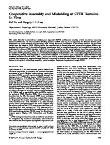
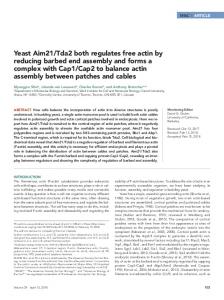
RESULTS A coiled-coil region adjacent to the F-BAR domain mediates the binding of Hof1 to septins To understand the recruitment of Hof1 to septin rings, we mapped the septin interaction domain of Hof1 using the yeast two-hybrid system (Figure 1A). We found that Hof1 constructs carrying either the F-BAR or SH3 domains but lacking codons 293–355 failed to interact with septins (Figure 1A). This indicated that amino acids 293–355 were critical for Hof1 binding to septins. In line with this, the Hof1 fragment 293–355 localized at the cell division site in a septin-like manner (Figure 1B). Domain prediction analysis identified one coiled-coil domain between amino acids 293 and 333 (Figure 1C). F-BAR domains comprise an FCH domain with an associated coiled-coil region (Aspenstrom, 2009). Three-dimensional (3D) structural modeling revealed that ∼280 amino acids at the N-terminus of Hof1 were sufficient to generate the typical banana-shaped F-BAR domain of F-BAR proteins (Figure 1D; Shimada et al., 2007). We therefore concluded that the coiled-coil region that mediates Hof1 binding to septins is not part of the F-BAR domain. Similar results were independently obtained by another group (Oh et al., 2013). We refer to this region between codons 293 and 355 as the Hof1–septin interaction domain (SID). Volume 24 May 1, 2013
We previously reported that the F-BAR domain was required for the association of Hof1 with the septin scaffold (Meitinger et al., 2011). This conclusion was based on a Hof1 N-terminal truncation that lacked amino acids 2–299 (∆2-299). We thus reasoned that either the F-BAR domain contributes to Hof1 septin localization in vivo or the truncation spanning amino acids 2–299 influenced the SID of Hof1. To discriminate between these possibilities, we compared the levels of septin-associated Hof1 truncated forms (Figure 1, E–G). We found that the deletion of either F-BAR domain (∆2279) or SID coiled coil (∆293-333) reduced the association of Hof1 with septins (Figure 1, E–G). The deletion of both F-BAR and coiledcoil regions (∆2-399) completely abolished Hof1 association with the septin complex (Figure 1, E–G). We therefore concluded that both the F-BAR and SID domains contribute to the association of Hof1 with the septin scaffold in vivo.
Phosphorylation of serine 313 in the SID decreases the binding of Hof1 to septins We previously reported that phosphorylation of Hof1 by the Dbf2Mob1 kinase complex leads to the premature release of Hof1 from the septin collar during metaphase (Meitinger et al., 2011). This suggests that phosphorylation of Hof1 by Dbf2 weakens the association between Hof1 and septins. To confirm this hypothesis, we performed in vivo pull-down assays using the established Hof1-4E mutant, in which four Dbf2 phosphorylation sites were mutated to glutamic acid to mimic phosphorylation (Figure 2A). For this analysis, we expressed HOF1-3 hemagglutinin (HA) and hof1-4E-3HA in cells harboring the septin CDC10 fused to the tandem and affinity purification (TAP) tag (Puig et al., 2001; Figure 2B). In comparison to Hof1-3HA, 2.5-fold less Cdc10-TAP could be coimmunoprecipitated with Hof1-4E–3HA (Figure 2B), indicating that Hof1 phosphorylation at Dbf2 sites decreases the binding of Hof1 to septins. We next asked whether Dbf2 specifically phosphorylated the SID region of Hof1. Of importance, one of the Dbf2 phosphorylation sites (RXXS) was present in the SID region (amino acids 310–313; Figure 2A). A single substitution of serine 313 to glutamic acid decreased the interaction between Hof1 and Cdc10 in the yeast two-hybrid system (Hof1-S313E) to a similar degree as the Hof1-4E mutations (Figure 2C; Meitinger et al., 2011). This indicated that serine 313 is critical for the Hof1–septin interaction. This possibility was further evaluated by in vitro pull downs by using a bacterially purified septin complex and Hof1 truncations comprising amino acids 200–355 (Hof1-SID; Figure 2D; Versele and Thorner, 2004). In support of our model, glutathione S-transferase (GST)–Hof1-SID bound to the purified septins (Figure 2D). The degree of binding between GST–Hof1SID-S313E and septins was significantly lower than to GST–Hof1-SID (Figure 2D). In line with this result, we observed that the levels of Hof1-S313E–green fluorescent protein (GFP) bound to septins were significantly diminished in metaphase-arrested cells (Figure 2E-G). In addition, Hof1-S313E–GFP prematurely shifted from septins to the AMR in anaphase-arrested cells. In contrast, in ∼30% of wild-type cells, Hof1-GFP persisted at septins during the anaphase arrest (Figure 2, H and I). We therefore concluded that phosphorylation of serine 313 by Dbf2 kinase decreases the association of Hof1 with the septin complex both in vitro and in vivo. The association of Hof1 with the septin scaffold was largely impaired upon deletion of the F-BAR domain in the hof1-S313E mutant (hof1-∆2-279-S313E; Figure 2, J and K). These data are in agreement with our previous observation that both the F-BAR and SID domains are required for Hof1 colocalization with septins in vivo (Figure 1, E–G). The levels of Hof1 associated with septins were also reduced by the inactivation of the SH3 domain (W637A) Hof1 phosphoregulation | 1291
septin empty
A
1
CC
SH3
P
669 FCH
CC
FCH
CC
300 CC
293
355
CC
P
334
506
P
506 SH3
501 SH3
CC
P
SH3
CC
P
334 293 CC
FCH
1
669
P
669 669 600
septin interaction domain (SID)
C
F-BAR
HOF1
-
hof1-1-300
+
hof1-1-355
+
hof1-293-506
-
hof1-334-506
*
hof1-501-669*
-
hof1-334-506
+
hof1-293-669
+
hof1-1-600
E
CC
1
Hof1GFP
Shs1Cherry
merged
strong
window 14 window 21 window 28
0,8
probability
+
septin collar
1
CC
GFP-Hof1293-355
split septin ring
1
phase
empty
F-BAR FCH
B
0,6
HOF1
0,4
∆293-333
100
200
300
400
500
600
∆2-279
amino acid residue
∆2-299 no signal
D
0 0
weak
0,2
Hof1 F-BAR aa1-285 Cip4 (2EFK) F-BAR aa1-288
∆2-399
116 97 66
GFP
200
45
unsp.
fluorescence intensity at bud neck [a.u.]
∆2-399
∆2-299
∆2-279
G ∆293-333
HOF1-GFP
F
6 5 4
***
***
***
3
***
relative fluorescence intensity bud neck/cytoplasm
metaphase arrest
2,5
***
2
***
3
1,5
2
1
1
0,5
0
HOF1- ∆293- ∆2-279 ∆2-299 ∆2-399 GFP 333
0
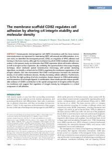
FIGURE 1: Mapping of the septin interaction domain of Hof1. (A) Yeast two-hybrid interactions between Hof1 fragments and septins (Cdc12). Blue color indicates interaction; asterisk, fragment is self-activating. (B) Localization of Hof1-293-355. Hof1-293-355 was overexpressed from the Gal1 promoter. Scale bar, 5 μm. (C) Graph shows probability of coiled-coil regions in Hof1. (D) In silico 3D model of the F-BAR domain of Hof1 in comparison to the resolved structure of the F-BAR domain of CIP4 as a reference. (E) Colocalization of Hof1-GFP, Hof1-∆293-333–GFP, Hof1-∆2279–GFP, Hof1-∆2-299–GFP, and Hof1-∆2-399–GFP with the septin Shs1-Cherry in metaphase-arrested cells. (F) Immunoblot shows the amount of Hof1-GFP, Hof1-∆293-333–GFP, Hof1-∆2-279–GFP, Hof1-∆2-299–GFP, and Hof1-∆2-399–GFP in metaphase-arrested cells. An unspecific signal of the GFP antibody served as loading control. (G) Quantification of fluorescence intensity of Hof1-GFP (n = 56), Hof1-∆293-333–GFP (n = 39), Hof1-∆2-279–GFP (n = 69), Hof1-∆2-299–GFP (n = 50), and Hof1-∆2-399–GFP (n = 34) at the bud neck in metaphase-arrested cells. Blue bars, relative fluorescence intensity at the bud neck. Values >1 indicate an accumulation of Hof1 at the bud neck, whereas values 100 per strain). Error bars, SD. (J) Quantification of fluorescence intensity of Hof1-GFP (n = 37), Hof1-S313E–GFP (n = 28), Hof1-W637A–GFP (n = 30), Hof1-S313E–W637A (n = 31), Hof1-∆2-279–GFP (n = 33), and Hof1-∆2-279-S313E–GFP (n = 30) at the bud neck in metaphase-arrested cells. Blue bars, relative fluorescence intensity at the bud neck. Values >1 indicate an accumulation of Hof1 at the bud neck, whereas values 100 per strain and experiment). Error bars, SD. (H) Localization of Hof1-4A–GFP, Hof1-4E–GFP, and Hof1-S313E–GFP in gin4∆ strains. Cells were grown at 30°C. Arrows indicate mislocalization of Hof1. (I) Quantification of H. Two independent experiments were quantified (n > 100 per strain and experiment). Error bars, SD. (J) Localization of Shs1-GFP in gin4∆ and hof1-4E gin4∆ mutants. Arrows indicate mislocalization of Shs1. (K) Quantification of J. Three independent experiments were quantified (n > 100 per strain and experiment). Error bars, SD. (J) Model for regulation of septin stability. Scale bars, 10 μm. Volume 24 May 1, 2013
Hof1 phosphoregulation | 1297
phase
GFP
Shs1Cherry merged enlarged
B
100
HOF1 -GFP
cells [%]
80
hof1-4A -GFP
40
S533E S563E
4E
hof1
Hof1 Shs1- merged -GFP Cherry enlarged phase s+
Hof1S313E Shs1- merged -GFP Cherry enlarged s-
E
septin structure
100
cells [%]
pGal1-clb2∆DB
phase
S421E
hof1-S313E -GFP
4A
0
S313E
20
hof1-4E -GFP
C
septin structure asymmetric symmetric
60
HOF1
A
symmetric asymmetric mis-localized
80 60 40 20
s+
s-
s+
s-
s+
a-
0
3h
F
HOF1 hof1 hof1- HOF1 hof1 hof1-4E S313E -4E S313E 3h 5h no cortex cortex
Hof1 localization
*-
60 40 20 0
G GFP Tub1
5h
I
***
***
***
***
***
3 2 1 0
∆293-332
∆293-332
∆2-399
∆2-299
∆2-279-S313E
∆2-279
S313E W637A
W637A
S313E
0
HOF1
20
4
2h 3h 4h 5h 6h 7h time of anaphase arrest
∆2-399
40
***
experiment 1 experiment 2
∆2-299
60
5
***
HOF1 hof1 hof1- HOF1 hof1 hof1-4E S313E -4E S313E 3h 5h
∆2-279-S313E
cells [%]
80
***
S313E
100
fluorescence intensity at bud neck [a.u.]
H
3h
W637A
log
1 0.8 0.6 0.4 0.2 0
S313E W637A
HOF1-GFP + - - + - - + - hof1-4E-GFP - + - - + - - + hof1-S313E-GFP - - + - - + - - +
HOF1
D
hof1-S313E-GFP dbf2-2 dbf20∆ relative survival rate of hof1-4E mutant
HOF1-GFP dbf2-2 dbf20∆
1,6 1,4 1,2 1 0,8 0,6 0,4 0,2 0
relative fluorescence intensity bud neck/cytoplasm
*+
80
∆2-279
5h
cells [%]
100
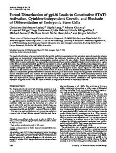
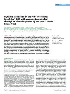
FIGURE 4: Hof1 phosphorylation induces septin rearrangement. (A) Localization of Shs1-Cherry in HOF1-GFP, hof1-4A-GFP, hof1-4E-GFP, and hof1-S313E mutants after prolonged anaphase arrest (pGal1-clb2∆DB, 6 h). (B) Quantification of asymmetric Shs1-Cherry localization in HOF1-GFP, hof1-4A-GFP, hof1-4E-GFP, hof1-S313E, hof1-S421E, and hof1-S533E-S563E mutants after prolonged anaphase arrest (pGal1-clb2∆DB, 6 h). Three independent experiments were quantified (n > 100 per strain and experiment). Error bars, SD. (C) Localization of Hof1-GFP and Hof1-S313E–GFP in comparison to Shs1-Cherry in anaphase-arrested cells (dbf2-2 dbf20∆). Cycling cells were shifted from 23 to 37°C to arrest them in anaphase for 5 h. Samples were taken after 3 and 5 h to analyze septin localization, Hof1 localization, and protein amounts. Red arrowhead indicates mislocalization of Hof1-S313E–GFP to the cell cortex; a, asymmetric septin structure, s, symmetric septin structure; *, mislocalization of septin structure; +, complete colocalization of Hof1-GFP and Shs1-Cherry; –, no complete colocalization of Hof1-GFP and Shs1-Cherry. (D) Immunoblot shows the protein levels of Hof1-GFP, Hof1-4E–GFP, and Hof1-S313E during the course of the 1298 | F. Meitinger et al.
Molecular Biology of the Cell
23°C
30°C
B
37°C galactose
hof1-4A
pGal1-CYK3 pGal1-CYK3 hof1-4A pGal1-CYK3 hof1-4E
glucose
hof1-4A pGal1-CYK3 pGal1-CYK3 hof1-4A pGal1-CYK3 hof1-4E
C
23°C HOF1 HOF1
hof1-S313A hof1-S517A hof1S533A-S563A
D
SC-complete
5-FOA
septum open
pGal1-3HA-CYK3 hof1-4A -GFP Gal Glu
pGal1-CYK3
A
closed
open
closed
0 19 0 0
0 19 0 0
hof1-4E -GFP Gal Glu
gal. (n=33) hof1-4A pGal1-3HA-CYK3 glu. (n=31)
anti-Tub1
gal. (n=17) hof1-4E pGal1-3HA-CYK3 glu. (n=21)
F
phase Hof1-GFP Shs1-Cherry merge 1
24 45 18 19
phase
76 16 82 81
Hof1-4A -GFP Shs1-Cherry merge
*
2
37 °C
1
37 °C (sick)
E
anti-GFP
cells [%]
anti-HA
2
*
3
*
5
23 °C (dead)
4
23 °C
3 4 5 HOF1-GFP (glucose)
hof1-4A-GFP pGal1-CYK3 (glucose)
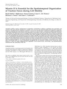
FIGURE 5: Hof1 phosphorylation is essential for AMR contraction and septum formation. (A) Genetic interactions are shown between the indicated Hof1 mutants with expression (galactose) or repression (glucose) of pGAL-CYK3 at different temperatures. (B) Genetic interactions of the indicated genotypes. Cells contain an URA3-based plasmid carrying wild-type CYK3 and were grown in glucose (2%)-containing medium at 23°C. Serial dilutions were spotted onto glucose (2%)-containing plates without or with 5-FOA. 5-FOA selects against the URA3-based plasmid carrying wild-type CYK3. Plates were incubated at 23°C. (C, D) Repression of pGAL-CYK3 causes cytokinesis defects in hof1-4A but not in hof1-4E cells. Quantification of cells with open and closed septa. Septa were analyzed with a TEM. (E, F) Localization of Hof1-GFP and Hof1-4A–GFP after repression of pGAL-CYK3. The septin Shs1 fused to Cherry served as cell cycle marker (collar, before mitotic exit; split rings, after mitotic exit). Asterisk, Hof1 disappears from the bud neck after septin splitting; arrows, Hof1-4A is stabilized between the split septin rings. Scale bars, 10 μm.
experiment (anaphase arrest). (E) Quantification of septin structures as indicated during prolonged anaphase arrest. Three independent experiments were quantified (n > 100 per strain and experiment). Error bars, SD. (F) Quantification of Hof1 localization as indicated during prolonged anaphase arrest. Three independent experiments were quantified (n > 100 per strain and experiment). Error bars, SD. (G) Graph shows the survival rate of hof1-4E-GFP mutant cells relative to HOF1-GFP wild-type cells (see Materials and Methods). (H) Quantification of asymmetric Shs1-Cherry localization in the indicated Hof1 mutants after prolonged anaphase arrest (pGal1-clb2∆DB, 6 h). Three independent experiments were quantified (n > 100 per strain and experiment). Error bars, SD. (I) Quantification of Hof1-GFP (n = 38), Hof1-S313E–GFP (n = 35), Hof1-W637A–GFP (n = 38), Hof1-S313E-W637A–GFP (n = 36), Hof1-∆2-279–GFP (n = 36), Hof1-∆2-279-S313E–GFP (n = 38), Hof1-∆2-299–GFP (n = 35), Hof1-∆2-399–GFP (n = 37), and Hof1-∆293-333–GFP (n = 38) at the bud neck after prolonged anaphase arrest (pGal1-clb2∆DB, 6h). Blue bars, relative fluorescence intensity at the bud neck. Values >1 indicate an accumulation of Hof1 at the bud neck, whereas values p>0.0001
pGal1-CYK3
p0.01
(1) (2) (3) (4) (5) (6) (7) (8) (9) (10) (11) (12) (13)
C3
HOF1 (1)
90 80 70 60 50 40 30 20 10 0
hof1-4A (2)
time [min]
A
t44
t
t46
F
C2
phase
C3
GFP
GFP
G
HOF1-GFP
phase
HOF1-GFP hof1-W637A-GFP
hof1-4A-GFP
HOF1-GFP pGal1-CYK3
HOF1-GFP hof1-4A-GFP hof1-W637A-GFP hof1-4A-W637A-GFP SC-complete
hof1-W637A-GFP hof1-S517A-GFP hof1-S517A-W637A-GFP hof1-S533A-S563A-GFP
5-FOA
hof1-S533A-S563A -W637A-GFP
H
pGal1-CYK3
C1
SC-complete
septin complex
CC
R
F-BA
Dbf2 Mob1 P S517S533 S563 P
AMR
SH3 mediated inhibition of AMR contraction
septin complex
P S313
PXXP SH3
CC
P S313
5-FOA
R F-BA
Dbf2 Mob1 P P P S517S533 S563 P
AMR
PXXP
SH3
Hof1 phosphorylation releases inhibitory effect of SH3 domain on AMR contraction
FIGURE 6: Hof1 inhibits AMR contraction and PS formation via its SH3 domain. (A) Quantification of the duration of Hof1 (wild type or indicated mutant) appearance at the AMR until completion of AMR contraction (see B for representative images). HOF1-GFP (n = 8), hof1-4A-GFP (n = 6), hof1-4E-GFP (n = 16), HOF1 pGal1-CYK3 (n = 10), hof1-4A-GFP pGal1-CYK3 (n = 11), hof1-4E pGal1-CYK3 (n = 7), hof1-W637A-GFP pGal1-CYK3 (n = 9), hof1-4A-W637AGFP pGal1-CYK3 (n = 9), hof1-S533A-S563A-GFP pGal1-CYK3 (n = 10), hof1-S533A-S563A-W637A-GFP pGal1-CYK3 (n = 13), hof1-S517A-GFP pGal1-CYK3 (n = 12), hof1-S517A-W637A-GFP pGal1-CYK3 (n = 6), and hof1-S313A-GFP pGal1-CYK3 (n = 7). Cells were grown in galactose medium (2%) for 24 h at 23°C. At 12 h before inspection, cells were transferred to glucose medium (2%) to repress CYK3 expression from the Gal1 promoter. (B) Representative kymographs quantified in A. Examples for category C1 (Hof1-GFP), C2 (Hof1-S533A-S563A–GFP Gal1-CYK3), and C3 (Hof1-S533A-S563A-W637A–GFP Gal1-CYK3). (C) Map showing the statistical significance of differences between the 1300 | F. Meitinger et al.
Molecular Biology of the Cell
A
B
hof1-4A pGal1-CYK3 (glucose)
hof1-4A CYK3
hof1-4A cyk3∆
SC-complete
5-FOA
cts1∆ egt2∆ dse2∆
0.2 µm
swc11∆ 1 µm
LEU2-2µ
C
hof1-4A cyk3∆ URA3-CYK3 CYK3 CHS2 INN1 SC-complete
5-FOA
SC-complete
23°C
5-FOA
SC-complete
30°C
5-FOA 37°C
D spindle pole body
bud neck
Tem1
Inn1
mitotic Cdk1 Ace2
Chs2 coordination?
Cts1 Egt2 Dse2 Swc11
cell separation
Dbf2-Mob1
Cbk1-Mob2
Cyk3
cell cycle progression
Hof1
AMR contraction septum formation
Dbf2-Mob1
Cdc15
Cdc14
hof1-S533Awild type S563A cyk3∆
viable
inviable
FIGURE 7: Hof1 phosphorylation defects can be bypassed by promoting PS formation or inhibiting PS degradation. (A) TEM images of cross sections of the bud neck region of the hof1-4A pGAL-CYK3 mutant after CYK3 repression. Arrows indicate the starting process of cell separation. (B) Growth rescue of the hof1-4A cyk3∆ mutant after deletion of chitinase or glucanases. (C) Growth rescue of hof1-4A cyk3∆ mutant after overexpression of CHS2 or INN1. (D) Model shows pathways controlling cytokinesis and cell separation. Blue proteins can rescue the lethality of hof1-4A cyk3∆ and are downstream of Hof1 and Cyk3 in the cytokinesis pathway. Red proteins are important for cell separation and cause lethality in hof1-4A cyk3∆ cells.
analyzed mutants (Student’s t test). (D) The relationship between the duration of Hof1/AMR contraction and cell death. Dead cells were counted after they were grown for 12 h in glucose as described in A. Numbers correspond to the genotypes as shown in A (n > 100 per strain). (E) Representative example for cell death during AMR contraction is shown. Left, cell before the onset of cytokinesis (t0), before cell lysis (t44), and after cell lysis (t46). Right, corresponding kymographs for the x- and y-axes as indicated (t, pictures were taken every 1 min). Scale bar, 5 μm. (F, G) Genetic interactions of indicated genotypes. Cells contain a URA3-based plasmid carrying wild-type CYK3 and were grown in glucose (2%)-containing medium at 23°C. Serial dilutions were spotted onto glucose (2%)-containing plates without or with 5-FOA, which selects against the URA3-based plasmid carrying CYK3. Plates were incubated at 23°C for 2–3 d. (H) Model for Dbf2-dependent Hof1 regulation during cytokinesis (see the text). Volume 24 May 1, 2013
Hof1 phosphoregulation | 1301
Of interest, the SID locates next to the F-BAR domain that we previously reported to contribute to the recruitment of Hof1 to the bud neck in vivo (Meitinger et al., 2011). This raises the interesting question as to whether the SID and F-BAR domains have interdependent roles in the stabilization of the septin complex. F-BAR domains are known for their membrane-binding ability, which, so far, has not been proven for Hof1. Nevertheless, 3D modeling of the Hof1–FBAR revealed that positively charged amino acid clusters, which are essential for membrane binding (Frost et al., 2008), are conserved (our unpublished observation). It therefore seems likely that the FBAR domain of Hof1 binds directly to the membrane. Of interest, the deletion of the F-BAR domain decreases the levels of septin associated with Hof1, which is comparable to the effect of the phosphomimetic Hof1-S313E mutant (Figure 2; Meitinger et al., 2011). In addition, deletion of the FCH domain phenocopies the S313E mutation in being synthetic sick with SHS1 deletion (our unpublished data). However, the F-BAR domain of Hof1 failed to associate with septins in the yeast two-hybrid system (Figure 1), indicating that the F-BAR domain alone might not be sufficient to mediate the binding to septins. These observations indicate that the F-BAR, together with the SID domain, might have a role in regulating septins in vivo, for example, by connecting septins with the membrane.
Hof1 regulates AMR contraction and septum formation After mitotic exit, Hof1 moves from septins to the AMR ring, where Hof1 now interacts with Cyk3 and Inn1 proteins to regulate AMR contraction and primary septum formation by an unknown mechanism (Nishihama et al., 2009). Our findings indicate that phosphorylation of C-terminal residues of Hof1 by Dbf2 is the most likely mechanism that facilitates the function of Hof1 in AMR contraction through inhibition of Hof1 protein interaction via its SH3 domain. We previously showed that the phosphoinhibitory hof1-4A mutant lacking CYK3 is unable to survive (Meitinger et al., 2011). A detailed analysis of these cells now shows that the AMR did not contract, remaining in a frozen state that could be reverted through additional inactivation of the SH3 domain (by substitution of tryptophan 637 to alanine; Figure 6; Meitinger et al., 2011). Thus we speculate that a protein with an inhibitory effect on AMR contraction might bind to the SH3 domain of Hof1. One obvious candidate is Cyk3, which is targeted to the bud neck in a Dbf2-dependent manner (i.e., at the same time that Hof1 moves to the AMR ring) and directly interacts with the SH3 domain of Hof1 (Labedzka et al., 2012; Tonikian et al., 2009). One could envisage that the binding of Cyk3 to the SH3 tail of Hof1 could compete with other interacting partners, thereby promoting AMR contraction and/or primary septum formation. However, the inactivation of the PXXP motif of Cyk3, which abrogates its interaction with Hof1 (Labedzka et al., 2012; Tonikian et al., 2009), does not phenocopy CYK3 deletion in hof1-4A cells in respect to cell viability (our unpublished data). Therefore it is unlikely that the frozen AMR in hof1-4A cyk3∆ cells result from the missing interaction between Hof1 and Cyk3. We propose that Dbf2-dependent phosphorylation of Hof1 outside the SID domain might dislodge an inhibitor of AMR contraction that specifically associates with the SH3 domain of Hof1. So far, the identity of this putative inhibitor is unknown. Inn1—one established interaction partner of the SH3 domain of Hof1—might form a stable complex with unphosphorylated Hof1 in the absence of Cyk3. Alternatively, other known interaction partners of the Hof1SH3 domain, such as Bnr1 and Vrp1, which both regulate actin dynamics, might block AMR contraction (Kamei et al., 1998; Naqvi et al., 2001). However, the inactivation of these proteins by deletion and/or PXXP mutation analysis reverted neither the growth lethality nor AMR contraction of hof1-4A cyk3∆ cells (our unpublished data). 1302 | F. Meitinger et al.
We therefore anticipate that the identification of Hof1 SH3 interaction partners and their subsequent molecular characterization will constitute important aspects of future research.
Linking cytokinesis with cell separation Cytokinesis (AMR contraction and septum formation) is followed by cell separation (abscission in mammalian cells), during which daughter and mother cells are physically resolved from one another. How cytokinesis and cell separation are coordinated at the molecular level is not fully understood. The ultrastructural analysis of hof1-4A cyk3∆ cells indicated that septum degradation by hydrolases initiates during the process of AMR contraction/primary septum closure. The promotion of primary septum formation via either deletion of septum hydrolases or the ectopic overproduction of Inn1 or Chs2 restored viability to hof1-4A cyk3∆ cells, strongly indicating that the onset of cell separation before completion of cytokinesis is deleterious for cell survival. Of interest, in fission yeast the inactivation of the Dbf2 homologue Sid2 leads to a growth lethal phenotype that could be rescued by impairing cell separation (Jin et al., 2006). In budding yeast, both cytokinesis and cell separation are under the control of MEN and Cdc14 (Figure 7D). Activation of Cdc14 by MEN signaling counteracts mitotic Cdk1 function and thereby enables the translocation of Dbf2-Mob1 to the bud neck and the recruitment and activation of Cyk3 and Hof1 (Hwa Lim et al., 2003; Meitinger et al., 2010, 2011). In parallel, Cdc14 activates another NDR kinase, Cbk1, which in turn promotes the function of the transcription factor Ace2 (Brace et al., 2010). Ace2 is responsible for the transcription of septum hydrolases, including chitinase and glucanases. The observation that Cdc14 activates both cytokinesis and cell separation leads to the exciting question as to whether a bud neck–associated mechanism exist that makes the activation of cell separation dependent on the successful completion of cytokinesis. If such a mechanism is in place, it must be inactive in hof1-4A cyk3∆ cells, as cell separation starts despite of the incomplete cytokinesis. Thus it is possible that Hof1 and/or Cyk3 are part of such a control mechanism. We therefore anticipate that the identification of the underlying mechanisms by which cell separation/ abscission is coordinated with cytokinesis/AMR contraction in a timely manner and the means by which Hof1 and Cyk3 are involved in these processes will provide important insights into the identification of novel cytokinetic control pathways contributing to cell survival.
MATERIALS AND METHODS Strains, plasmids, growth conditions, and genetic methods Yeast strains and plasmids used in this study are listed in Supplemental Tables S1 and S2. Yeast growth conditions and media were as described (Sherman, 1991). Gene deletions and epitope tagging were performed using PCR-based methods (Knop et al., 1999; Janke et al., 2004). Yeast strains were grown in yeast/peptone/ dextrose medium containing 0.1 mg/l adenine (YPAD). Instead of dextrose, either 3% raffinose (YPAR) or a mixture of 3% raffinose and 2% galactose (YPARG) was used in experiments involving expression of genes under control of the Gal1 promoter. For time-lapse analyses in Figure 6, strains with or without pGal1-3HA-CYK3 were grown in galactose medium and shifted 12 h before the experiment started into glucose medium to repress CYK3 expression. Synthetic complete (SC) media lacking corresponding amino acids were used to grow strains carrying plasmids. Loss of URA3-containing plasmids was tested using plates containing 1 mg/ml 5-fluoroorotic acid (5-FOA). To test the viability of double mutants, we used a plasmid shuffle strategy. Briefly, mutant strains containing the corresponding wild-type gene on a URA3-based plasmid were analyzed for growth on 5-FOA plates (select against URA3). At least six individual Molecular Biology of the Cell
transformants were analyzed per double mutant, and one represen tative mutant is shown. HOF1-GFP-hphNT1, hof1-4A-GFP-hphNT1, hof1-4E-GFP-hphNT1, hof1-S313E-GFP-hphNT1, hof1-S313A-GFPhphNT1, hof1-4A-W637A-GFP-hphNT1, hof1-S517A-GFP-hphNT1, hof1-S533A-S563A-GFP-hphNT1, hof1-S517A-W637A-GFP-hphNT1, and hof1-S533A-S563A-W637A-GFP-hphNT1 fragments were cut out from the corresponding plasmids with XbaI and StuI and integrated into the genomic HOF1 locus.
Cell culture synchronization Cells were synchronized/arrested in metaphase (Figures 1, F and G, and 2, E–G, J, and K) and in anaphase (Figures 2, H–I, and 4). To arrest cells in metaphase, we added 15 μg/ml nocodazole (SigmaAldrich, St. Louis, MO) to the culture media and incubated for 2–4 h until >90% of the cells arrested with large buds and one DNAstained region (4′,6-diamidino-2-phenylindole staining). To provide late-anaphase arrest, 2% galactose was added to the log-phase culture of pGal1-clb2∆DB cells grown in YPAR medium. Alternatively, we arrested cells in late anaphase at 37°C using the temperaturesensitive dbf2-2 dbf20∆ double mutant. For the survival test cells were arrested in late anaphase by shifting dbf2-2 dbf20∆ cells to 37°C. For the survival test, ∼100 cells were plated at time point zero in triplicates on YPD plates. After each hour the same volume was plated as at time point zero. The number of growing colonies gives the number of surviving cells. The survival rate of hof1-4E mutant was plotted relative to wild-type cells (Figure 4G). The survival test was repeated twice, with similar outcomes.
Protein detection methods Yeast protein extracts and Western blotting were performed as described (Janke et al., 2004). Antibodies were rabbit anti-GFP antibody, mouse anti-tubulin (Tub1), mouse anti-HA (clone 12CA5; Sigma-Aldrich), and rabbit anti-Clb2 (Maekawa et al., 2007). Secondary antibodies were goat anti-mouse, goat anti-rabbit, and goat anti-guinea pig immunoglobulins coupled to horseradish peroxidase (Jackson ImmunoResearch Laboratories, West Grove, PA).
reaction. Radioactivity was detected using a Bas 1800 II imaging system (Fujifilm, Tokyo, Japan). The detected phosphorylation (P32) was quantified and corrected for background, input of the substrate, and input/activity of Dbf2-Mob1 kinase.
In vitro binding assay Purified septin complex (6His-Cdc12, Cdc10, Cdc11, and Cdc3) bound to Ni2+ nitriloacetic acid agarose beads (Qiagen, Valencia, CA) was washed three times with binding buffer (50 mM Tris-HCl, pH 7.4, 10% glycerol, 100 mM NaCl, 1 mM EDTA, 1 mM DTT, and 0.5% NP-40). Septin complex was mixed with ∼2 μg GST, GSTHof1-CC, and GST-Hof1-CC-S313E and incubated in binding buffer for 1 h at 4°C. The beads were washed eight times with binding buffer and 1% NP-40 and analyzed by SDS–PAGE and Western blotting. Bound GST-Hof1 fragments were calculated as (IHof1-GST – Ibackground)/(Iseptins-6His – Ibackground), where I is the mean gray value of the protein bands measured using ImageJ (National Institutes of Health, Bethesda, MD).
Immunoprecipitation experiments For the immunoprecipitation experiment shown in Figure 2B, dbf2-2 dbf20∆ CDC10-TAP cells carrying hof1-4E-3HA or HOF1-3HA were arrested for 3h at 37°C. Pellets from a 100-ml yeast culture (107 cells/ ml) were lysed in a FastPrep FP120 Cell Disturber (MP Biomedicals, Solon, OH) using acid-washed glass beads (Sigma-Aldrich). Lysis buffer contained 50 mM Tris-HCl, pH 7.5, 150 mM NaCl, 10% glycerol, 1 mM EDTA, 1 mM DTT, 350 μg/ml benzamidine, 100 mM βglycerophosphate, 50 mM NaF, 5 mM NaVO3, and complete EDTAfree protease inhibitor cocktail (Roche, Indianapolis, IN). Cell lysates were incubated with 1% Triton X-100 for 15 min. Total extracts were clarified by centrifugation at 10,000 × g for 10 min. Hof1-3HA was immunoprecipitated from total extracts using anti-HA coupled protein A–Sepharose beads (GE Healthcare). Bound Cdc10-TAP was calculated as (ICdc10-TAP – Ibackground)/(IHof1-3HA – Ibackground), where I is the mean gray value of the protein bands measured using ImageJ.
Yeast two-hybrid assay Microscopic techniques For fluorescence still-image analysis, cells carrying GFP or Cherry fusion proteins were fixed in 4% formaldehyde for 10–30 min before inspection. Live-cell imaging and quantification of fluorescence still images were performed as described (Meitinger et al., 2011). Specimens for electron microscopy were prepared as described (Maier et al., 2008).
Protein purifications Expression of GST, GST-HOF1-SID (amino acids [aa] 200–355), GSTHOF1-SID-S313E (aa200–355, S313E), GST-HOF1 (aa521-586), GST-HOF1 (aa521-586)-S533A, and GST-HOF1 (aa521-586)-S563A was induced in Escherichia coli BL21 (DE3) at 30°C and purified according to manufacturer’s instructions (GE Healthcare, Piscataway, NJ). 6His-Cdc12, Cdc3, Cdc10, and Cdc11 (plasmid pMVB128/133; generous gift from Jeremy Thorner, University of California) were purified as described (Versele and Thorner, 2004). Dbf2-Mob1 was purified from yeast cells as described (Geymonat et al., 2007).
In vitro kinase assay In vitro kinase assays of purified Dbf2-Mob1 were performed in a kinase reaction buffer containing 50 mM Tris-HCl, pH 7.5, 1 mM dithiothreitol (DTT), 10 mM MgCl2, and 0.1 mM ATP. GST-Hof1 purified from E. coli served as substrate. Reactions were held for 30 min at 30°C. We used 5 μCi of γ-[32P]ATP (0.05 nM) per radioactive kinase Volume 24 May 1, 2013
Indicated genes or gene fragments were cloned into pMM5 (fusion to LexA) and/or pMM6 (fusion to Gal4) and transformed into the yeast strains YPH500 (MATalpha) and SGY37 (MATa), respectively. After mating of YPH500 and SGY37 strains the yeast two-hybrid assay was performed as described (Geissler et al., 1996). Interacting proteins activate the expression of β-galactosidase, which converts X-Gal into a blue detectable product.
Amino acid sequence alignment, protein modeling, and coiled-coil prediction The 3D structure of the Hof1 F-BAR domain was performed by homology-modeling using SWISS-MODEL (Arnold et al., 2006; Kiefer et al., 2009). Protein structures were visualized with the SwissPdb-Viewer (Guex and Peitsch, 1997). Coiled-coil regions were predicted using COILS (Lupas et al., 1991).
ACKNOWLEDGMENTS We thank Erfei Bi for communicating results before publication; Jeremy Thorner for plasmids; and Elmar Schiebel, Iain Hagan, Fouzia Ahmad, and members of G.P.’s lab for comments on the manuscript. F.M. and S.P. were funded by Marie Curie Grant MEXT-CT-042544. This work was also funded by Helmholtz Young Investigator Grant HZ-NG-111 to G.P. S.P. is funded by the Graduate School Landesgraduiertenförderung, University of Heidelberg, Heidelberg, Germany. Hof1 phosphoregulation | 1303
REFERENCES
Arnold K, Bordoli L, Kopp J, Schwede T (2006). The SWISS-MODEL workspace: a web-based environment for protein structure homology modelling. Bioinformatics 22, 195–201. Aspenstrom P (2009). Roles of F-BAR/PCH proteins in the regulation of membrane dynamics and actin reorganization. Int Rev Cell Mol Biol 272, 1–31. Balasubramanian MK, Bi E, Glotzer M (2004). Comparative analysis of cytokinesis in budding yeast, fission yeast and animal cells. Curr Biol 14, R806–R818. Bardin AJ, Amon A (2001). Men and sin: what’s the difference? Nat Rev Mol Cell Biol 2, 815–826. Barr FA, Gruneberg U (2007). Cytokinesis: placing and making the final cut. Cell 131, 847–860. Blondel M, Bach S, Bamps S, Dobbelaere J, Wiget P, Longaretti C, Barral Y, Meijer L, Peter M (2005). Degradation of Hof1 by SCF(Grr1) is important for actomyosin contraction during cytokinesis in yeast. EMBO J 24, 1440–1452. Brace J, Hsu J, Weiss EL (2010). Mitotic exit control of the Saccharomyces cerevisiae Ndr/LATS kinase Cbk1 regulates daughter cell separation after cytokinesis. Mol Cell Biol 31, 721–735. Brace J, Hsu J, Weiss EL (2011). Mitotic exit control of the Saccharomyces cerevisiae Ndr/LATS kinase Cbk1 regulates daughter cell separation after cytokinesis. Mol Cell Biol 31, 721–735. Cabib E, Mol PC, Shaw JA, Choi WJ (1993). Biosynthesis of cell wall and septum during yeast growth. Arch Med Res 24, 301–303. Colman-Lerner A, Chin TE, Brent R (2001). Yeast Cbk1 and Mob2 activate daughter-specific genetic programs to induce asymmetric cell fates. Cell 107, 739–750. Demeter J, Sazer S (1998). imp2, a new component of the actin ring in the fission yeast Schizosaccharomyces pombe. J Cell Biol 143, 415–427. Dobbelaere J, Gentry MS, Hallberg RL, Barral Y (2003). Phosphorylationdependent regulation of septin dynamics during the cell cycle. Dev Cell 4, 345–357. Frenz LM, Lee SE, Fesquet D, Johnston LH (2000). The budding yeast Dbf2 protein kinase localises to the centrosome and moves to the bud neck in late mitosis. J Cell Sci 113, 3399–3408. Frost A, Perera R, Roux A, Spasov K, Destaing O, Egelman EH, De Camilli P, Unger VM (2008). Structural basis of membrane invagination by F-BAR domains. Cell 132, 807–817. Geissler S, Pereira G, Spang A, Knop M, Soues S, Kilmartin J, Schiebel E (1996). The spindle pole body component Spc98p interacts with the gamma-tubulin-like Tub4p of Saccharomyces cerevisiae at the sites of microtubule attachment. EMBO J 15, 3899–3911. Geymonat M, Spanos A, Sedgwick SG (2007). A Saccharomyces cerevisiae autoselection system for optimised recombinant protein expression. Gene 399, 120–128. Guex N, Peitsch MC (1997). SWISS-MODEL and the Swiss-PdbViewer: an environment for comparative protein modeling. Electrophoresis 18, 2714–2723. Holt LJ, Tuch BB, Villen J, Johnson AD, Gygi SP, Morgan DO (2009). Global analysis of Cdk1 substrate phosphorylation sites provides insights into evolution. Science 325, 1682–1686. Hwa Lim H, Yeong FM, Surana U (2003). Inactivation of mitotic kinase triggers translocation of MEN components to mother-daughter neck in yeast. Mol Biol Cell 14, 4734–4743. Janke C et al. (2004). A versatile toolbox for PCR-based tagging of yeast genes: new fluorescent proteins, more markers and promoter substitution cassettes. Yeast 21, 947–962. Jin QW, Zhou M, Bimbo A, Balasubramanian MK, McCollum D (2006). A role for the septation initiation network in septum assembly revealed by genetic analysis of sid2–250 suppressors. Genetics 172, 2101–2112. Kamei T, Tanaka K, Hihara T, Umikawa M, Imamura H, Kikyo M, Ozaki K, Takai Y (1998). Interaction of Bnr1p with a novel Src homology 3 domain-containing Hof1p. Implication in cytokinesis in Saccharomyces cerevisiae. J Biol Chem 273, 28341–28345. Kiefer F, Arnold K, Kunzli M, Bordoli L, Schwede T (2009). The SWISSMODEL Repository and associated resources. Nucleic Acids Res 37, D387–D392. Knop M, Siegers K, Pereira G, Zachariae W, Winsor B, Nasmyth K, Schiebel E (1999). Epitope tagging of yeast genes using a PCR-based strategy: more tags and improved practical routines. Yeast 15, 963–972. Labedzka K, Tian C, Nussbaumer U, Timmermann S, Walther P, Muller J, Johnsson N (2012). Sho1p connects the plasma membrane with proteins of the cytokinesis network through multiple isomeric interaction states. J Cell Sci 125, 4103–4113.
1304 | F. Meitinger et al.
Lippincott J, Li R (1998). Dual function of Cyk2, a cdc15/PSTPIP family protein, in regulating actomyosin ring dynamics and septin distribution. J Cell Biol 143, 1947–1960. Luca FC, Mody M, Kurischko C, Roof DM, Giddings TH, Winey M (2001). Saccharomyces cerevisiae Mob1p is required for cytokinesis and mitotic exit. Mol Cell Biol 21, 6972–6983. Lupas A, Van Dyke M, Stock J (1991). Predicting coiled coils from protein sequences. Science 252, 1162–1164. Maekawa H, Priest C, Lechner J, Pereira G, Schiebel E (2007). The yeast centrosome translates the positional information of the anaphase spindle into a cell cycle signal. J Cell Biol 179, 423–436. Maier P, Rathfelder N, Maeder CI, Colombelli J, Stelzer EH, Knop M (2008). The SpoMBe pathway drives membrane bending necessary for cytokinesis and spore formation in yeast meiosis. EMBO J 27, 2363–2374. Meitinger F, Boehm ME, Hofmann A, Hub B, Zentgraf H, Lehmann WD, Pereira G (2011). Phosphorylation-dependent regulation of the F-BAR protein Hof1 during cytokinesis. Genes Dev 25, 875–888. Meitinger F, Palani S, Pereira G (2012). The power of MEN in cytokinesis. Cell Cycle 11, 219–228. Meitinger F, Petrova B, Lombardi IM, Bertazzi DT, Hub B, Zentgraf H, Pereira G (2010). Targeted localization of Inn1, Cyk3 and Chs2 by the mitotic-exit network regulates cytokinesis in budding yeast. J Cell Sci 123, 1851–1861. Mortensen EM, McDonald H, Yates J 3rd, Kellogg DR (2002). Cell cycle-dependent assembly of a Gin4-septin complex. Mol Biol Cell 13, 2091–2105. Naqvi SN, Feng Q, Boulton VJ, Zahn R, Munn AL (2001). Vrp1p functions in both actomyosin ring-dependent and Hof1p-dependent pathways of cytokinesis. Traffic 2, 189–201. Nishihama R et al. (2009). Role of Inn1 and its interactions with Hof1 and Cyk3 in promoting cleavage furrow and septum formation in S. cerevisiae. J Cell Biol 185, 995–1012. Oh Y, Chang KJ, Orlean P, Wloka C, Deshaies R, Bi E (2012). Mitotic exit kinase Dbf2 directly phosphorylates chitin synthase Chs2 to regulate cytokinesis in budding yeast. Mol Biol Cell 23, 2445–2456. Oh Y, Schreiter J, Nishihama R, Wloka C, Bi E (2013). Targeting and functional mechanisms of the cytokinesis-related F-BAR protein Hof1 during the cell cycle. Mol Biol Cell 24, 1305–1320. Palani S, Meitinger F, Boehm ME, Lehmann WD, Pereira G (2012). Cdc14dependent dephosphorylation of Inn1 contributes to Inn1-Cyk3 complex formation. J Cell Sci 125, 3091–3096. Puig O, Caspary F, Rigaut G, Rutz B, Bouveret E, Bragado-Nilsson E, Wilm M, Seraphin B (2001). The tandem affinity purification (TAP) method: a general procedure of protein complex purification. Methods 24, 218–229. Ren G, Wang J, Brinkworth R, Winsor B, Kobe B, Munn AL (2005). Verprolin cytokinesis function mediated by the Hof one trap domain. Traffic 6, 575–593. Schmidt M, Bowers B, Varma A, Roh DH, Cabib E (2002). In budding yeast, contraction of the actomyosin ring and formation of the primary septum at cytokinesis depend on each other. J Cell Sci 115, 293–302. Sherman F (1991). Getting started with yeast. Methods Enzymol 194, 3–21. Shimada A et al. (2007). Curved EFC/F-BAR-domain dimers are joined end to end into a filament for membrane invagination in endocytosis. Cell 129, 761–772. Surana U, Amon A, Dowzer C, McGrew J, Byers B, Nasmyth K (1993). Destruction of the CDC28/CLB mitotic kinase is not required for the metaphase to anaphase transition in budding yeast. EMBO J 12, 1969–1978. Tonikian R et al. (2009). Bayesian modeling of the yeast SH3 domain interactome predicts spatiotemporal dynamics of endocytosis proteins. PLoS Biol 7, e1000218. Tully GH, Nishihama R, Pringle JR, Morgan DO (2009). The anaphasepromoting complex promotes actomyosin-ring disassembly during cytokinesis in yeast. Mol Biol Cell 20, 1201–1212. Vallen EA, Caviston J, Bi E (2000). Roles of Hof1p, Bni1p, Bnr1p, and myo1p in cytokinesis in Saccharomyces cerevisiae. Mol Biol Cell 11, 593–611. Versele M, Thorner J (2004). Septin collar formation in budding yeast requires GTP binding and direct phosphorylation by the PAK, Cla4. J Cell Biol 164, 701–715. Versele M, Thorner J (2005). Some assembly required: yeast septins provide the instruction manual. Trends Cell Biol 15, 414–424. Yang X, Yu K, Hao Y, Li DM, Stewart R, Insogna KL, Xu T (2004). LATS1 tumour suppressor affects cytokinesis by inhibiting LIMK1. Nat Cell Biol 6, 609–617. Yoshida S, Toh-e A (2001). Regulation of the localization of Dbf2 and mob1 during cell division of Saccharomyces cerevisiae. Genes Genet Syst 76, 141–147. Young BA, Buser C, Drubin DG (2010). Isolation and partial purification of the Saccharomyces cerevisiae cytokinetic apparatus. Cytoskeleton (Hoboken) 67, 13–22. Molecular Biology of the Cell