parasites because they must replicate inside a host cell, utilizing its ... Probably the best-studied example of a nonenveloped ..... The third domain is internal.
Viral Capsids and Envelopes: Structure and Function William Lucas, Harvard Medical School, Boston, Massachusetts, USA David M Knipe, Harvard Medical School, Boston, Massachusetts, USA
Secondary article Article Contents . Introduction . Viral Capsids . Viral Envelopes . Capsid and Envelope Function: Viral Entry into Host Cells
Virus particles contain the viral genome packaged in a protein coat, called a capsid, and sometimes a lipid coat called the envelope. These structures play many roles in viral infection, including virus entry into cells and spread from one cell to another.
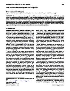
Introduction Viruses are infectious agents that are obligate intracellular parasites because they must replicate inside a host cell, utilizing its macromolecular machinery and energy supplies for their replication process. The infectious form of a virus, the virus particle or virion, replicates itself by entering a host cell, disassembling itself and copying its components, which are then assembled into progeny virus particles. These progeny virus particles can then infect additional cells. Survival of the virus requires transport of the genetic material from an infected cell to an uninfected cell in either the same or a new host organism. To accomplish this, viruses have evolved mechanisms of packaging their genomic nucleic acids, ribonucleic acid (RNA) or deoxyribonucleic acid (DNA), along with any other components necessary for replication, within protein coats comprised of repeating protein subunits. The protein coat is called the capsid, and the complex of the genome plus capsid is called the nucleocapsid. Capsids can be classified into three general classes based on the symmetry of the protein arrangement within the capsid. The first form of symmetry is helical, where the subunits are wound around a central axis (Figure 1a). The second is icosahedral, resulting in a spherical particle with 2-, 3- and 5-fold axes of symmetry (Figure 1b). The third general class contains more complex virion structures, such as those of poxviruses. In some viruses this complex is surrounded by a lipid membrane and associated proteins, a structure called the viral envelope. Thus, we can broadly divide viruses into five classes based on structure: helical nonenveloped virions (e.g. tobacco mosaic virus), helical enveloped virions (e.g. rabies virus: Figure 1a), icosahedral non-enveloped virions (e.g. adenovirus), icosahedral enveloped virions (e.g. herpes simplex virus), and other more complex structures (e.g. pox viruses such as smallpox and vaccinia virus) (Sander et al., 2002). Despite the fact that these viral structures are made from only a few different proteins (in some cases only one), they must be able to perform a wide variety of functions. The virus must successfully package all of the viral nucleic acids
and proteins necessary for a stable, infectious virus particle. This particle must exit the cell and survive in the extracellular environment until it encounters a suitable host cell. The virion must then bind to and enter a new host cell for replication. Thus, the virus particle must be stable extracellularly but readily disassembled upon entry into a new host cell. Some of the best-understood examples of these structure–function relationships will be reviewed in this article.
Viral Capsids Helical capsids Probably the best-studied example of a nonenveloped helical capsid is tobacco mosaic virus (TMV). TMV is a plant virus with a single-stranded RNA genome of 6390 nucleotides (nt). The capsid of TMV is comprised of 2130 copies of a single protein. The overall structure of TMV is a hollow tube with the genomic RNA attached to the internal face of the tube, 3 nt held in the cleft of each capsid subunit. The capsid contains 1613 subunits per helical rotation. This results in a final structure that is a rigid rod approximately 300 nm in length and 18 nm in diameter. Because the capsid protein units assemble on to the RNA molecule, the size of the TMV particle is defined by the size of the genome. The direct relationship between genome size and capsid size is an important advantage for helical viruses. If additional genetic material is inserted into the genome of a helical virus, the capsid size can expand to allow incorporation. Icosahedral viruses have a rigidly defined amount of internal space to package nucleic acid and thus their genomes have a predetermined maximum size. Many examples of enveloped viruses with helical nucleocapsids also exist. One example of such viruses is vesicular stomatitis virus (VSV), a rhabdovirus (structure similar to rabies virus, Figure 1a (Sander et al., 2002). VSV is approximately 180 nm long and 75 nm wide. The nucleo-
ENCYCLOPEDIA OF LIFE SCIENCES / & 2002 Macmillan Publishers Ltd, Nature Publishing Group / www.els.net
1
Viral Capsids and Envelopes: Structure and Function
2-fold symmetry
3-fold symmetry
Glycoprotein Envelope (membrane)
Ribonucleoprotein
Matrix protein
5-fold symmetry
(a)
(b)
f=1
f=2
f=3
Figure 1 Helical and icosahedral symmetry. (a) The structure of rabies virus, an example of a virus containing a helical nucleocapsid, labelled here as ribonucleoprotein. From http://www.cdc.gov/ncidod/dvrd/rabies/the_virus/virus.htm (b) Icosahedral symmetry, with axes of 2-, 3-, and 5-fold symmetry indicated. From http://www-micro.msb.le.ac.uk/335/335Structure.html with permission of Dr Shaun Heaphy.
capsid has a bullet-shaped morphology characteristic of rhabdoviruses, consisting of 35 helical turns of the ribonucleoprotein complex. Closely associated with the N protein is the viral matrix protein, which bridges the membrane and nucleocapsid. This close association between the nucleocapsid and the membrane results in a very uniform bullet-shaped structure for the viral particles. This can be contrasted with the overall structure of influenza A virus, another helical enveloped virus. While the nucleocapsid of influenza has an ordered helical structure, the association of membrane and matrix protein is not uniform. This results in particles that can appear in any form from roughly sphere-shaped blobs to highly pleomorphic rod-shaped particles.
Icosahedral capsids The second form of symmetry used in capsids is the icosahedral or spherical symmetry. An icosahedron is a 20faced structure made up of triangular faces, each of which is built from three subunits (Figure 1b). Thus, the smallest number of protein subunits found in icosahedral virions is 60 (triangulation number or T 5 1), with larger viruses built from multiples of 60 subunits (T 4 1). In a 60-subunit virus, each protein subunit will have identical shape and bonding to the other subunits surrounding it. This structure has symmetry around 2-fold, 3-fold and 5-fold axes (Figure 1b). An example of a 60-subunit capsid is the parvoviruses, which package a relatively small genome of approximately 5.3 kb of single-stranded DNA (ssDNA). In fact, only a few viruses are able to fit their genetic material in a 60-subunit capsid. Because most viruses require more space inside the capsid structure than is afforded by a 60-subunit icosahedron, a slight modification of the structure is necessary. In viruses that use multiples of 2
60 subunits, the particles contain pentameric and hexameric assemblies. Because the same groups of proteins are used to build both pentamers and hexamers, the bonding between proteins and the angles of joining are not exactly equivalent. This concept of quasiequivalence of subunit packing in virus capsids was first described by Caspar and Klug (1962). X-ray crystallography is used to define the atomic structure of virus capsids. The first crystal structure for an icosahedral virus was for tomato bushy stunt virus (TBSV) solved by Stephen Harrison and his colleagues (Harrison et al., 1978). The T 5 3 structures like TBSV and poliovirus are often built up of wedge-shaped b-barrel subunits, sometimes linked together by arm-like extensions from the individual subunits.
Capsid function: packaging of nucleic acids The viral capsid performs a variety of specialized functions, which are directed toward the goal of disseminating the viral genes to suitable hosts. In this section we will discuss the mechanisms of packaging of nucleic acids into capsids, a process often called encapsidation. For viruses without envelopes, this would result in a complete virion particle. Additional assembly processes are involved in the final assembly of enveloped virions. Capsid– envelope interactions will be discussed in a later section. In infected cells, viruses produce many copies of their genomes for assembly into new virions. The capsid proteins must be able to recognize the viral genome and efficiently package it into the virion particle at the correct time after infection. It is important that this packaging reaction does not occur too early in infection, because this would deplete the pool of genomes that serve as templates for replication. Many viruses avoid this potential problem
ENCYCLOPEDIA OF LIFE SCIENCES / & 2002 Macmillan Publishers Ltd, Nature Publishing Group / www.els.net
Viral Capsids and Envelopes: Structure and Function
by temporally regulating gene expression such that the capsid proteins are expressed only after the proteins needed for replication have been made. This results in a readymade pool of genomes available for packaging as soon as the capsid proteins are produced. In general, viral genomes have packaging signals in their genome to which capsid proteins bind to initiate encapsidation, followed by the progressive assembly of the capsid around the nucleic acid or the insertion of the nucleic acid into the capsid. Encapsidation of viral genomes into helical nucleocapsids is generally thought to begin by attachment of one or more of the nucleocapsid proteins to a specific recognition site on the viral genome. Following this initial attachment, other single nucleocapsid proteins or preassembled complexes of nucleocapsid proteins polymerize away from the first complex. The sequential addition of nucleocapsid proteins continues until the nucleic acid is fully encased. This is illustrated well by the packaging of TMV RNA. This virus has an assembly signal sequence at nucleotides 5444–5518 (of 6390) that serves as the site to which a small preformed disc of capsid protein attaches. During the association of RNA and capsid protein a conformational change occurs in the capsid–protein complex, resulting in a helical nucleocapsid structure. Complexes of capsid protein of various sizes then attach and extend the helical nucleocapsid structure until the genome is completely encased. For icosahedral viruses, the exact mechanism of packaging nucleic acid into the capsid is still a topic of debate. Two general models have emerged. In the first model the viral nucleic acid associates with single capsid proteins or small preformed capsid subunits. The capsid then builds itself by polymerization around the nucleic acid. The second model requires assembly of nearly complete particles, which then draw the genome inside through a pore or a gap in the particle. A final structural rearrangement is then required to yield a structurally sound virus particle. Both of these mechanisms have been invoked for the assembly of poliovirus. Poliovirus has a 60-subunit icosahedral shell, each subunit being comprised of four proteins known as VP1–VP4. Prior to final maturation of the capsid, VP2 and VP4 exist as a single protein called VP0. Early in capsid formation VP0, VP1 and VP3 join together to form a single subunit, five of which join together to form a free pentamer. A favoured model of poliovirus assembly has the RNA genome first associating with one pentamer, followed by the stepwise addition of 11 more pentamers to the structure. This results in a completed provirion, which then autocatalyses the proteolytic cleavage of VP0 to VP2 and VP4. This final step results in the production of an infectious poliovirus particle. However, there are also data to suggest that the order of assembly of poliovirus may be different. Like other icosahedral viruses, poliovirus produces many particles that are ‘empty’, i.e. they do not have a viral genome packaged inside the particle. The first model involving nucleation around an RNA–pentamer complex
would predict that these particles would begin forming with RNA inside the particle. The nearly completed particle would then lose the RNA shortly before the final penton attaches to the rest of the shell, resulting in an empty particle. However, it has been shown that empty capsids can dissociate into pentamers and that these pentamers can reform into empty capsids without the need for genomic RNA. The formation of these higher order structures in the absence of RNA allow for the possibility that nearly complete capsids form and only then does the genome enter the particle. Certain bacteriophages and animal DNA viruses do form preformed shells of capsid proteins into what is called an empty capsid or procapsid. These particles contain an internal protein network called the scaffolding. Viral DNA binds to a portal complex at one vertex of the procapsid, and the DNA is drawn into the capsid concomitant with the expulsion of the scaffolding protein. The capsid shell expands during this process to form the final nucleocapsid structure. An even more complicated task is performed by viruses that package multisegmented genomes. An infectious influenza A virus must contain at least one copy of each of eight different RNA molecules, and a mammalian reovirus must package 10 double-stranded RNA (dsRNA) segments. The reovirus particle has icosahedral symmetry with an internal cavity predicted to be large enough to hold not many more than the 10 dsRNA genomic segments. Clearly there must be a selection process to package one copy of each segment without duplication. As the capsid subunits are repeating units of the same proteins, it is unlikely that the capsid alone selects the genome segments. One proposed mechanism that solves this problem is a chain-like selection method in which one segment (A) attaches to the capsid. Segment A is also able to associate with only one other genome segment (B). Segment B is involved in selecting segment C and so on, until a chain containing only one of each genome segment is selected for packaging. While this model has also been suggested for influenza A virus packaging, the helical nucleocapsid structure allows enough flexibility in genome packaging size to permit another theory. By packaging a number of genome segments larger than the number needed for a complete genome, the virus can remove the need for selection during packaging. It has been estimated that less than one in 10 influenza particles is actually infectious. This level of infectivity could be achieved by random packaging of approximately 12–13 segments in the virion. While it has been demonstrated that influenza particles can contain at least nine segments, proof for this, as well as other packaging theories, is still lacking. Unenveloped virions may be infectious once the capsid has assembled around the viral genome. For these viruses, surface proteins play roles in entry of these viruses into host cells, as described below in the section on viral entry.
ENCYCLOPEDIA OF LIFE SCIENCES / & 2002 Macmillan Publishers Ltd, Nature Publishing Group / www.els.net
3
Viral Capsids and Envelopes: Structure and Function
Viral Envelopes Many types of virus particles exist not as naked nucleocapsids but as nucleocapsids surrounded by lipid membranes. These membranes contain various viral encoded proteins and perform some subset of the functions that are required for successful viral spread. We refer to these structures comprised of lipid bilayer and associated proteins as the viral envelopes, and many examples exist of both helical and icosahedral viruses that have envelopes.
proteins, E1 and E2, are regularly arrayed in the viral envelope. There are 80 of these trimers, and each is postulated to interact with capsid proteins at a three-fold axis. However, this relationship between icosahedron and envelope protein disappears in more complex viruses. Herpes simplex virus, for example, is a large enveloped icosahedral virus that has at least nine different envelope proteins. Additionally, the capsid is physically separated from the envelope membrane by a layer of proteins known as the tegument. Thus, other methods for envelope protein selection must exist, such as attachment to matrix proteins that bridge the nucleocapsid to membrane.
Envelope structure The major component of viral envelopes is one or more lipid bilayers. Nearly all enveloped viruses have one lipid bilayer, but one form of poxvirus virion has two membrane layers. The lipid bilayers in virions are generally derived from pre-existing membranes of the host cell; therefore, the lipid components are taken from the cellular membrane. In many cases the acquisition of an envelope occurs as the nucleocapsid buds out from the cytoplasm to the extracellular milieu. Other membranes that may contribute to viral envelopes include the nuclear envelope, the Golgi apparatus and the endoplasmic reticulum. The site of virus budding is determined, at least in part, by the sites of localization of the envelope glycoproteins. In addition to targeting to specific organelles, viral glycoproteins may target to specific membrane sites within an organelle, such as lipid rafts or glycosphingolipid-enriched microdomains within the membrane. Thus, viral envelope lipids may reflect the composition of local regions within a membrane or organelle. While the lipid portion of viral membranes is comprised of cellular products, viruses have developed ways to select their own outer membrane proteins for inclusion in the envelope. Viral outer membrane proteins are usually transmembrane proteins and therefore consist of three major functional domains. There is an extracellular domain, often glycosylated, that may perform various functions including receptor binding, receptor destruction, membrane fusion, or binding to and anchoring other viral proteins to the membrane. There is usually one or more transmembrane domains, which are needed to hold the protein in the viral envelope. The third domain is internal to the viral membrane and may be used to select the protein for inclusion into the viral envelope. Incorporation of these proteins into budding virus particles occurs through specific interactions with other viral proteins. Icosahedral viruses have highly ordered structures with a fixed number of contact sites on the capsid. Based on this structure, one prediction would be that the outer membrane proteins would attach to capsid proteins, resulting in a fixed number of outer membrane proteins that are held in a regular array. Indeed, this structure is seen with some simple viruses such as Sindbis virus. Trimers of the Sindbis virus envelope 4
Assembly of enveloped viruses As previously mentioned, viral envelopes perform some of the functions that would otherwise need to be performed by the viral capsid proteins. However, the addition of a membrane to the viral capsid also offers some distinct advantages. We will explore these functions and advantages using influenza A virus as a model system. Influenza A viruses have a negative-sense, segmented single-stranded RNA genome surrounded by a nucleocapsid protein (NP) that results in a helical virus structure. The virus envelope contains three proteins. The haemagglutinin protein (HA) is the protein used to bind to sialylated proteins that serve as receptors on the target cells. The influenza HA protein was the first transmembrane protein and viral envelope protein whose crystal structure was solved by the late Don Wiley and his colleagues (Wilson et al., 1981). To be infectious, the HA must be proteolytically cleaved into parts HA1 and HA2. The neuraminidase protein (NA) is able to remove sialic acid from proteins, and thus can destroy the viral receptors. This receptor-destroying enzyme function is thought to help prevent the virus from sticking to cells during egress as well as preventing the clumping of virions. The third membrane protein is the viral M2 protein, which forms a channel in the membrane that allows the transmission of protons across the viral envelope. During infection these three proteins are targeted to the plasma membrane of the infected cell (Figure 2, right half). The proteins are assembled in patches on the cell surface from which host proteins have been largely excluded. Organization of these patches of viral proteins is thought to occur by interaction of the cytoplasmic tails of the membrane proteins with the viral matrix (M1) protein. The M1 protein can also interact with viral nucleocapsids. The process of viral budding is thought to occur by the movement of nucleocapsids to the ‘patched’ areas of the cellular membrane. The nucleocapsids are then surrounded by matrix protein. Because the matrix proteins are attached to the envelope proteins, wrapping of M1 around the nucleocapsids results in a bulging of the cellular membrane. As the M1 completely surrounds the nucleo-
ENCYCLOPEDIA OF LIFE SCIENCES / & 2002 Macmillan Publishers Ltd, Nature Publishing Group / www.els.net
Viral Capsids and Envelopes: Structure and Function
a Respiratory tract Influenza virus Matrix
Neuroaminidase
k
Haemagglutinin RNA
M2 Protein Neuraminidase
Nucleoprotein b and polymerases c Virus
j
Endosome Viral messenger RNA
d e Viral RNA Sialic Endosome acid Haemagglutinin Infected Cell nucleus cell
g
h
i
Viral proteins Ribosome from cell
f Viral RNA copies
Figure 2 The replication cycle of influenza virus, showing entry (left) and budding (right). Redrawn after http://www.sciam.com/1999/0199issue/ 0199laverbox4.html with permission of Dr Robert Webster.
capsid, the membrane pinches off to form a complete viral envelope. The virus is now free from the cell and able to infect cells of this or other host organisms. Following assembly of a complete infectious virus particle, the particles exit the cell, either through cell lysis or budding from the cell. With naked capsid virions, capsid proteins play an important role in protecting the virus from damaging chemical or physical conditions until a suitable host cell is located. Successful completion of this task requires a high degree of stability from the capsid structure. Genetic mapping has identified proteins that protect viruses from hazards such as drying, heat and chemical disinfectants, such as ethanol. Indeed, stable capsid properties are greatly desired by manufacturers of livevirus vaccines so that these vaccines can be used in locations of the world where refrigeration is unreliable. Enveloped virions are often less stable in the environment because they are often susceptible to drying and other environmental conditions; therefore, they are usually spread by direct contact between host organisms. Enveloped virions are highly efficient in entering a host cell, if they come into contact with a new host cell within the appropriate time.
Capsid and Envelope Function: Viral Entry into Host Cells Assuming that the capsid and envelope are successful in protecting the virus from the environment, the next major
task to be performed is entry into new host cells. Entry involves the attachment to a receptor molecule on the surface of the host cell, crossing the plasma membrane to gain entry into the host cell, and uncoating of the genomic nucleic acid within the host cell. Icosahedral viruses often have one of two different structures that are used for attachment to host cells. The first is a cleft on the surface of the subunits that can recognize the receptor. Again we shall use poliovirus as a model for this type of receptor. Each of the 60 external faces of the poliovirus capsid is comprised of VP1, VP2 and VP3 (with VP4 buried below). These three external proteins sit at the vertices of the triangular-shaped subunits. In the centre of this triangle is a cleft or ‘canyon’, which binds to the cellular receptor. It has been proposed that having the receptor-binding site buried in a canyon offers an advantage to the virus. Because the canyon is too narrow to allow antibodies access to the amino acid residues that contact the receptor, the residues that bind to the receptor are protected from immune attack. The second method of icosahedral virus attachment utilizes a single specialized receptor-binding protein that is exposed on the outer surface. Often these proteins are attached to the capsid at the vertices of the five-fold axes of symmetry. Examples of this type of receptor-binding protein arrangement can be found with the adenovirus fibre protein or the reovirus s1 protein. In the case of reovirus, a region on the stalk of the s1 protein binds to sialylated glycoproteins, which are very common viral receptors. A second region of the s1 protein more distal to the virus, in the globular head of the structure, is thought to bind to a protein receptor and give some neuronal specificity to certain strains of the virus. Thus, in the case of reovirus, a single protein can provide the means for the virus to attach to two different classes of receptor protein. The multiple receptor functions for adenovirus require two receptor proteins on both the virus and cell. Adenovirus attachment is thought to begin with binding of the fibre protein to a cellular receptor. However, adenovirus attachment through the fibre protein is not sufficient to ensure entry into the cells. Further research has revealed that a second interaction between the penton base proteins of the capsid and a family of proteins on the cell surface, known as integrins, is required for efficient infection of the cells. Attachment to the cellular surface can be considered the first step in viral entry. The virus is now in close proximity to the cellular membrane and can interact with this membrane at the cell surface or after the virus has been endocytosed to form a virus-containing intracellular compartment. This interaction with cellular membranes requires structural alterations in the viral capsid that result in delivery of the viral payload of nucleic acids and/or proteins into the cytoplasm. Note that the ability to uncoat and discharge the contents of the virion is in direct opposition with the requirement that capsids provide protection and stability to the virus. Viruses have solved this problem by requiring that a specific signal must be
ENCYCLOPEDIA OF LIFE SCIENCES / & 2002 Macmillan Publishers Ltd, Nature Publishing Group / www.els.net
5
Viral Capsids and Envelopes: Structure and Function
HIV-1
gp120 variable loops
Core
CD4
gp120 CXCR4
gp41
CCR5 Cell
Figure 3 The stages of entry of human immunodeficiency virus (HIV). gp, glycoprotein. Redrawn after http://www.brown.edu/Courses/Bio_160/ Projects1999/hiv/infect.html with the permission of Dr Joseph Sodroski.
delivered by the cell to the virus to start uncoating, essentially informing the virus that it has successfully reached its final destination. Most often this signal is delivered by either contact with the receptor protein or acidification of the environment inside of the viruscontaining endosome. This is illustrated by the current model for HIV entry into its host cell, the CD4+T lymphocyte (Figure 3). The virus attaches to the cell by binding of the virion glycoprotein gp120 to the CD4 protein on the cell surface. This binding causes a conformational change in gp120 that allows it to bind to a cellular coreceptor molecule, a CXCR4 or CCR5 chemokine receptor. This brings the gp41 to the cellular plasma membrane and allows it to insert and promote fusion between the viral envelope and the cellular plasma membrane, releasing the nucleocapsid into the cytoplasm. For poliovirus, binding to the cellular receptor initiates a sequence of events that results in delivery of the RNA genome into the cytoplasm. This sequence is thought to begin with a structural rearrangement of the capsid that results in the loss of the most internal of the four capsid proteins, VP4. This alteration also changes the structure of the VP1 protein such that the N-terminal portion of VP1 is repositioned to interact with the cellular membrane. Although not proven, it is believed that the RNA then enters the cell through a pore made from the VP1 and possibly VP4 proteins. For many viruses, this step marks the final capsid function. In others, the capsid or a subassembly of the capsid may have additional functions. For example, the capsid may provide the function of transport of the viral genome to the nucleus. More complicated functions may also be performed by some capsids. For example, a form of the reovirus capsid functions as the viral transcriptase/replicase. During the extracellular journey from one cell to another, the advantages and disadvantages of having a viral envelope become apparent. The major disadvantage 6
is that a lipid–protein structure is generally far less stable in the environment than is a protein shell. Many enveloped viruses can exist for only minutes or hours in the environment. This instability is balanced by the advantages of having an envelope to shield the virion from antibody neutralization and to enhance entry into cells by fusion. The proteins found in viral envelopes are often glycoproteins. The carbohydrate structures attached to these proteins can help prevent the binding of antibodies to the proteins, protecting the virus from the immune system. The presence of a lipid envelope also helps to shield the internal proteins from detection by the immune system. For influenza virus, protective immunity can only be achieved if antibodies are generated to the HA and NA proteins. Antibodies to the internal proteins have little or no effect on the course of disease, presumably because the internal proteins are protected by the viral envelope. From a biochemical point of view it should be easy to accommodate additional proteins in the envelope. Proteins only need the intracellular recognition sequence on their cytoplasmic domain. The same modification would very difficult for nonenveloped viruses, as major structural rearrangements of the capsid would be necessary. Entry of enveloped viruses into cells is also easier to envision. The virus need only bring its envelope into close contact with the cellular membrane and provide a catalyst for fusion. In addition to fusion at the cell surface, membrane fusion between the viral envelope and cell membrane may also occur in internal vesicles. For example, the entry process for influenza virus begins with the attachment of the HA protein to receptors on the cell membrane (Figure 2, lower left). The virus is then internalized in an endosome, which is acidified by the cell. This acidification causes the HA to change shape and a hydrophobic portion of HA2 interacts with the vesicle membrane. This interaction is thought to lead to fusion of the viral envelope and the vesicle membrane. Acidification of the vesicle also leads to acidification of the internal components of the virus, as protons can pass freely through the M2 channel in the viral envelope. This acidification dissociates the nucleoproteins from the matrix protein, so that upon membrane fusion the nucleocapsid structures can release from the other viral components and move to the nucleus. The viral replication cycle can then begin. In summary, the viral capsid and envelope are highly specific structures that promote the transmission of the viral genomic material from one host cell to another. Antiviral strategies are beginning to target the molecules in these structures and the processes they mediate to block viral infection and prevent or treat viral disease. One example of such an antiviral drug is amantidine, which blocks the M2 ion channel protein of influenza virus and blocks uncoating and infection by influenza virus. A second example is the class of drugs called fusion inhibitors, which are being tested against human immunodeficiency virus (HIV). We can hope that further study of
ENCYCLOPEDIA OF LIFE SCIENCES / & 2002 Macmillan Publishers Ltd, Nature Publishing Group / www.els.net
Viral Capsids and Envelopes: Structure and Function
the biochemistry of viruses will lead to drugs specific for all classes of viruses.
References Caspar D and Klug A (1962) Physical principles in the construction of regular viruses. Cold Spring Harbor Symposium on Quantitative Biology 27: 1–24. Harrison SC, Olson A, Schutt CE et al. (1978) Tomato bushy stunt virus at 2.9 Angstrom resolution. Nature (London) 276: 368–373. Sander DM (2002) The Big Picture Book of Viruses. http://www.Virology.net/Big_Virology Wilson IA, Skehel JJ and Wiley DC (1981) Structure of the haemagglutinin membrane glycoprotein of influenza virus at 3Angstrom resolution. Nature (London) 289: 366–373.
Further Reading Flint J, Enquist LW, Krug RM, Racaniello VR and Skalka AM (2000) Principles of Virology, Washington, DC: ASM Press. Harrison SC (2001) Principles of virus structure. In: Fields BN, Knipe DM, Howley PM et al. (eds) Fields Virology, 4th edn, pp. 53–85. Philadelphia: Lippincott Williams and Wilkins. Hunter E (2001) Virus assembly. In: Fields BN, Knipe DM, Howley PM et al. (eds) Fields Virology, 4th edn, pp. 171–197. Philadelphia: Lippincott Williams and Wilkins. Laver WG, Bischofberger N and Webster RG (1999) Disarming flu viruses. Scientific American 280: 78–87. Young JAT (2001) Virus entry and uncoating. In: Fields BN, Knipe DM, Howley PM et al. (eds) Fields Virology, 4th edn, pp. 87–103. Philadelphia: Lippincott Williams and Wilkins.
ENCYCLOPEDIA OF LIFE SCIENCES / & 2002 Macmillan Publishers Ltd, Nature Publishing Group / www.els.net
7