Index Terms â Biomedical Image processing, Virtual ... Augmented reality allows then to superimpose that virtual .... virtual instrument is in contact with tissues.
VIRTUAL REALITY AND AUGMENTED REALITY APPLIED TO LAPAROSCOPIC AND NOTES PROCEDURES Luc Soler1, Stéphane Nicolau1, Jean-Baptiste Fasquel1, Vincent Agnus1, Arnaud Charnoz1, Alexandre Hostettler1, Johan Moreau1, Clément Forest2, Didier Mutter1, Jacques Marescaux1 1:IRCAD, 1, place de l’hôpital, 67091 Strasbourg cedex, France 2: Digital Trainers, 4 rue boussingault, 67000 Strasbourg, France ABSTRACT Computer-assisted surgery led to a major improvement in medicine. Such an improvement can be summarized in three major steps. The first one consists in an automated 3D modelling of patients from their medical images. The second one consists in using this modelling in surgical planning and simulator software offering then the opportunity to train the surgical gesture before carrying it out. The last step consists in intraoperatively superimposing preoperative data onto the real view of patients. This Augmented Reality provides surgeons a view in transparency of their patient allowing to track instruments and improve pathology targeting. We will present here our results in these different domains applied to laparoscopic and NOTES procedures. Index Terms – Biomedical Image processing, Virtual Reality, Simulation, Surgery.
1.
The use of stereoscopic vision however allowed to lessen that perception limit, compensating it by a 3D view of the operative scene filmed by two cameras. But this technique will be difficult to implement for transluminal endoscopic surgery since it requires the extreme miniaturization of cameras while maintaining a high image resolution. Another solution consists in using virtual reality and augmented reality. Indeed, virtual reality allows to provide a preoperative 3D view of patients, operated from their medical image (CT scan or MRI). This virtual copy of patients can then be used in a preoperative simulator, what provides a realistic three-dimensional view of patients. Augmented reality allows then to superimpose that virtual image onto the real patient view. It is thus possible to make up for the lack of sense of touch thanks to an improved visualized image augmented with virtual information, thus making the patient transparent. This virtual transparency will therefore allow to find tumours or vessels not by locating them thanks to touch but simply by visualizing them thanks to augmented reality.
INTRODUCTION
Introducing an optical device into the abdomen of a patient so as to carry out the surgical procedure via a miniaturized camera, represented the major change the surgical world experienced during the 20th century: the “minimally invasive” surgery era was born. This revolution is about to experience a new twist linked to the appearance of a new original technique called Natural Orifice Transluminal Endoscopic Surgery (NOTES), that could replace traditional laparoscopic surgery for a large set of procedures. By replacing the rigid optic that is introduced through the skin by a flexible optic that is introduced through a natural orifice such as stomach, vagina or colon, this new technique should eliminate all visible incisions. If the benefits for patients have clearly been proved for laparoscopic surgery, and whatever the result for NOTES, such minimally invasive techniques bring up new difficulties for surgeons, thus reducing their gesture capacity. The first difficulty is the loss of several senses such as the sense of touch and a modification of the force feedback feeling. In NOTES this loss is greatly amplified due to the length of instruments making it difficult to feel a contact between an instrument and an organ. This lack of force feedback is also featured by current robotic systems such as the Da Vinci robot from the Intuitive Surgical company, currently most used surgical robot worldwide.
978-1-4244-2003-2/08/$25.00 ©2008 IEEE
A second well-known limitation of these techniques is the gesture complexity due to the loss of orientation and inversion of movement. In laparoscopy, this inversion is due to the fixed point localized at the instrument entry port on the skin. In NOTES, the loss of orientation is due to the flexibility of the endoscope. Such difficulties can be solved thanks to augmented reality. Indeed, augmented reality techniques combined with instrument tracking can provide location and internal orientation of instruments. We have developed a set of tools providing such improvements for laparoscopic and NOTES procedure. The first step consist in realizing a 3D model of patients from their medical image. This patient-specific model is then used in our preoperative surgical planning and surgical simulators. The last step consists in superimposing the preoperative models onto the video view of the patient thanks to specific augmented reality techniques and efficient instrument tracking. We will present here all these steps. 2.
3D MODELLING OF PATIENTS
From a medical image of patients, standard software can provide an efficient direct volumetric rendering. This technique, available on all current imaging systems (MRI or CT-scan), can be sufficient for a good 3D visualization of anatomical and pathological structures. It consists in
1399
ISBI 2008
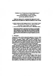
replacing the standard slice view by the visualisation of all slices together in 3D. In order to see internal structures, the initial grey level is replaced by an associated colour and transparency. This transparency gives the feeling of seeing delineated organs which are not delineated in reality. Indeed, with this technique organ and pathology volume are not available and it is not possible to perform a virtual resection of an organ without cutting the entire surrounding structure. To overcome this limit, each anatomical and pathological structure in the medical image has to be delineated. Such a delineation is a long and difficult manual task for radiologists with a standard software. Therefore, several research teams have developed automated 3D patient modelling software tools that can interactively provide the main visible organs from CT-scan or MRI [1-5]. We have also developed several 3D patient modelling software tools, that automatically or interactively provide the main visible organs from CT-scan or MRI for the digestive area [6-7]. Our latest software, 3D Virtual Patient Modelling (3D VPM), allows to model the thoracic or pelvis area in 3D thanks to an improved management of medical knowledge combined with an interactive process [8] (Figure 1).
Figure 1 : 3D modelling of thoraco-abdominal area using 3D VPM ©IRCAD 2006. Our method is based on a hierarchical segmentation of visible organs, from the simplest to the most complex one. The first step respectively detects and delineates skin, lungs, aorta, kidneys, spleen and bones. These organs are automatically delineated using thresholding, gradient, shape and morphology-based methods. All defined constraints are based on simple localization and shape rules used by radiologists and surgeons when they analyze images. From this segmentation, delineated organs are then removed from the initial image which allows to reduce the image to a smaller one containing other not yet segmented organs. Moreover, segmented organs can be used to provide useful information such as location for not yet segmented organ segmentation. The second step then performs the delineation of veins, liver and its internal tumours with our method described in [6] or by using more recent techniques [9-11]. Several companies propose today such 3D modelling as an online distant service by delivering a DICOM image mainly for dental pathologies but also more recently for the digestive area (MeVis Distant Services AG, PolyDimensions GmbH). In the same way, since 2003 we have set up several free cooperations with distant university hospitals (Geneva, Montreal, Lausanne and Strasbourg) in order to offer a similar experimental 3D modelling service named MEDIC@. More than 500 clinical cases have thus been modelled in 3D for the thoraco-abdominal area, 60%
being reconstructed for liver pathologies. As the MeVis Distant Services, this distant 3D modelling service showed its efficiency in providing an accurate 3D model of patient anatomy and pathology in a short delay (between one and three days). Such web-based services could represent in the future the first step of any patient-specific surgical simulation. 3.
FROM VIRTUAL PATIENT TO PATIENTSPECIFIC SURGICAL SIMULATOR
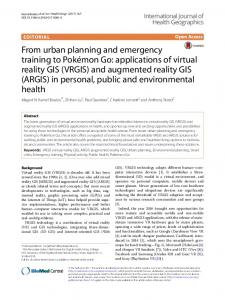
Existing 3D delineation software platforms are usually linked to surgical planning tools [12-14]. For instance, our 3D Virtual Surgical Planning software allows to put each structure in transparency, to interact on them, to navigate anywhere and thus to simulate any kind of minimally invasive procedure. It also allows virtual resections defined by interactively positioned cutting planes and provides the volume of all visualized structures. Surgical planning allows to define the best operative strategy, but it does not provide the possibility to evaluate a surgeon’s ability to perform this strategy. This is the objective of realistic surgical simulators. There is today a large number of commercial surgical simulators for abdominal surgery, urology, gynaecologic or arthroscopic procedures (Surgical Science, SimbionixTM, SimSurgery, Haptica or Karl Storz) . Various scenarios are then available as separate modules. The increasing realism of visual rendering, due to the use of textures obtained from real images, and the progress in force feedback mechanisms enabled those products to acquire some maturity. Though they are attractive, these simulators are limited to the simulation of restricted and determined virtual models within a set database. The main idea of a patient specific simulator is to offer surgeons the opportunity to carry out preclinical training on a virtual copy of the patient. In this objective, we have developed a first patient-specific simulator, the Haptic Operative Realistic Ultrasonographic Simulator called HORUS [15]. HORUS is dedicated to ultrasound-guided percutaneous interventions for training or for preoperative simulation from CT or MRI images of patients. To simulate a realistic ultrasonographic image, we have developed an automatic process that converts a 3D volumetric medical image (CT-scan or MRI) into a realistic US image [16] and simulated thermal ablation effect (figure 2).
Figure 2: HORUS allows a preoperative patient specific simulation of thermal ablation, the burning effect being also simulated in the virtual ultrasonographic image (right). The system has successfully been tested with a 1mm CTscan of a foetus at 36 weeks gestation, a 2mm MRI image of
1400
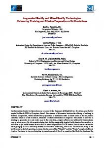
a foetus at 29 weeks gestation, 4 patients for hepatic biopsy and 2 patients for hepatic thermal ablation. From HORUS, we developed in partnership with the Karl Storz Company a laparoscopic simulator called ULIS (Unlimited Laparoscopic Immersive Simulator). The objective of its first version is to be the first patient-specific laparoscopic basic skill training simulator using the Karl Storz Force Feedback System allowing to introduce real surgical instruments. This simulator uses 3D modelled patients extracted from CT-scan reconstructions that can be deformed locally with force feedback rendering when a virtual instrument is in contact with tissues. A photorealistic texturing of tissues is performed manually, which required more than a week of work. ULIS includes camera and surgical instrument manipulation, and simulation of suction and coagulation procedures. The next step of this work will consist in developing a preoperative version, which means an automatic texturing of 3D modelled organs and the integration of the complete simulator engine SOFA (Simulation Open Framework Architecture) [17] allowing multiple interactions and including resection developed in our first patient-specific simulator for laparoscopic liver surgery.
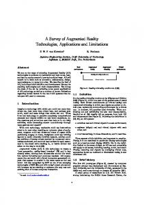
correct all visible mistakes or any wrong registration. Such an augmented reality is necessarily user-dependant due to manual positioning. Therefore, it is impossible to ensure accuracy for this first interesting image-guided surgical tool, that can only be used for an easier understanding of patient anatomy using preoperative data. This limitation can be overcome by automatically superimposing preoperative data onto the real patient during the intervention and by automatically tracking surgical tools in real-time. Automatic registration is the first problem to be solved. Indeed, preoperatively, during the CT-scan acquisition, patients hold their breath, which is not the case during a surgical procedure. Organ movements due to breathing create differences between the patient-specific model and the real organ during surgery. To solve this problem, we have developed a predictive deformation of organs and tissues during a breathing cycle from a single CT image and a video sequence of the patient. Results are very encouraging and show the feasibility of such predictive simulation control by the real breathing of the patient [19]. They are due to the use of volume deformation fields that are defined from skin movements tracked thanks to the use of structured light and to the preliminary segmentation of thorax-abdomen regions and organs. The use of two cameras allows then an efficient surface reconstruction of skin, and also provides a real vision of the patient on which we can superimpose the virtual patient model (Figure 4). Registration is then done using our 3D/2D registration method described in [20]
Figure 3 : laparoscope manipulation (left), virtual blood injury in the bowel area (centre) and coagulation including smoke rendering (right) in ULIS ©IRCAD 2007. 4. VIRTUAL REALITY FOR INTRAOPERATIVE ASSISTANCE : AUGMENTED REALITY
Figure 4: Use of structured light combined with breathing movement simulation allows a real-time augmented reality of organs in movement without any skin marker.
Preoperative surgical planning and simulation will mainly improve the efficiency of MIS procedures thanks to a better pre-operative learning of the patient’s anatomy and a real pre-operative training. But pre-operative use of virtual reality is not sufficient to ensure safety during the surgical procedure. Such an improvement can be provided by an intra-operative use of virtual reality through the Augmented Reality (AR) concept. Indeed, AR consists in superimposing the pre-operative 3D patient modelling onto the real intraoperative patient view. AR provides a transparency view of patients and can also guide surgeons thanks to the virtual improvement of their real surgical tools that are tracked in real-time during the procedure. We have developed two kinds of AR software tools : Interactive Augmented Reality and Fully Automatic Augmented Reality.
Our objective being to use such a system during laparoscopic and NOTES surgery, our next work will be to adapt this system by taking the deformation due to the insufflation of gas inside the abdomen into account.
Interactive Augmented Reality [18] is composed of two steps. The first step consists in registering the virtual patient view with the real patient view. The second step consists in moving virtual tools in real-time, putting them in the same position as the real ones. These two steps are performed manually using an interactive software, what allows to
Automatic surgical instrument tracking is a second problem to be solved. There is today a large set of taker systems essentially based on optics and electromagnetic fields. In the laparoscopic application, we chose not to use a second system by using the same two cameras used for patient registration to track instruments [21]. Such a solution allows to reduce errors that will be linked to calibration of the two tracking systems. Such a solution can not be efficient for NOTES. We have thus developed and patented a new automatic tracking system called METRIS 3D, providing the 3D shape and location of the flexible endoscope in realtime. This system is based on the Aurora tracking system developed by NDI. It is composed of a 1.20 m long and 2.2 mm diameter tube. It can thus be introduced into the operating channel of a flexible endoscope. This tube contains seven 8mm x 1mm miniature electromagnetic coils distributed on its length and which can be located by a
1401
tracking system commercialized by the NDI company under the name Aurora (©NDI). Locating these various coils like a GPS, the software developed in link with that device allows both to reconstruct the tube shape and location in three dimensions and to measure the distance between two interactively selected positions of the tube. Our preclinical validation performed on pigs shows an accurate precision of 1 mm for distance measurement (see table 1). Distance were computed on liver through a transgastric approach with the METRIS system, and compared in a second step with a millimetre ruler measurement. Distance 1 Distance 2 Distance 3 Distance 4 METRIS 7.1 mm 15.2 mm 5.2 mm 13.4 mm Ruler 8 mm 16 mm 6 mm 14.3 mm Table 1 : METRIS accuracy for distance measurement After a calibration phase and thanks to the tracking of the endoscopic camera, it is then possible to provide a real-time automatic augmented reality endoscopic view [21]. 5.
CONCLUSION
We presented a set of tools for diagnosis support and surgical intervention planning. They also allow to use preoperative information during intervention, in particular by superimposing the virtual image of internal organs and pathologies onto the abdomen of patients. These systems, at an experimental stage, are progressively being tested clinically for laparoscopic and NOTES surgery, with the objective of eventually being used in clinical routine. They represent the first essential phase for surgical gesture automation, which will allow to reduce surgical mistakes. REFERENCES [1] W Seong, E-J Kim, and J-W Park: “Automatic Segmentation Technique Without User Modification for 3D Visualization in Medical Image”, CIS 2004, LNCS 3314, 2004, pp. 595–600. [2] A. Schenk, G. Prause and H.-O. Peitgen: “Efficient SemiAutomatic Segmentation of 3D objects in Medical Images”, MICCAI 2000, LNCS 1935, 2000, pp. 186-195.
[7] M. Simone, D. Mutter, F. Rubino, E. Dutson, C. Roy, L. Soler, J. Marescaux: “Three-Dimensional Virtual Cholangioscopy : A Reliable Tool for the Diagnosis of Common Bile Duct Stones”, Annals of Surgery, 240(1), pp. 82-88, 2004. [8] Fasquel JB, Brocker G, Moreau J, Agnus V, Papier N, Koehl C, Soler L, Marescaux J: “A modular and evolutive component oriented software architecture for patient modelling”, Computer Methods and Programs in Biomedicine, 83(3), pp. 222-233, September 2006. [9] Seong-Jae Lim, Yong-Yeon Jeong, and Yo-Sung Ho : “Automatic liver segmentation for volume measurement in CT Images”, Journal of Visual Communication and Image Representation, 17(4), pp. 860-875, August 2006. [10] Yuan-Nan Young, Doron Levy : “Registration-Based Morphing of Active Contours for Segmentation of CT Scans”, Math Biosciences and Engg, 2(1), 79-96, January 2006. [11] Heimann T, Meinzer HP, Wolf I: “A statistical Deformable Model for the segmentation of Liver CT volumes”, MICCAI 2007, Workshop proceedings on 3D Segmentation in the clinic – a grand challenge, pp. 161-166. [12] Kirsti Numminen, Outi Sipilä, Heikki Mäkisalo : “Preoperative hepatic 3D models: Virtual liver resection using three-dimensional imaging technique”, European Journal of Radiology, 56, pp. 179–184, 2005. [13] A. Radtke, S. Nadalin, G. C. Sotiropoulos, E. P. Molmenti, T. Schroeder, C. Valentin-Gamazo,H. Lang, M. Bockhorn, H.O. Peitgen, C. E. Broelsch, M. Malago: “Computer-Assisted Operative Planning in Adult Living Donor Liver Transplantation: A New Way to Resolve the Dilemma of the Middle Hepatic Vein”, World Journal of Surgery, 31, pp. 175–185, 2007. [14] C. Koehl, L. Soler, J. Marescaux: “A PACS Based Interface for 3D Anatomical Structures Visualization and Surgical Planning”, SPIE proceeding 4681, 2002, pp. 17-24. [15] Soler L, Forest C, Nicolau S, Vayssiere C, Wattiez A, Marescaux J : “Computer-assisted operative procedure: from preoperative planning to simulation”, European Clinics Obstetric and Gynaecology, 2:201–208, 2007. [16] Hostettler A, Forest C, Forgione A, Soler L, and Marescaux J : “Real-time ultrasonography simulator based on 3D CT-scan images.”, Stud Health Technol Inform, 111, pp. 191-193, 2005.
[3] Yuan-Nan Young, Doron Levy: “Registration-Based Morphing of Active Contours for Segmentation of CT Scans”, Mathematical Biosciences and Engineering, 2(1), pp. 79-96, January 2006.
[17] Jérémie Allard, Stéphane Cotin, François Faure, Pierre-Jean Bensoussan, François Poyer, Christian Duriez, Hervé Delingette and Laurent Grisoni : “SOFA – an Open Source Framework for Medical Simulation”, Studies in health technology and informatics, 125: 13-18, 2007.
[4] O. Camara, O. Colliot and I. Bloch: “Computational Modeling of Thoracic and Abdominal Anatomy Using Spatial Relationships for Image Segmentation”, Real Time Imaging, 10(4), pp. 263-273, august 2004.
[18] J. Marescaux , F. Rubino, M. Arena and L. Soler: “Augmented Reality Assisted Laparoscopic Adrenalectomy”, JAMA, 292 (18), pp. 2214-2215, 2004.
[5] T. Kitasaka, H. Ogawa, K. Yokoyama, K. Mori, Y. Mekada, J.I. Hasegawa, Y. Suenaga and J. Toriwaki: “Automated extraction of abdominal organs from uncontrasted 3D abdominal X-Ray CT images based on anatomical knowledge”, Journal of Computer Aided Diagnosis of Medical Images, 9(1), pp. 1-14, 2005. [6] Soler L, Delingette H, Malandain G, Montagnat J, Ayache N, Koehl C, Dourthe O, Malassagne B, Smith M, Mutter D and Marescaux J: “Fully automatic anatomical, pathological, and functional segmentation from CT scans for hepatic surgery”, Computer Aided Surgery, 6(3), pp. 131-142, 2001.
[19] Hostettler A, Nicolau S, Soler L, Remond Y : “Real Time Simulation of Organ Motions Induced by Breathing: First Evaluation on Patient Data”, ISBMS 2006, LNCS 4072, pp.9-18. [20] Nicolau S, Pennec X, Soler L et al: “Evaluation of a new 3D/2D registration criterion for Liver Radio-frequencies Guided by Augmented reality”, IS4TM 2003, LNCS 2673, pp.270-283. [21] Nicolau S, Goffin L and Soler L : “A Low Cost and Accurate Guidance System for Laparoscopic Surgery: Validation on an abdominal phantom”, VRST 2005, proceeding of ACM Symposium on Virtual Reality Software and Technology, pp. 124-133.
1402