William N. McFarland & Christina M. Wahl'. Friday Harbor Marine Laboratory, 620 University Road, Friday Harbor, WA 98250, U .S.A.. ' Department of Anatomy ...
Environmental Biology of Fishes 46:109-122,1996 . ©1996 Kluwer Academic Publishers. Printed in the Netherlands .
Visual constraints on migration behavior of juvenile French grunts* William N . McFarland & Christina M . Wahl' Friday Harbor Marine Laboratory, 620 University Road, Friday Harbor, WA 98250, U .S.A . ' Department of Anatomy, Cornell University, Ithaca, NY 14853, US.A . Received 21 .2.1995
Key words :
Accepted 19 .7 .1995
Retina, Fish, Behavior, Development, Haemulon flavolineatum, Haemulidae
Synopsis Retinomotor movements of three size classes of juvenile French grunts, Haemulon flavolineatum, are characterized during the periods of their daily evening and dawn migration . Differences in the extension and withdrawal of the cones correlate with the earlier departure and later return to the daytime schooling sites of the smallest juveniles as compared to the larger juveniles . For each size class changes in the dimensions and retinal distribution of the cone cells and optical properties of the eye are used to estimate differences in photosensitivity under conditions of equal radiant intensities . For a target subtending any given angle, calculation indicates that the largest juveniles should be capable of discrimination at one-thousandth the light intensity of the smallest juveniles. The differences in potential photosensitivity correlate with the different intensities at which they migrate. The likely trigger to initiate their daily migrations probably involves circadian factors at dawn and extension of the single cones from the focal region of the retina prior to the evening migration .
Introduction Most species of fishes rely on vision as a primary sensory mode to guide varied aspects of behavior . Vision serves a fundamental role in predator-prey interactions, territorial defense, social interactions such as courtship and agonistic bouts, orientation, navigation, and, as considered in this study, daily migratory movements (see Munz & McFarland 1977, Lythgoe 1979, Douglas & Dj amgoz 1990, Crescitelli 1991, Heuter & Cohen 1991, McFarland 1991 for reviews) . A few studies relate specific visual properties of fishes to broad behavioral events ; such as movement about the primary visual axes (Tamura & Wisby 1963), visual acuity related to prey choice (Hairston et al . 1992, Wahl et al . 1993),
and retinomotor activity correlated with general activity levels (Ali 1975, Douglas & Wagner 1982) . Compelling experimental demonstrations indicate that predation influences natural selection in different ambient light environments, including spectral shifts at dawn and dusk, where slight variations in visibility of a prey can mean its life or death (Munz & McFarland 1973, Endler 1991) . Specific daily migratory behaviors in several coral reef and freshwater fishes are known to be closely timed to changing light levels (Hobson 1965, 1972, 1973, McFarland et al . 1979, Helfman 1986). Stereotyped visually-linked behaviors are demonstrated by juvenile members of the grunt family Haemulidae . These fish form daytime resting schools within defined territories over coral formations . At dusk the schools depart the daytime
* This paper is dedicated to the memory of John Lythgoe, whose insight served as inspiration for this study .
110 schooling sites and individuals swim in close file along specific migration routes into surrounding grass and/or sand flats . During the night the larger juveniles forage as solitary individuals, returning along the same migratory routes to their schools at dawn . Smaller juveniles undergo similar but shorter migrations and are solitary at night, but are strictly diurnal plankton feeders (Ogden & Ehrlich 1977, McFarland et al . 1979, Helfman et al . 1982, McFarland & Kotchian 1982) . This stereotyped behavior is associated with a high degree of individual site fidelity (McFarland & Hillis 1982) . Migrations of the larger juveniles occur at specific light intensities during the twilight periods at dawn and dusk and correlate with photomechanical changes in retinal rods and cones, known as retinomotor movements (RM) . A combination of circadian pacemaker activity and light levels are known to drive RM (for review see Burnside & Nagle 1983) . In this paper, ontogenetic changes in the eye of the French grunt Haemulon flavolineatum, including the development of RM, visual acuity, and dimensions of the retina pertaining to photosensitivity, are described . These morpho-physiological changes are shown to closely correlate with the times of migration of juvenile grunts .
and isolate three juvenile size classes (large juveniles = LJ's, medium juveniles = MJ's, and early juveniles = EJ's, Table 1) .
Collections
Juvenile French grunts of each size class were collected from patch reefs in Tague Bay, which is located inside the barrier reef on the north-eastern shore of the island of St . Croix, U .S . Virgin Islands . Groups of fish were guided into transparent plastic nets anchored at the bases of the patch reefs next to schooling sites . Because the larger juveniles were difficult to capture with this method, a small beach seine was set adjacent to their schooling sites and across their evening migration routes . The LJ's were guided into the cod end of the seine during their evening migrations to the sand and/or grass beds .
Experimental protocol
To evaluate the effect of light intensities on RM, juveniles were separated by size class into three aquaria where they remained exposed to natural light throughout each 24 hour experimental period . Ambient light levels were recorded in lux with a LUNAPRO photometer. The spectral quality of light in the atmosphere changes dramatically around twilight . This change is reflected usually in a reduction in yellow-orange photons just before sunset
Materials and methods Because grunts typically school during the daytime in groups of similar size (Helfman et al . 1982, McFarland & Hillis 1982), it was possible to identify Table 1 .
Morphometric and behavioral characteristics of juvenile French grunts. EJ, MJ and U refer to early, middle and late juveniles . EJ
MJ
LJ
Mean SL (mm) ± 1 SD (n, rg)
13 .7 ± 2 .7 (16, 10-19) 23-40 diurnal planktivore
21 .8 ± 1 .9 (16, 19-26) 40-56
43 .0 ± 8 (15, 28-57)
Age in days* Feeding mode
diurnal planktivore
87-150 transitional diurnal planktivore, grading into nocturnal benthic omnivore
Diadema;
Schooling site
sand, grass beds ; small structures small coral heads :
Color patterns
near base of coral patches larval-like, translucent grading
usually adjacent to patch reefs silvery body with two
with growth into silvery body
longitudinal black stripes
with a single black stripe * Otolith age, based on number of daily otolith growth increments (Brothers & McFarland 1981) .
coral patches or main reefs of Porites or Montastrea adult livery with many distinct yellow stripes
1 11
LJ 2 - 10' ~1h••}}SSS}SS ~~SS : ~Sh; - 10 .3 - 10 .4 10 . 5 -30
0
+10
-10
0
Sunrise
+30 Sunset
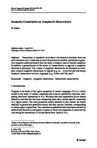
Time in minutes Fig . 1 . Correlation of daily migrations of intervals of juvenile French grunts with sunrise and sunset . Black circles are horizontal light intensities at = 3 meters depth . Time and intensities over which the size classes return and depart from their daytime schooling sites on or near the reefs are indicated by different stippling . EJ, MJ, and U refer to early, middle and late juvenile intervals (see Table 1) ; QP refers to the end and commencement of the quiet period, times of transition when neither diurnal nor nocturnal coral fishes venture into the water column (Hobson 1972) . The graph is a summary of field measurements from the experience of the authors and others (Ogden & Ehrlich 1977, McFarland et al . 1979, Helfman et al . 1982, McFarland & Hillis 1982, McFarland & Kotchian 1983) .
until 30 to 40 minutes postsunset, and again at 30 to 40 minutes prior to dawn (McFarland & Munz 1975) . Thus the spectral light that grunts are exposed to during the critical twilight periods was dominated by blue and green photons . Large juveniles are photopically sensitive to these wavebands because they possess green and blue-green cone visual pigments (McFarland 1991) . The Lunapro photometer, although maximally sensitive in the yellow region of the spectrum, retains sufficient sensitivity in the blue-green to yellow-green bands to provide a reasonable index of the light intensities to which the grunts were exposed . Individuals from each group were sampled during midday and at midnight to establish the position of cone photoreceptors and
melanin screening pigment during periods of light and dark adaptation. Samples were taken at 10 minute intervals through twilight, beginning at one hour before sunset and sunrise . Replicate experiments were performed on four occasions : October 1983, July 1984, August 1985, and January 1986 .
Circadian experiment
To assess the possible influence of circadian rhythms on RM, a group of LJ's were held in either continuous light or darkness. Their eyes were fixed for retinal examination at intervals during 24 hours .
112
OLM-, v m . CL
•T m o • p a
~
m
•
C O
(A
•m rr a° Y Q
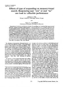
Base PE 1830 Fig. 2. Relationship of the extension of the twin cones from the focal region of the retina in different size classes of juvenile French grunts during evening twilight. OLM is the outer limiting membrane where the cones reside during light adaptation ; RPE is the retinal pigment epithelium base to which the melanin screening pigment withdraws during dark adaptation (Figs 3, 4) . EJ = closed circle, MJ = open circle, U = closed triangle . Each symbol represents the position of the cones within the photoreceptor layer of an individual grunt . Shading represents the period of time for evening migration over which each size class de-
1840
1850 1900 1910 Time in hours
1920
2200
Fig. 3. Relationship between the migration of the twin and single cones of large juvenile French grunts to the time of departure from the reefs during the evening migrations . Vertical axis scale as in Fig . 2 . Open triangles are twin cones, open circles the leading edge and black circles the trailing edge for extension of the single cones across the photoreceptor layer . Vertical shading is the time over which departure from the reefs occurs (from Fig . 1) .
the difference in minutes when the twin cones of each size class extend from the OLM toward the RPE .
graphed to determine cone densities using a microscope equipped with Nomarski interference optics . Only the central posterior part of the retina was counted for cone cell densities .
Sampling was increased during the periods of dusk and dawn .
Histology
parts the reef (from Fig. 1) . Numbers refer to the approximate minutes between the beginning of the evening migrations and
Examination of whole eyes and retinae
Standard lengths, lens diameters, and cone dimensions were measured from unfixed specimens . Intraocular distance of the lens to the focal plane of the retina was measured from photographs of frozen sections of whole eyes . Retinae of individual fish were dissected away from the pigment epithelium (PE) and floated, photoreceptor side up, onto clean glass slides, using the fluid of the eye's own vitreous. Two slips of paper were placed at either side of the retina, to avoid distortion by coverslip pressure. The retinal cone mosaic was photo-
Eyes were fixed in 10% formalin made up in seawater diluted 1 :1 with deionized water, and buffered with coral sand . The preserved eyes were paraffin embedded . Cross-sections were obtained at six micrometers, then stained by the Heidenhain-azan technique to highlight the cone photoreceptor cells . Retinomotor movements were evaluated using the method devised by Ali et al . (1961) . In 1985 and 1986, a less labor intensive method developed by Burnside (personal communication) was used to section and visualize fixed retinae . With this technique, embedding and staining are circumvented by stabilization of the tissue using cetylpyridinium chloride. Retinae are chopped transversely to ap-
Fig. 4. Differential movement of the single and twin cones during the twilight period : a - Period just before evening migration of EJ French grunts showing the withdrawal of the melanin screening pigment within the RPE toward the base of the RPE, and the positioning of both single and twin cones in their light adapted position at the OLM ; b - Period when the evening migration commences showing the extension of the single cones from the focal region and the twin cones still positioned within the focal region at the OLM .
1 13
114 proximately 15 micrometer sections and examined using Nomarski interference contrast optics for the analyses as described above .
fig . 8 in McFarland et al . 1979) . Movement of the RPE melanin granules precedes elongation of the cones at sunset . At sunrise extension of the melanin follows cone contraction (McFarland et al . 1979) . The identical twin cones and the single cones of the
Results
LJ's independently initiate and complete RM (Figs 3, 4b) . A notable difference in RM between the
Size classes were identifiable in the field by their
three sizes, in addition to differences with light in-
coloration, size, and types of daytime schooling
tensity, is the rate at which twin cones move from
sites (Table 1) . As the young fish grow, they aban-
the focal plane to their dark adapted position near
don early sites and join older conspecifics at other
the RPE. The twin cones move as a single wave
schooling sites . Thus, the discrete differences ob-
across the photolayer in LJ's, but are splayed out as
served in behaviors and retinal physiology would
they move in the MJ's, and especially the EJ's . Cone movements in the LJ's are more organized and
probably resemble intergradations, if groups clearly identifiable as EJ's, MJ's and LJ's had not been
tightly coupled with respect to time and light inten-
treated as averaged pools . Consistent and distinct
sity than those observed in MJ's, which are in turn
differences occur between the age classes of French
more organized than the RM in EJ's . The total time
grunts and the light intensity levels at which each
required in the evening for RM in EJ's is greater
developmental stage departs a schooling site during
than that required in LJ's (50 versus 20 minutes) .
dusk and returns to that site during dawn (Fig . 1) .
Whether this relates specifically to inherent differences in properties of the `young' cone cell (as ex-
Timing of retinomotor movements between size
pressed chiefly by twin cones), or is due to another factor is unknown. Nevertheless, the difference cor-
classes
relates with the greater range of time and light intensities over which EJ's migrate as compared to
RM in fishes involve three processes : (1) retinal pig-
LJ's (Figs 1, 2) .
ment movement within the pigmented epithelium
At dawn, RM begin approximately 10 minutes
(RPE), (2) cone cell myoid contraction and exten-
before first light. Both cone types begin contraction
sion across the width of the photoreceptor layer,
together, but in the MJ's and especially the LJ's the
and (3) rod cell myoid extension and contraction
twins are fully contracted about five minutes prior
across the photolayer, which occurs in the reverse
to the single cones . Light adaptation at dawn is rap-
direction to the movements exhibited by the cones .
id for both cone types in all size classes, full contrac-
Direction of RM is linked to increasing or decreas-
tion occurring within 15 minutes of RM onset, a circumstance noted in the RM of photoreceptors of
ing light levels, respectively (Burnside & Nagle 1983) . Juvenile French grunts displayed RM 1 and 2 movements, but very limited movement of the rods .
other species (Douglas 1982a, Burnside & Nagle 1983) .
Large juveniles possess 4 to 5 tiers of stacked rod
Regional differences in cone migration were
cells which constrain their total presence at the fo-
noted in all size classes of fish . However the differ-
cal plane . Differences in the timing of cone RM rel-
ences are most marked in older juveniles . The dorsal half of the retina is first to initiate RM, which
ative to sunset for the three size juvenile classes correlate with light intensity (Fig . 2) . In each size class,
correlates with the lower intensity of horizontal and
the evening migrations are initiated before the twin
upwelling light versus downwelling light underwa-
cones withdraw from their daytime focal positions .
ter (Loew & McFarland 1990) . Our measurements were taken from the central posterior retinal re-
Migrations of the melanin granules within the retinal pigment epithelium (RPE) in LJ's achieved the fully dark or light adapted position prior to sunset and well before first light at dawn (Fig . 4a, and see
gion, as this area fixates targets along the forward visual axis and is most consistently examined in other studies .
115
Timing of retinomotor movements between cone types
Discussion The results established two general conclusions : (1)
As indicated, as well as differences in RM between
retinal morphology changes dramatically within the
size and age classes, there are differences in the RM
juvenile growth intervals and (2) the RM of cones become more tightly coupled and occur at lower
between twin and single cones . These differences become more noticeable as the juveniles grow . Ini-
light intensities with increasing age . This correlates
tially, cones move essentially en masse across the
with the light dependent daily migrations of differ-
photolayer . However this is a relatively lengthy
ent sized French grunts (Fig . 1) . The precision of
process, requiring up to 50 minutes . As a grunt
these daily migrations implies that retinal attributes
grows a clear delineation in the onset of cone elon-
differentially constrain the timing of the migrations
gation between single and twin cones becomes evi-
of EJ's, MJ's and LJ's . For example, do EJ's depart
dent . Cone movements become more rapid, espe-
their daytime schooling sites at sunset (^- 10 lux) be-
cially when considering that the photolayer doubles
cause they would be visually disadvantaged at the
in thickness (Table 2) .
lower postsunset intensities at which the larger ju-
Single cones are first to initiate elongation away
veniles actually depart (- 0.01 lux)? Certainly di-
from the focal plane at the onset of the evening migration (Fig . 3) . This is most clearly evident in the
minishment of vision at twilight influences the timing of the migrations and, especially so, because
more organized RM of U's (Fig . 4b) .
predation tends to increase during twilight (Hobson 1972, Munz & McFarland 1973, Major 1977, Hobson et al . 1981) . To what extent is a French grunt
Circadian cycle contribution to RM
EJ visually constrained compared to an MJ and LJ? A partial answer issues from calculations of poten-
Fish that were held in absolute darkness through
tial photon capture for different sizes of juvenile
normal dawn exhibited sporadic cone RM, indicat-
grunts under equal radiant conditions .
ing that cone movements in French grunts are intrinsically cyclic as in many other fishes (Burnside & Nagle 1983) . However, no cone RM occurred
Photon capture as a function of size
when grunts were exposed to constant room light (fluorescent illumination at 300 lux) .
Evaluation of photon capture per individual cone cell and per unit area of retina requires consider-
Cone dimensions and visual acuity
ation of several optical properties, e .g . eye size, lens diameter, focal length, retinal illumination, and cone dimensions and densities (Fig . 5) . In addition,
Cones increase in overall size and change propor-
assumptions must be made about visual pigment
tions with growth (Table 2, Fig . 5b, c) . The absolute
absorption(s) and how light is transmitted through
length and volume of cone outer segments (OS) de-
the eye and into the cone cells .
creased with growth, but the inner segments (IS)
The amount of light cast per unit area of retina
grow fourfold in volume and nearly tripled in cross-
(i .e . illumination) is independent of eye size for an
sectional area . The ontogenetic changes in lens size, changes in cone density, and potential visual acuity
equal radiance source, if the f-number is constant (f-no . = f/A, where f = focal distance, and A = area
(MSA) for French grunts (Table 2) resembles similar data from other fish species (Otten 1981, Cam-
of lens, or diameter of the aperture), since illumination = 1/f-no . 2 (Lythgoe 1979) . Retinal illumina-
eron 1982, Wahl 1991, Hairston et al . 1992, Wahl et
tion (RI) diminishes in French grunts with growth,
al . 1993) .
however, because the focal length of the eye relative to lens diameter increases (Table 2) . As a result, illumination of the retina is similar for EJ's and
0.34 (6) 2.0 (6)
0.34 (6) 0.17 (6) 2.87 (6)
30 1 .7 1 .97 2.31 140 28 0.10 ± 0.008 (12)
U
3 .08 ± 1 .25 ± 7 .60 ± 29 6 .18 ± 13 .19 ± 396
2.5 ± 0 .16 (25) 1 .0 ± 0 .01 (25) 6.6 ± 0 .55 (25) 17 .4 5.45 ± 0.43 (25) 11 .56 ± 0.72 (25) 270
135 6 .5 8 .98 2.76 57 10
3 .11 ± 1 .42 ± 7.31 ± 30.7 12 .67 ± 16.73 ± 1055
86 4 .2 5 .26 2.6 74 15
2.44 ± 0 .12 (43) 0.99 ± 0.09 (43) 7.99 ± 0 .49 (43) 18.9 8.76 ± 0.31 (43) 13.8 ± 0 .8 (43) 416
0.11
55*
Adult
0 .38 (24) 0 .77 (24)
0 .17 (24) 0 .10 (24) 0 .52 (24)
0 .91 (24) 0 .65 (24)
0 .17 (24) 0 .17 (24) 0 .54 (24)
* Values for cone dimensions of this specimen yielded low values . This is probably due to the section being obtained from a more peripheral area of the retina as compared to the other sections .
0.74 (9) 1 .49 (9)
0.36 (9) 0.32 (9) 1 .8 (9)
0.26 (44) 0.54 (44)
3 .03 ± 1 .57 ± 10 .61 ± 45 .6 5 .13 ± 9 .72 ± 201
2.94 ± 0.78 (9) 1 .48 ± 0.27 (9) 10.16 ± 1 .83 (9) 40 .3 7 .25 ± 0.89 (9) 12 .2 ± 1 .47 (9) 252
3 .42 ± 1 .61 ± 12 .76 ± 66 .1 7 .38 ± 9 .98 ± 215
3 .22 ± 1 .45 ± 11 .23 ± 50.4 4 .53 ± 9 .68 ± 156
22 1 .2 1 .3 2 .36 178 37 0.09 ± 0 .008 (14)
10 0.5 0.53 2.13 288 76 0.07 ± 0 .01 (13)
SL (mm) Lens dia. (mm) Focal length (mm) Matthieson's ratio Cones/0 .01 mm' MSA (min arc) PL thickness (mm) Cone dimensions (gm) Twin cones WOS ba~ WOS ape% HOS Vol . OS (µm3) ;, Wisp HIS,; ng le Vol . IS (gm) Single cones WOSb_ WOSaPe „ HOS Vol . OS (gm') WIS HIS Vol . IS (gm') 0 .13 (45) 0.12 (45) 0 .6 (14)
MJ
EJ
Characteristic
Table 2. Eye dimensions of juvenile and adult French g runts . SL = standard length, Matthieson's ratio = focal length/lens radius, MSA = minimum separable visual angle, PL = photoreceptor layer, WOS and HOS =width and height of cone outer segments, WIS and HIS =width and height of cone inner segment, volumes are calculated for individual cone outer segments. Cone dimensions are given as the mean ± 2 SE's (n), where n is the number of individual cells measured .
117
200
150 6
50
100
1500
Standard length (mm) 03
18
(c)
80
'S
A \o L
A
L rn 0-4 0
10
40
a3 c 0
v
O 0
20
OS I 1 t l I 1 1 1 I I i 1 1 1
0 0
50
100
150
Standard length (mm)
0 0
50
100
150
Standard length (mm)
Fig . 5. Optical and retinal parameters for different sized French grunts: a - Open squares are retinal illumination (RI), black circles the minimum separable visual angle (MSA) ; b - cross sectional area of outer segments (OS) and inner segments (IS), open squares individual twin cone IS, black squares individual single cone IS, open circles individual twin cone OS, black circles individual single cone OS ; c percentage of cross sectional area of twin and single cone OS and IS per 1 mm 2 of retina ; d - number of cones illuminated for targets subtending equal visual angles .
MJ's, but for LJ's and adult grunts it is diminished
cones can vary considerably in size (Ali & Anctil
(Table 3, Fig . 5a) . Also decreasing with growth are the densities of cone cells (Table 2) . By inference,
1976, Wahl 1991), and vary ontogenetically as well,
should operate under a visual disadvantage . But in
as displayed in French grunts as they grow (Table 2) . These enlarged IS's may act as 'light-guides' to concentrate photons and direct them into the OS's
nature the opposite prevails ; large juveniles migrate
that contain the visual pigment (Winston & Enoch
at lower intensities and also feed at night (Table 1,
1971, Snyder 1975,1979, Ali & Anctil 1976, Welford
Fig . 1) . An approximation of photon capture re-
& Winston 1989) . The number of photons that can
quires consideration of cone cell structure . Among different species of fish the inner segments of the
be absorbed in French grunts consequently depends upon the validity of this hypothesis . Two cal-
therefore, larger grunts compared to smaller grunts
118 culations of photon capture are necessary : Assump-
ues) . The relative number of photons absorbed per
tion 1- cone IS's do not enhance photon collection, and Assumption 2 - cone IS's do act to concentrate
OS is obtained by dividing the number of photons
photons .
absorbed per 1 mm 2 by the appropriate number of single or individual twin cones per mm 2 of retina
tion : (1) the number of photons captured per mm 2
(values in Table 2) . We emphasize that the solutions provide relative values of photon capture .
retina and, (2) the relative number of photons absorbed per single cone OS and per individual twin
vide insight into how retinal changes during devel-
Calculations yield two results for each assump-
The calculations presented in Table 3 and 4 pro-
cone OS (Table 3) . We assumed that 10 6 photons were cast on 1 mm 2 of the posterior retina of an EJ .
smaller grunts (EJ's) absorb from 2 to 4 times more
Using the normalized RI values for each size class
photons per OS and per unit area of retina than
of grunt the number of photons cast per mm 2 was
MJ's and LJ's (assumption 1) . If the IS's concentrate
established by multiplication (Table 3) . The total
light then the differences in absorption per OS van-
photons absorbed per mm2 becomes RI * 10 6 pho-
ish (assumption 2) . If assumption 2 prevails, then the capacity of a cone cell to absorb photons would
tons * Absorption * A, where A is x-sectional area of cone OS's and IS's per 1 mm 2 (Tables 2, 3 for val-
opment can benefit each size class . Consider that
be independent of fish size . There still remains,
Table 3. Relative photon catch by individual cones and photon catch by total twin and single cones per unit area of retina in different sizes of French grunts . RI = retinal illumination, OS = outer segment and IS = inner segment of a cone photoreceptor . Assumption 1 : IS of twin and single cones do not act as light concentrators ; Assumption 2 : IS of twin and single cones do act as light concentrators . Characteristic SL (mm) RI (1/f-no. 2) RI normalized Relative absorption* Twin OS Single OS X-section area OS & IS per mm' retina Twins OS Singles OS Twins IS Singles IS Photons impinging per mm 2 of retina**
EJ
Mi
LJ
Adult
10 0 .88 1 .00
22 0 .85 0.97
55 0.63 0.72
135 0 .52 0 .59
0 .55 0 .51
0 .48 0 .49
0.40 0.44
0 .37 0 .38
0 .27 0 .12 0 .62 0 .23 106
0.16 0 .04 0.49 0 .12 970000
0.08 0.03 0.51 0.10 720000
0 .06 0 .01 0 .48 0 .06 590000
Assumption 1 . Absorption by twins per mm2 retina Absorption per twin OS Absorption by singles per mm 2 retina Absorption per single OS
148506 5 .1 61200 4 .3
74496 3 .1 18934 3 .2
23040 1 .4 9504 2.3
13098 1 .7 2248 1 .2
Assumption 2. Absorption by twins per mmZ retina Absorption per twin OS Absorption by singles per mm Z retina Absorption per single OS
341000 11 .8 117300 8 .2
228144 9 .6 57030 9 .6
146880 8.8 31680 7 .6
104784 13 .8 13452 7.1
* The relative absorption per OS decreases with increased size because the HOS and volume of OS declines with increased size . The absorption values assume an optical density of 0 .2 for the smallest OS (see Table 2) . ** Calculations of photon capture assume that 1 x 10⁶ photons per cm2 per sec impinge on 1-mm' of the retina in a 10 mm SL grunt . Pre-retinal absorption and scattering have been excluded because of the high transmission typical of the ocular media of teleosts (Kennedy & Milkman 1956) .
119 however, a reduction in photon capture per unit ar-
is mostly filled by rods, then the rods of U's have
ea of retina in larger fish (Table 4) . To arrive safely
more than twice the collection area per mm z of reti-
at their evening feeding sites or the daytime school-
na for photons as EJ's (0.36 mmz for U's, 0 .15 mm2
ing sites the migrations must be initiated when vi-
for EJ's) . In addition, the photoreceptor layer
sual sensitivity of each fish is sufficient to recognize
thickens with growth (Table 2) and the rods become
and maintain proximity to school mates and to de-
stacked in tiers of 4 to 5 . The resultant increase in
tect predators . How, then, do U's manage to mi-
absorption is undetermined, but we suspect it is at
grate at light intensities that are - 3 orders of mag-
least 4-fold . As an approximation scotopic vision
nitude lower than the intensities at which EJ's mi-
would improve 10-fold per mm' of retina and 100-
grate? Several visual factors, such as size of a tar-
fold for targets subtending equal visual angles . The
get's image, rod vision, and the degree of neural
combined photon capture by cones and rods in U's
development of the retina are probably involved . Photopic sensitivity will depend partly on the fact
infers a potential photosensitivity equal to or slightly greater than the 1000-fold difference in light in-
that images of targets subtending a given angle are
tensities over which the actual migrations occur .
cast on fewer total cone cells in smaller fish (Fig .
What the calculations emphasize is not just the
5d) . Calculations for similar targets show that the
ability of larger grunts to absorb sufficient photons
retinal image will be approximately 122 times
to `see' at lower light intensities, but also how pho-
larger, and will illuminate 32 times as many cone
tons absorbed in cones and rods may be integrated
cells for an U as an EJ . If neural processing of an
and processed differently in juvenile fishes . Perti-
image is similar in EJ's and U's then an U should
nent questions for different sizes of grunts remain .
`see' targets subtending equal visual angles at approximately 3 percent of the radiant intensity re-
How large are the receptive fields? What are the
quired by the EJ . Given the differences in growth, we infer that retinal processing of images in EJ's is
are the retinal neural networks that process absorbed photons in each size class?
dimensions of rod summation? And, how different
less developed than in U's . To adequately discriminate objects EJ's would have to migrate at `brighter'
Triggers of migration
light intensities than our simplified calculations of relative photon capture reveal, which of course they do .
Differences in the ability to visually discriminate
John Lythgoe suggested during an earlier investi-
objects that result from the described differences in
gation, that a critical aspect in the migrations was
photosensitivity with size, underlie the stereotyped
the simultaneous exposure of cones and rods to
differences in initiation of the daily migrations of
light in a focal position and, insightfully, long after withdrawal of the melanin screening pigment
EJ's, MJ's and U's . What mechanisms might trigger
(McFarland et al . 1979) . What might the rods con-
trigger the time of migrations then one would infer
tribute? If the area of retina not occupied by cones
that differences in absolute light sense occur for
the migrations? If differences in photosensitivity
Table 4. Comparison of the relative absorption of photons per mm z of retina in different sizes of French grunts . Table is based on data from Table 3 . Category
Twin cones only Single cones only
Assumption
Percent absorption EJ/EJ
EJ/MJ
100
50
15 .6
67
43
31
1
100 100
2
100
45 62 .5
15 .6 38 .5
7 .3 25 .6
1 2
EJ/LJ
EJ/Adult 8 .8
120 each size class . Although possible, an alternative trigger might be the movement of cones away from the focal region.
MJ's show intermediate development of the retina and extension of the single cones, and correspondingly migrate earlier at dusk and later at dawn than the U's .
Single versus twin cone RM
Varying rates of cone elongation among cone types have been reported in fishes (Mueller 1954) . The green-sensitive twin cones do not extend from the focal region at dusk until 10 to 15 minutes after the evening migrations begin (Fig . 2) . This provides sufficient time for individual grunts to reach their nocturnal destinations on the sand and grass beds. Thus, extension of the twin cones cannot trigger the evening migrations . However, extension of the single cones from the focal region prior to movement of the twin cones may signal individual grunts that it is time to migrate (Fig . 3) . The movement of the single cones would have at least three effects : (1) a diminishment in visual acuity, (2) a decrease in photopic sensitivity and (3) a loss of chromatic sensitivity expressed at short wavelengths because the single cones are blue-green sensitive and the identical twin cones green-yellow sensitive (McFarland 1991) . In MJ's single cones also extend prior to the twin cones, but their movements are not as crisply defined as in LJ's . Clear separation of single and twin cone RM, however, does not occur in EJ's where there is little temporal distinction btween twin and single cone movements . We infer for the smallest grunts that vision is primarily photopic, the rods having not achieved the developmental state observed in the larger juveniles and adults . Their smaller eyes must limit even their photopic sensitivity. In contrast the largest juveniles and adults benefit because their photopic and scotopic systems are well defined anatomically and function mesopically when they migrate during twilight . Extension of their single cones from the focal region of the retina could trigger migration because it would initiate a diminishment in visual sensitivity to shorter wavelengths of light (i .e. blue-wavebands) . We stress that predators and divers can threaten and do delay the evening departure of LJ's from their daytime schooling sites, but only briefly (McFarland et al . 1979) . No matter what the threat LJ's depart and stream from the reefs, a behavior that correlates with single cones moving from the focal region. The
Circadian factors The sporadic but incomplete movement of the cone cells in grunts held in CD prior to and following natural dawn are suggestive of an intrinsic daily rhythm . In CL the total suppression of RM in French grunts, at least at and above 300 lux, emphasizes the dominant role of light on RM (Douglas 1982b, Burnside & Nagel 1983) . The regrouping of juvenile grunts prior to dawn from their solitary overnight stance on the sand/grass beds is executed only when first light has occurred (McFarland & Hillis 1982) . A circadian rhythm synchronized by dusk and/or dawn may prime grunts to anticipate and prepare for migration, but the actual trigger to move certainly involves a visual process . Chromatic triggers
Spectral shifts associated with twilight may serve to synchronize circadian locomotor activities . Blue light has been shown to excite, and yellow light inhibit, the `on' phase of the circadian clock in mammals and birds (Nuboer et al . 1983) . A prerequisite for such a chromatically driven circadian-behavioral system is the presence of blue- and green-sensitive photoreceptors (Nuboer 1971) . The antagonistic interplay between the blue and green photic mechanisms of rabbits, for example, has been used to explain the onset of their nocturnal activities on the basis of the relative reduction in yellow light that occurs during dusk and its relative increase during dawn (Seliger & Fastie 1968, Munz & McFarland 1973, McFarland & Munz 1975, Geusz & Page 1991) . Because French grunts possess blue-sensitive single cones, green-sensitive twin cones and rods that absorb maximally at 497-498 nm, they possess the requisites of an antagonistic visual mechanism that, as in rabbits, might serve to synchronize daily activities . In MJ's and U's, but not EJ's, the blue cones elongate first at dusk, and, therefore, a fish presumably must become more green-sensitive, even though the spectrum underwater shifts to shorter wavelengths (Munz & McFarland 1973,
121 McFarland & Munz 1975) . The actual role of shifts in the twilight spectrum and potential changes in
Douglas, R.H . & M. Djamgoz (ed.) . 1990 . The visual system of fish. Chapman & Hall, London . 526 pp. Douglas, R . & H .J. Wagner . 1982 . Endogenous patterns of pho-
chromatic sensitivity of grunts, that result from the different retinomotor movements of the cones dur-
tomechanical movements in teleosts and their relation to activity rhythms. Cell Tiss . Res . 226 :133-144 .
ing this period, remains obscure . The fact that their
Endler, J . 1991 . Variation in the appearance of guppy color pat-
chromatic abilities must be changing during the critical periods over which they migrate presents an
terns to guppies and their predators under different visual
experimental challenge for behavioral and ecologically oriented physiologists, because it is at this very time that they are most subject to predation .
conditions . Vision Res. 31 : 587-608 . Geusz, M . & T. Page . 1991 . An opsin-based photopign-ent mediates phase shifts of the Bulla circadian pacemaker. J . Comp. Physiol. 168A: 565-570 . Hairston, N ., K. Li & S. Easter, Jr. 1992. Fish vision and detection of planktonic prey. Science 218 :1240-1242 . Helfman, G. 1986 . Fish behavior by day, night, and twilight . pp. 366-387 . In : T.J . Pitcher (ed .) The Behavior of Teleost Fishes,
Acknowledgements This work was funded in part by Cornell University, NSF Grant OCE 79-18569, and by NIH grant EY06401-02 . The staff of the West Indies Laboratory, where the experiments were carried out, were most helpful . Drew Noden facilitated write-up of the work . This is contribution no . 169 of the Wrigley Marine Science Center .
Croom-Helm, London . Helfman, G ., J . Meyer & W.N. McFarland . 1982. The ontogeny of twilight migration patterns in grunts (Pisces : Haemulidae) . Anim . Behav. 30 :317-326 . Hobson, E . 1965 . Diurnal-nocturnal activity of some inshore fishes in the gulf of California . Copeia 1965 : 291-302 . Hobson, E . 1972. Activity of Hawaiian reef fishes during the evening and morning transitions between daylight and darkness . U .S . Fish . Bull. 70:715-740. Hobson, E . 1973 . Diel feeding migrations in tropical reef fishes . Helgolander Meeresunter . 24 : 361-370 . Hobson, E ., W.N. McFarland & J .R. Chess . 1981 . Crepuscular and nocturnal activities of Californian nearshore fishes, with
References cited
consideration of their scotopic visual pigments and the photic environment . U.S . Fish . Bull . 79:1-30 .
Ali, M .A. 1975 . Retinomotor responses. pp. 313-355 . In : M .A . Ali (ed .) Vision in Fishes, Plenum Press, New York .
Hueter, R .E . & J . Cohen (ed .) . 1991 . Vision in elasmobranchs : a comparative and ecological perspective . (Symposium 1990,
Ali, M .A . & M . Anctil . 1976 . Retinas of fishes . Springer Verlag, Berlin. 284 pp .
Kennedy, D . & R.H . Milkman . 1956 . Selective light absorption
Ali, M .A ., W.R. Stevenson & J .S . Press. 1961. Histophysiological studies on the juvenile Atlantic salmon (Salmo salar) retina . 1 .
by the lenses of lower vertebrates and its influence on spectral sensitivity. Biol . Bull . 111 : 375-386 .
Rates of light and dark adaptation . Can . J . Zool. 391 :123-128. Brothers, E .V. & W.N . McFarland . 1981. Correlations between
Loew, E .R. & WN. McFarland. 1990 . The underwater visual en-
Sarasota, Florida) . J . Exp . Zool . Suppl . 5 .182 pp.
otolith microstructure, growth, and life history transitions in
vironment . pp. t-43 . In: R . Douglas & M . Djamgoz (ed .) The Visual System of Fish, Chapman and Hall, London .
newly recruited French grunts (Haemulon flavolineatum) (Desmarest), Haemulidae) . Rapp . Cons. Explor . Mer 178:
Lythgoe, J .N. 1979 . The ecology of vision . Clarendon Press . Oxford. 244 pp .
369-374 .
Major, PF. 1977 . Predator-prey interactions in schooling fishes
Burnside, B. & B. Nagle. 1983 . Retinomotor movements of photoreceptors and retinal pigment epithelium : mechanisms and regulation . Progr . Retinal Res . 2: 67-109 . Cameron, N . 1982 . The photopic spectral sensitivity of a dichromatic teleost fish (Perca fluviatilis) . Vision Res . 22:1341-1348. Crescitelli, F. 1991 . Adaptations of visual pigments to the photic environment of the deep sea . J . Exp. Zool . Suppl . 5 : 66-75 . Douglas, R .H . 1982a . The function of the photomechanical movements in the retina of rainbow trout (Salmo gairdneri) . J . Exp . Biol . 96 : 389-403 . Douglas, R .H . 1982b . An endogenous crepuscular rhythm of rainbow trout (Salmo gairdneri): photomechanical movements . J . Exp. Biol . 96 : 377-388 .
during periods of twilight: a study of the silverside Pranesus insularum in Hawaii . U.S. Fish . Bull . 75 :415-426. McFarland, W.N. 1991 . The visual world of coral reef fishes . pp . 16-38 . In: P. Sale (ed.) The Ecology of Fishes of Coral Reefs, Academic Press, New York . McFarland, W.N. & Z .M . Hillis. 1982. Observations on agonistic behavior between members of juvenile French and white grunts - Family Haemulidae . Bull. Mar . Sci . 32 : 255-268 . McFarland, W.N. & N . Kotchian . 1982 . Interaction between schools of fish and mysids . Behav. Ecol . Sociobiol . 11 : 71-76 . McFarland, WN. & FW. Munz . 1975 . The visible spectrum during twilight and its implications to vision . pp. 249-370. In : G .C . Evans, R . Bainbridge & O. Rackham (ed .) Light as an Ecolog-
122 ical Factor: II, The 16th Symposium of the British Ecological Society, Blackwell Scientific Publ ., Oxford. McFarland, W .N., J .C . Ogden & J .N . Lythgoe. 1979 . The influence of light on the twilight migrations of grunts . Env. Biol . Fish . 4: 9-22 . Mueller, H . 1954 . Die Dunkeladaptation beim Guppy (Lebistes reticulatus P) . Z. Vergl . Physiol . 37 :1-18.
Seliger, H .H. & W.G . Fastie. 1968 . Studies at Oyster Bay in Jamaica, West Indies . Measurements of underwater-sunlight spectra . J . Mar. Res . 26: 273-280. Snyder, A .W. 1975 . Photoreceptor optics - theoretical principles. pp . 38-55 . In: A .W. Snyder & R . Menzel (ed.) Photoreceptor Optics, Springer-Verlag, Berlin . Snyder, A .W. 1979 . The physics of vision in compound eyes . pp.
Munz, EW. & W.N. McFarland . 1973. The significance of spectral
225-314 . In: H . Autrum (ed .) The Handbook of Sensory Physi-
position in the rhodopsins of tropical marine fishes . Vision
ology, Vol . VII/6A, Springer-Verlag, Berlin . Tamura, T. & W.J. Wisby. 1963 . The visual sense of pelagic fishes, especially the visual axis and accommodation . Bull . Mar. Sci .
Res. 13 :1829-1874 . Munz, FW & WN . McFarland . 1977 . Evolutionary adaptations of fishes to the photic environment . pp . 193-274 . In: E Crescitelli (ed .) The Handbook of Sensory Physiology, Vol . V/7, Springer-Verlag, Berlin . .W . J F 1971 . Spectral discrimination in a rabbit. DocuNuboer, . menta Ophthalmologica 30: 279-298 .
13 :433-448 . Wahl, C.M . 1991 . Ontogeny of the retina in two species of percid, the yellow perch Perca flavescens, and the walleye, Stizostedion vitreum. Ph .D. Dissertation, Cornell University, Ithaca . 182 pp.
Nuboer, J .F W., W.M. van Nuys & J .C. van Steenbergen . 1983 . Color changes in a light regimen as synchronizers of circadian
Wahl, C .M ., E .L . Mills, W.N. McFarland & J . DeGisi . 1993 . Ontogeny of visual acuity in the yellow perch, Perca flavescens. Can .
activity. J . Comp . Physiol . 151A : 359-366. Ogden, J.C . & P. Ehrlich . 1977 . The behavior of heterotypic rest-
Winston, R. & J .M . Enoch. 1971 . Retinal cone receptor as an ide-
ing schools of juvenile grunts (Pomadasyidae) . Mar . Biol. 42: 273-280. Otten, E. 1981 . Vision during growth of a generalized Haplochromis species, H. elegans . Netherland J . Zool . 31 : 650-700.
J . Fish . Aquat . Sci . 50 : 743-749. al light collector. J. Opt . Soc. Am . 61 :1120-1121. Welford, W.T. & R . Winston. 1989 . High collection nonimaging optics . Academic Press, San Diego . 284 pp.