American Society for Parenteral and Enteral Nutrition,. Silver Spring ... Multiple
swallowings, peristaltic failure, gastric. Safety Issues in the ..... of choice in many
institutions. Feeding ... of placement.12 If there is any question of tube migration
or ...
Safe Practices in Patient Care
Volume 1, No. 1
Advisory Board Janet Barber MSN, RN
Col. US Air Force (Ret’d) Editor Critical Care Nursing Quarterly Greensburg, IN
Lisa Caffery MS, RN, BC, CGRN Epidemiology Specialist Genesis Medical Center Davenport, IA.
Jan Foster RN, PhD, MSN, CCRN Chair-elect, American Association of Critical Care Nurses Certification Corporation Houston Baptist University, TX
Peggi Guenter PhD, RN, CNSN
Managing Editor, Special Projects American Society for Parenteral and Enteral Nutrition, Silver Spring, MD
Barbara J. Holtzclaw PhD, RN, FAAN Professor Emeritus, School of Nursing University of Texas Health Science Center San Antonio, TX
M. Terese Verklan PhD, RNC
Assoc. Professor of Nursing University of Texas Health Science Center Houston, TX
���� �������������� �������������������������������������������� �������������������������������������������� ������������������������������������������������ ������������������������� ���������������������� ��������������������� ��������������������������������
� ��
��������
�
������� ��
It is with this goal that we welcome you to the inaugural issue of Safe Practices in Patient Care. This serial publication is dedicated to helping nurses caring for the critically and chronically ill to minimize medical errors by presenting clinically and evidenced-based safe practices. Our first issue focuses on safe practices in enteral feeding among critically ill adults and neonates. Safe Practices is an accredited home-study program and is underwritten by an educational grant from Kendall Healthcare.
Safe Nursing Practices for Enteral Nutrition in the Acutely Ill
��
M
edical errors are responsible for injury in as many as 1 out of every 25 hospital patients and an estimated 48,000 to 98,000 patients die from medical errors each year. Errors in health care have been estimated to cost more than $5 million per year in a large teaching hospital, and preventable health care-related errors cost the economy from $17 to $29 billion each year. Most of the medical errors are systemsrelated and not attributable to individual negligence or misconduct. The key to reducing medical errors is to focus on improving the systems of delivering care and not to blame individuals.
�������� �
Helping to promote a culture of safety
By Peggi Guenter, PhD, RN, CNSN
E
nteral nutrition can be defined as nutrition that is provided via the gastrointestinal (GI) tract, usually through a tube, catheter, or stoma that delivers nutrients distal to the oral cavity.1 Tube feeding and enteral nutrition are terms often used interchangeably. Enteral nutrition is indicated when a patient will not, should not, or cannot eat but has a functional GI tract. It should be used in appropriate patients who are or will become malnourished and in whom oral feedings are inadequate to maintain nutritional status.2 Advantages of enteral nutrition over parenteral (intravenous) nutrition include:3,-5 1. Enteral nutrition is less expensive than parenteral. 2. Enteral nutrition is considered safer than parenteral nutrition. 3. Enteral nutrition is considered more physiological, in that it is digested and absorbed via the GI tract, which may better maintain gut structure and function, as opposed to being infused directly into the bloodstream. 4. Enteral nutrition is associated with better clinical outcomes, as compared to parenteral nutrition, in a variety of clinical circumstances. Major considerations when selecting the type of nutritional support for patients include gastrointestinal function, expected duration of therapy, potential risk for aspiration, and level of organ function. Tube feeding, although a relatively safe method of nutritional support, is associated with major and minor complications, many of which can be prevented or treated. Complications are generally classified into three main types: mechanical, gastrointestinal, and metabolic (Table 1).6 This article will discuss a few selected procedures, complications, and practice guidelines to increase patient safety.
Tube feeding-related complications Mechanical tube placement injuries Tubal displacement injury/rupture insertion site breakdown occlusion aspiration Gastrointestinal nausea/vomiting gastroparesis diarrhea constipation Metabolic dehydration elevated serum electrolytes depressed serum electrolytes hyperglycemia Table 1
Safe practices The nurse’s role in enteral nutrition delivery is to provide safe, effective therapy. The 1999 Institute of Medicine report on medical errors concluded that most medical errors are caused by faulty systems, processes, and conditions that lead people to make mistakes or fail to prevent them. These mistakes can be prevented by designing a safer health system that makes it harder for people to do something wrong and easier to do it right.7 In 2002, the Joint Commission on Accreditation of Healthcare Organizations (JCAHO) published the 2003 National Patient Safety Goals (NPSG).8 They are to: ● improve accuracy of patient identification. ● improve effectiveness of communication between caregivers. Continued on page 4
Supported by an educational grant from Kendall, a business unit of Tyco Healthcare Group LP
Safe Practices
Safety Issues in the Enteral Feeding of Neonates By M.T. Verklan, RN, PhD, CCNS, and S. Premji, RN, PhD
E
nteral feeding is the only source of nutrients that influences gastrointestinal regulation after birth.1 The early start of enteral feedings, including trophic feedings, can prevent border brush atrophy, encourage gut colonization with appropriate flora, and improve nutrient absorption in neonates. This paper discusses gastrointestinal motor function, nutritive sucking, safety issues, and strategies to minimize death and injury related to enteral feeding.
Pathophysiology Ontogeny of esophageal sphincters, esophageal motor function, gastric emptying, intestinal transit, and coordinated sucking, swallowing, and breathing reflexes are important factors to consider, when reviewing the safety of enteral feeding. They underscore the importance of watchful vigilance for complications in neonates who are fed by this method.
Esophageal sphincters Upper esophageal sphincter sphincter: The upper esophageal sphincter relaxes and opens for normal swallowing.2 Healthy infants from 33 to 37 weeks of gestational age have a highpressure zone in this area that decreases in response to dry pharyngeal swallow. Premature infants generate lower intraoral pressures when sucking.3,4 They have a diminished capacity to propel a bolus beyond the upper esophageal sphincter, which often causes feeding difficulties.2 Furthermore, premature neonates are unable to protect the airway by glottal closure and central inhibition of the diaphragm during swallowing, so they often experience episodes of apnea, bradycardia, and oxygen desaturation and are at risk for aspiration.5,6 Lower esophageal sphincter sphincter: Recent studies suggest that all very premature infants have a high-pressure zone at the lower esophageal sphincter.7,8,9 Transient relaxation of the lower esophageal sphincter (TRLES), which occurs in the absence of primary or secondary peristalsis, is the primary mechanism that triggers the acidic or non-acidic reflux of gastric contents across the lower esophageal sphincter.8,9 TRLES occurs more often in infants of lower gestational age probably because of impaired gastric function, which may delay gastric emptying and prolong gastric distension.8 Multiple swallowings, peristaltic failure, gastric 2 www.safe-practices.org
The provision of enteral nutrition by tube feedings is a common intervention in North American neonatal intensive care units distension, or vomiting may trigger TRLES by way of vagal nerve pathways.8,10,11 TRLES may be induced by esophageal stimuli, such as an improperly placed feeding tube.12
Esophageal motor function At ≥26 weeks of gestational age, control of dry swallow-induced peristalsis is fully developed and pressure waves can effectively promote the esophageal clearance of gastroesophageal refluxate.8,9 However, prolonged esophageal exposure to refluxate may be associated with obstructive apnea or aspiration and, over time, acid-induced mucosal damage.
Gastric emptying and intestinal motility The human fetus begins to swallow at 12 to 16 weeks of gestation.13 After 28 weeks, fetal gastrointestinal motility and the rate of propagation of gastric contents increase.14,15 Changes in the coordinated motor function of the fundus, antrum, pylorus, and duodenum can delay gastric emptying.16,17 After a bolus feeding, immediate maximal antral distention occurs. There is a rapid, then slower phase of gastric emptying. This pattern is similar, qualitatively, to the curvilinear pattern of gastric emptying of liquid meals in older preterm and term infants.15,18,19 The number and amplitude of duodenal contractions increase markedly with gestational age.20 Bisset et al have observed a four-fold increase in the amplitude of gastric antral contractions between 28 and 38 weeks of gestation.21 In premature infants of 25 to 35 weeks of gestation, coordination of antral and duodenal activity occurs five times less often than in term
infants. Lack of antroduodenal coordination has been implicated in delayed gastric emptying.22 Infants with feeding intolerance have longer times to maximal antral distention.15 Intestinal motor activity, specifically the migrating motor complex, seen during fasting, is responsible for the aboral movement of milk.17 The migrating motor complex involves irregular contractions (phase II), followed immediately by regular phasic contractions (phase III). The cycle repeats with a silent phase of motor quiescence (phase I). These three phases recur every 60 to 90 minutes.23 Feeding intolerance reflects a delay in the development of complete interdigestive cycles during fasting and an inability to change the pattern of motor activity in response to feedings.24 When food is ingested, simultaneous, sporadic, repetitive contractions of variable amplitude occur at multiple levels of the intestine. Nutrients are mixed and churned within various gastric sections.17 In 40% of preterm infants who are fed over a 15-minute period, contractions increase to interrupt the fasting pattern; in others, motor activity decreases.25 Gastrointestinal motility, gastric emptying, and intestinal transit are affected by the caloric content, osmotic load, pH, fat content, and fiber content of nutrients.26,27 Infants who receive higher-density formula have less motor activity;26 however, more energy is emptied into the duodenum during the first hour after meal intake. Differences in motor activity decrease over time, suggesting that it may be possible to introduce higher-density formula later in life.26 Motor activity is also influenced by associated disease.26-28
Coordinated sucking, swallowing, and breathing Nutritive sucking, which occurs only with oral fluids, consists of a continuous rhythmic pattern of slow mean rates of sucking and short pauses.29 The act of swallowing is coupled and coordinated with airway protective reflexes. They simultaneously transport fluid from the oral cavity to the stomach and prevent aspiration into the respiratory tract.30 Although components of sucking and swallowing are evident by 28 weeks of gestational age,31,32 the sucking and swallowing reflex is not fully developed until 32 to 34 weeks of gestation.33 Lack of synchrony between breathing and swallowing probably explains episodes of apnea, bradycardia, and oxygen desaturation during swallowing.2,5,6 Frequency of swallows is significantly greater during apnea than episodes of periodic breathing.6
Application and safe practices The provision of enteral nutrition by tube feedings is a common intervention in North American neonatal intensive care units (NICUs). However, little research has compared different types of gastric-feeding regimens, such as continuous, intermittent, gastric, duodenal,
Safe Practices and jejunal, in the critically ill neonate.34 Due to a lack of evidence-based guidelines, no uniform feeding method is used by clinicians.31,35-38 Early initiation of enteral feeding is associated with a shorter time to full feedings, better weight gain, and earlier hospital discharge.39,40 Tube feeding bypasses the issues of intubation, immaturity of coordinated sucking, swallowing, and breathing reflexes, lower esophageal pressure, and gastric emptying. Delaying enteral feedings leads to gut atrophy and may make feedings less safe when introduced later.40 Whether the substrate is delivered continuously or intermittently, the route of delivery may be orogastric (OG)/ nasogastric (NG) or transpyloric (duodenal or jejunal). The intragastric route is preferable, as it promotes gastric emptying and the release of hormones that may improve peristalsis. The transpyloric route has greater morbidity and mortality without proven benefit.41 However, prolonged OG/NG feedings may lead to oral aversion and damage the esophageal sphincter, causing gastroesophageal reflux.42
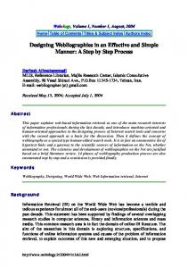
Equipment The type and size of feeding tube depends on placement and the amount and delivery rate of feedings. A general guideline is 4-French (Fr), 5/6-Fr, and 8-Fr tubes for infants 1kg, and >2.5 kg, respectively.41 Short-term neonatal and pediatric enteral feeding tubes range from 3.5 to 10 Fr and have a finish for easy insertion. The tubes are radiopaque with numerical depth marks at every centimeter from 5 to 25 cm to facilitate safer insertion as well as enhanced tube position surveillance. Polyurethane feeding tubes, designed for prolonged enteral feeding, have the same features as short-term tubes, which are composed of PVC. The rigidity of the PVC tube requires that di-2-ethylexyl-phthalate (DEHP) be added to the material to make it flexible. DEHP is a compound known to leach from PVC tubes and may produce chronic side effects in patients with protracted exposure.43,44 Research in rodent and laboratory animals have demonstrated that DEHP is a reproductive and developmental toxicant that may cause cellular abnormalities and impaired proliferation of the Sertoli cells of the testes in addition to general adverse effects on the structure and function of the male reproductive system.45 The long-term feeding tube is made from
Figure 1: ARGYLE Indwell Polyurethane Feeding Tube (Kendall/ Tyco Healthcare)
DEHP is a compound known to leach from PVC tubes and may produce chronic side effects in patients with protracted exposure. polyurethane materials that do not have DEHP. Short-term tubes should be changed every 24 to 72 hours, while long-term tubes should be changed once per month.
Tube insertion After hand-washing, the nurse determines the depth of tube insertion by placing the tip of the feeding tube at the tip of the neonate’s nose, then drawing the tube to the base of the ear, then halfway between the xiphoid process and umbilicus. The nurse notes the depth mark on the tube at the corner of the mouth or edge of the nose. For oral placement, the tube is inserted into the oropharynx and esophagus, using gentle pressure in a downward arc, until the tube reaches the pre-measured depth. For nasal placement, the lubricated tip is inserted into one nostril and advanced slowly with gentle pressure, until the predetermined depth is reached. If, during insertion, the infant gags, develops bradycardia, or becomes pale or cyanotic, the nurse removes the tube or pauses to allow the infant to recover. Tube placement is verified by aspiration of stomach contents. If no gastric secretions are obtained, the nurse advances the tube by 1 cm and aspirates again. Another technique is to inject 2 cc of air, while auscultating over the abdomen with a stethoscope. The same type of “swoosh”, indicating air entry, may be heard, even if the tube is not in the ideal location. Once tube placement is verified, the nurse notes the tube depth and securely tapes the tube in place, leaving the landmark visible. The nurse places notes on the Kardex and patient’s progress report, so that others can verify the landmark before each feeding. Transpyloric feeding tubes can either be made from PVC, soft silicone tubes that require a stylet for insertion or polyurethane. If the tube is made from silicone it should be inspected visually and flushed with water before insertion to ensure that it has not been perforated by the stylet. The depth of insertion is estimated, as described above for OG/NG placement. Using gentle pressure, the nurse advances the tube, until the estimated
depth is reached. Polyurethane tubes would be preferred, given the benefits of the material, including better flow rates, larger inner diameter, and less porous material than silicone. Whatever material is used, tube placement is verified by radiography.
Feeding Before each feeding, the nurse checks the landmark to verify that the tube has not moved and remains taped securely in place. A syringe is then connected to the tube. The nurse instills 1 to 2 cc of air into the feeding tube to ensure that tube holes are not lying directly against the stomach mucosa. When the tube is aspirated, direct contact can irritate or damage the stomach lining. Aspiration of stomach contents verifies that the tube is in place. The nurse notes the amount, color, and consistency of aspirate. No randomized controlled trial has defined the volume or characteristics of residuals that indicate feeding intolerance.46 Although it is common to find small aspirates, regardless of volume, excessive gastric residuals have been defined as 20% to 50% of the bolus-feeding volume, 1.5 times the hourly rate of a continuous feeding, or a consistent 2 to 3 cc residual.47 Feeding practices vary about returning the aspirate to the infant; however, an aspirate that appears excessively mucousy, green, bright yellow, bloody, or like “coffee-grounds” should never be refed. The nurse notes the characteristics of aspirate and discusses them with the neonatologist and nurse practitioner. Other observations to evaluate the tolerance of feedings are the appearance of the abdomen, measurement of abdominal girth, and stooling pattern. Tube feedings may be delivered by gravity or feeding pump. Gravity feedings should never be “pushed”, although a gentle shove may be needed to start the flow. The nurse positions the syringe about 8 inches over the neonate’s abdomen and allows it to run in slowly.41 After use, the gavage set-up may be rinsed with water and allowed to air dry or be discarded, depending on manufacturer’s instructions and hospital policy. The nurse draws the correct volume into the syringe and sets the period of time, e.g., 30 to 60 minutes. If the feeding is breast milk the syringe is positioned with the tip in the up position to prevent the loss of milk fat by settling. A pump specifically designed for the delivery of enteral feeds should be used as opposed to an intervenous fluid pump. Depending on the rate of delivery, the administration of the enteral feedings may approximate the drip rate of the intravenous fluid. The availability of certain newer needleless IV system connection ports makes it possible to actuate the valve of these connectors with an oral syringe and thereby inject the fluid into an IV line. To decrease the possibility of inadvertent systemic administration of enteral feeds, tube feeding administration sets should only be accommodated by specific feeding pumps. Both the 3 www.safe-practices.org
Safe Practices tubing and the pump should specify that they are for oral/enteral use only. Severe side effects as well as organ damage may result from oral feedings given intravenously. The nurse notes any gagging, bradycardia, tachycardia, and color changes. These signs indicate that tube position has changed or the neonate is not tolerating feedings. The nurse stops the feeding and watches the infant. If recovery occurs quickly, feeding may slowly resume. If the infant continues in distress or becomes distressed with subsequent feedings, the nurse stops all feedings and notifies the neonatologist and nurse practitioner. After the feeding volume is delivered, the nurse empties the tube with air to ensure that the infant has the full complement of food and to clear the tube in case of dislodgement and aspiration. For continuous tube feedings, a stopcock is placed between the feeding tube and an extension stubbing, so that residuals can be checked every 2 to 4 hours. The correct amount of formula or breast milk is drawn into the syringe and delivered at a specific rate. No more than 4 hours of breast milk or 4 to 8 hours of formula should be prepared in advance to decrease bacterial growth in the food.
16.
Assessment and documentation
38.
The nurse documents tube placement, size, and depth as well as the neonate’s response to enteral feeding. Before each feeding, the nurse notes the depth landmark visually and aspirates the tube to obtain and assess gastric aspirates. Any subtle changes in the neonate’s condition should be evaluated, as feeding intolerance is a presenting symptom of complications, such as necrotizing enterocolitis and sepsis. The Kardex should contain the target date for changing the tube. If the tube is inserted nasally, subsequent insertions should use the opposite naris.
39.
References 1. 2. 3. 4. 5. 6. 7. 8. 9. 10. 11. 12. 13. 14. 15.
Read L, Upton F, et al. Changes in the growth-promoting activity of human milk during lactation. Pediatric Research 1984,8:133-9. Omari T, Snel A, et al. Measurement of upper esophageal sphincter tone and relaxation during swallowing in premature infants. Am J Physiol - Gastrointestinal and Liver Physiology 1999,277:G862-G866. Bernbaum J, Pereira G, et al. Nonnutritive sucking during gavage feeding enhances growth and maturation in premature infants. Pediatrics 1983,71: 41-5. Medoff-Cooper B, Weininger S, et al. Neonatal sucking as a clinical assessment tool: preliminary fi ndings. Nursing Research 1989,38:162-165. Matthew O. Respiratory control during nipple feeding in preterm infants. Pediatric Pulmonology 1988,5:220-4. Miller M, DiFiore J A comparison of swallowing during apnea and periodic breathing in premature infants. Pediatric Research 1995,37:796-9. Omari T, Miki K, et al. Characterization of lower esophageal sphincter relaxation in healthy preterm infants. Gut 1997,268:G1-8. Omari T, Barnett C, et al. Mechanism of gastroesophageal reflux in healthy premature infants. Journal of Pediatrics 1998,5:650-4. Omari T, Benninga M, et al. Characterization of esophageal body and lower esophageal sphincter motor function in the very premature neonate. Journal of Pediatrics 1999,135:517-21. Ornstein S, Dent J, et al. Esophageal motor response to distension with graded volumes of air in infants. Gastroenterology 1994,106: A551. Martin C, Patrikios J, et al. Abolition of gas reflux and transient lower esophageal sphincter relaxation by vagal blockade in the dog. Gastroenterology 1986,106:890-6. Orenstein S, DiLorenzo C, et al. Isolated lower esophageal sphincter relaxation as ”wave-suppressed” secondary peristalsis. Dysphagia 1997,12: 207-11. Gupta M, Brans Y. Gastric retention in neonates. Pediatrics 1978,62:26-9. McLain C. Amniography studies of the gastrointestinal motility of the human fetus. Am J Obstet Gynecol 1963,86:1079-87. Carlos M, Babyn P, et al. Changes in gastric emptying in early postnatal life. Journal of Pediatrics 1997,130:931-7.
4 www.safe-practices.org
17. 18. 19. 20. 21. 22. 23. 24. 25. 26. 27. 28. 29. 30. 31. 32. 33. 34. 35. 36. 37.
40. 41. 42. 43. 44. 45. 46. 47.
Houghton L, Read N, et al. Motor activity of the gastric antrum, pylorus, and duodenum under fasted conditions and after a liquid meal. Gastroenterology 1988,94:1285-91. Berseth C. Effect of early feeding on maturation of the preterm infant’s small intestine. Journal of Pediatrics 1992,120:947-53. Cavell B. Gastric emptying in preterm infants. Acta Pediatrica Scandinavica 1979,68:725-30. Ewer A, Durbin G, et al. Gastric emptying in preterm infants. Archives of Disease in Childhood 1994,71:F24-7. Milla P, Fenton T. Small intestinal motor patterns in the perinatal period. J Pediatr Gastroenterol Nutrition 1983,2(suppl I):S141-4. Bisset W, et al. (1986). The ontogeny of small intestinal motor activity. Pediatric Research 1986,20:692. Ittman P, Amrnath R, et al. Maturation of antroduodenal motor activity in preterm and term infants. Digestive Diseases and Sciences 1992,37:14-9. Rhoades R, Tanner G, eds. Medical Physiology. Toronto: Little, Brown, 1995. Berseth C, Nordyke C. Manometry can predict feeding readiness in preterm infants. Gastroenterology 1992;103:152-36. Al-Tawil Y, Berseth C. Gestational and postnatal maturation of duodenal motor responses to intragastric feeding. J Pediatr 1996,129:374-81. Jadcherla S, Berseth C. Acute and chronic intestinal motor activity responses to two infant formulas. Pediatrics 1995,96:331-5. Wisen O, Hellstrom P, et al. Meal energy density as a determinant of postprandial gastrointestinal adaptation in man. Scand J Gastroenterol 1993,28:737-43. Hack M, Horbar J, et al. Very low birth weight outcomes of the national institute of child health and human development neonatal network. Pediatrics 1991,87:587-97. Herbst J. Development of suck and swallow. J Pediatr Gastroenterol Nutrition 1983,71:41-5. Altschuler S. Laryngeal and respiratory protective reflexes. Am J Med 2001,111:90S-94S. Benda G. Modes of feeding low birth weight infants. Seminary in Perinatology 1979,3:407-15. BuLock F, Woolridge M, et al. Development of co-ordination of sucking swallowing and breathing: Ultrasound study of term and preterm infants. Developmental Medicine and Child Neurology 1990,32:669-78. Dumont R, Rudolph C. Development of gastrointestinal motility in the infant and child. Gastroenterology Clinics of North America 1994,23:655-71. Horn D, Chaboyer W. Gastric feeding in critically ill children: a randomized controlled trial. Am J Crit Care 2003,12(5):461-468. Caille MV, Powell G. Nasoduodenal versus nasogastric feeding in the very low birth weight infants. Pediatrics 1975,56:1065-72. Beddis I, McKenzie S. Transpyloric feeding in the very low birth (1,500 oz and below) infant. Archives of Disease in Childhood 1974,54:213-7. Davies P. Low birth weight infants; immediate feeding recalled. Archives of Disease in Childhood 1991,66:551-2. Churella H, Bachhuber W, et al. Survey: methods of feeding low birth weight infants. Pediatrics 1985,76:342-9. Tyson J, Kennedy K. Minimal enteral nutrition for promoting feeding tolerance and preventing morbidity in parenterally fed infants. Cochrane Database of Systematic Review Review, 2000. Ziegler EE, Thureen PJ, Carlson SJ. Aggressive nutrition of the very low birthweight infant. Clinics in Perinatology 2002,29:225-244. Anderson MS, Johnson CB, Townsend SF, Hay WW. Enteral nutrition. In: Merenstein GB and Gardner SL (eds). Handbook of Neonatal Intensive Care, 5th ed. St. Louis: Mosby, 2002:314-340. Periera GR, Ziegler E. Nutritional care of the surgical neonate. Clinics in Perinatology 1989,16:233. Tickner JA, Schettler T, Guidotti T, McCally M, Rossi M. Health risks posed by use of di-2-ethylhexyl phthalate (DEHP) in PVC medical devices: a critical review. Am J Industrial Medicine 2001,39:100-111. Latini G. Potential hazards of exposure to di-(2-ethylhexyl)-pthalate in babies. Biology of the Neonate 2000,78:269-276. Schettler T. DEHP exposures during the medical care of infants. A cause for concern. Going Green: A Resource Kit for Pollution Prevention in Health Care. www.noharm.org . October 15, 2001. Anderson DM. Nutritional assessment and therapeutic interventions for the preterm infant. Clinics in Perinatology 2002,29:313-326. Mihatsch WA, von Schoenaich, Fahnenstich H, et. al. The significance of gastric residuals in the early enteral feeding advancement of extremely low birth weight infants. Pediatrics 2002,109:457-459.
M. Terese Verklan, PhD, CCNS, RNC, is an associate professor at the University of Texas Health Sciences Center at Houston, TX, and a neonatal intensive care nurse at Hermann Hospital, Houston, TX. She is also Director of Clinical Research, Memorial Hermann Hospital/Memorial Hermann Children’s Hospital, Houston, TX. S. Premji, RN, PhD is Assistant Professor of Nursing at the University of Calgary, Canada. She is also a neonatal nurse practitioner at Foothills Medical Centre, Calgary Health Region. Dr. Premji’s current research interests include high-risk neonates, nutritional management of premature infants, clinical practice guidelines, and feeding behaviors.
Safe Nursing Practices for Enteral Nutrition in the Acutely Ill — Continued from page 1 improve the safety of high-alert medications. ● eliminate wrong-site, wrong-patient, wrong-procedure surgery. ● improve the safety of infusion pumps. ● improve the effectiveness of clinical alarm systems. For 2004, an additional goal was added: ● to reduce the risk of healthcare-acquired infections. The delivery of enteral nutrition has direct relevance to several of these goals. Although many think of enteral nutrition as food, it must be considered as specialized infusion therapy and treated like a medication, particularly in acutely ill patients. Building systems to promote the safe administration of tube feeding is essential for nursing practice and optimal patient care. ●
Order sets or protocols Many institutions have established enteral nutrition order sets and protocols. They often are preprinted order sheets that include tube placement and verification orders, formula and flush administration rates and methods, monitoring procedures, and special treatment orders for complications, such as clogged tubes, diarrhea, or suspected aspiration. These types of protocols can improve delivery of enteral feeding.9 Most complications can be prevented with close monitoring and timely and accurate assessment of patients’ tolerance to feedings. Clinical pathways and care algorithms are often helpful in determining monitoring patterns. Clinical pathways allow clinicians to track patient outcomes related to enteral therapy and to document variances as they occur.10 The American Society for Parenteral and Enteral Nutrition (www.nutritioncare.org) has a clinical pathway that outlines monitoring guidelines for adult patients receiving enteral nutrition.11 Nurses are responsible for initiating tube feeding and monitoring tolerance for the duration of therapy. To assess tolerance, a nurse measures several subjective and objective parameters. Patients who are alert and oriented are asked to describe their symptoms of tolerance to enteral feeding. The nurse must examine the abdomen, auscultate for presence of bowel sounds, and evaluate stool patterns, while increasing feeding volume to goal. Weight changes are monitored and documented for review. Tube-fed patients are observed for any signs of aspiration, such as coughing during feedings. Initial serum labs typically requested include: electrolytes, liver and kidney function tests, prealbumin, albumin, glucose, calcium, magnesium, and phosphorus. Lab monitoring will vary, increasing in frequency for a patient
Safe Practices in the intensive care unit (ICU) and occurring less often for a stable home patient.
Feeding tube placement An enteral-feeding access device is defined as the tube or device placed directly into the gastrointestinal (GI) tract for the delivery of nutrients and drugs.2 These tubes are placed by a variety of techniques into the GI tract and have many important characteristics. Feeding-access device selection is based on several patient-related factors. They include patient diagnosis, functional status, and the anticipated length of time that tube feedings will be needed. The feeding formula can be administered into the stomach, i.e., gastric or prepyloric feedings, or small intestine, i.e., post-pyloric, duodenal, or jejunal feedings. It is essential that the nurse know what type of tube the patient has and exactly where the end or tip of the tube is located at the time of feedings, e.g., stomach or small intestine. Gastric feedings are generally preferred over small-intestinal feedings, because they are more physiological for digestion and absorption and it is often easier to gain access to the stomach, as compared to the duodenum or jejunum. However, there are conditions when small-intestinal feeding is preferable, e.g., with delayed gastric emptying, gastric outlet obstruction, or high aspiration risk. The anticipated length of time that tube feeding will be needed dictates selection of the type of enteral-feeding access device. If the patient requires tube feeding for less than 3 to 4 weeks, a short-term, less invasive, nasoenteric tube can be placed. If the patient will need tube feeding for longer than 3 to 4 weeks, a longterm feeding access device should be placed, e.g., a gastrostomy or jejunostomy tube.12 Nasoenteric small-bore feeding tubes are most common in acute-care settings. They are the easiest to place and least expensive.13 These tubes are placed by nurses, physicians, or dietitians. All tubes should be radiopaque for easy identification on X-ray and have outside markings to aid in placement and checks for migration. The tube tip position needs to be determined initially after tube placement and serially to monitor for tube migration. The ideal method for determining tube placement is X-ray verification. It ensures that the tube tip is indeed in the GI tract as opposed to the lung and defines where it lies in the upper GI tract, i.e., stomach vs. small intestine. Fluoroscopic (radiologic) placement of the tube tip into the small intestine is the practice of choice in many institutions. Feeding tubes can also be placed through the pylorus with the aid of an endoscope. Both techniques are very successful but require physician time, particular equipment, special scheduling, and are more expensive than bedside insertion techniques. Complications reported in the literature with small-bore nasoenteric feeding tube place-
ment include placement into the trachea, lung, brain, and through the esophagus or GI wall.14 These nasoenteric feeding tubes must be placed cautiously by experienced professionals.
Mechanical complications Mechanical complications can be categorized as: tube displacement, tube injury, tube clogging, injury due to presence of the tube, and pulmonary aspiration. Tube displacement, tube occlusion, and pulmonary aspiration will be discussed here in detail. Tube displacement Displacement can occur when the tube slides in or is pulled out of the GI tract. If a gastrostomy tube slides down into the lower stomach, the distal end can block the gastric outlet, causing nausea and vomiting. An unsecured jejunal tube can be pulled by peristalsis further into the small bowel, causing obstruction. It can also be pulled out. If the internal gastric balloon becomes deflated or the external tube suture, bumper, or disk is inadvertently removed, the tube may slide out. The tube tract will quickly close. This loss of enteral access will often precipitate a return to the operating, endoscopy, or radiology suite for tube replacement. When the tube tip is out of position, formula may be delivered into the wrong anatomical area, e.g., esophagus or peritoneal cavity, where it may cause aspiration or peritonitis.15 The auscultation method of listening for insufflated air over the epigastrum to check for tube placement is not always reliable. Bronchial sounds can be transmitted to the epigastric area and a pulmonary-placed tube can sound like a gastrically placed tube.16 A combination of this technique and checking the tube for gastric or intestinal contents is a fairly reliable predictor of placement.12 If there is any question of tube migration or displacement and the nurse is unable to determine tube placement, an X-ray should be requested. Causes of displacement include intense coughing, nasotracheal suctioning or vomiting, accidental pulling on the tube by patients or staff, or loss of a securing device, such as tape, suture, gastrostomy disk, or balloon. The incidence of accidental tube removal varies from less than 1% in one large series of surgically placed jejunostomies to up to 68% with small-bore nasoenteric feeding tubes, the most commonly displaced.13,15 Prevention of tube displacement can be accomplished by using a combination of measures. Using the external marks on the tube, the nurse assesses the length of tube outside of the body. If no marks exist on the tube, the nurse can place a mark at the level of the exit site and document this length in the nursing record. The external length needs to be verified by a nurse on each subsequent shift. The nurse should check that the disk, suture, or attachment device holding the tube externally is
secure and that the attached feeding-set tubing is not being pulled by the patient or staff. A number of tube-anchoring devices are available to secure the tubes. Signs of displacement include difficulty in infusing formula or flushing, leakage of fluid around the exit site, or change in length of external portion of the tube.6 It is essential for the feeding tube to be replaced or repositioned within a few hours to ensure adequate caloric intake and prevent closure of the tract. Depending on practice area, practice act or privileges, and type of tube, the nurse may be able to replace or reposition it. Otherwise, the physician should be notified immediately and replacement arranged. Patient and staff must be reeducated on methods to prevent further tube displacement. Tube occlusion Tube occlusion or clogging is one of the most frequent complications of enteral nutrition. Occlusions are generally caused by inappropriate administration of medications, poor flushing techniques, thick formulas, formula contamination leading to coagulation, or even reflux of gastric or intestinal contents into the tube. This change in pH, from digestive enzymes mixing with intact protein in the tube tip, causes protein denaturation (similar to curdling), clogging the tube.17 A recent development in pump technology is the automatic water flush system, designed to decrease clogging and provide additional water.18 Several clinical trials of the automatic flush pump have shown less tube clogging, as compared to manual flushing.19-21 In addition to feeding formula, pumps can provide up to hourly water flushes, which often help to meet the patient’s daily fluid requirements. When the patient is fluid-restricted, the automatic water flush feature can be turned off and a manual water flush (30 ml per shift) performed to maintain tube patency. The three automatic flush pumps currently on the market are the KANGAROO™ Entriflush Pump (Kendall Healthcare, Mansfield, MA), the COMPAT ™ DualFlo (Novartis Nutrition), and the FLEXIFLO™ Quantum Pump (Abbott Laboratories). The KANGAROO™ Entriflush Pump allows caregivers to program a flush rate from 10 cc to 250 cc in 10-cc increments. In an unpublished cost analysis, the author found that the use of more expensive flush bag sets and the automatic flush pump system was highly cost-effective, compared to the standard non-flush system. In this study, clogging complications cost over $2,000 per month, which included new tubes, nursing and physician time to replace clogged tubes, X-rays to verify tube position, and operating-room costs to replace clogged permanent jejunostomies. This cost far outweighed the difference in the cost of flush pump sets vs. standard sets.22 Many nurses use a variety of flush fluids 5 www.safe-practices.org
Safe Practices to prevent clogging or restore the patency of occluded tubes. Cranberry juice and carbonated cola beverages have been used and noted in the literature. These beverages are acidic and may contribute to tube clogging from protein denaturation. Water has been shown thus far to be the best flush solution.23 Flushing with 20 to 30 ml of water before and after checking for residuals, administering medications or intermittent feedings, and every 4 to 6 hours during continuous feedings are ideal for preventing tube occlusion. Other clogging preventive measures include choosing the appropriate size tube to maximize formula flow (usually >8 Fr), selecting a less calorically dense formula, and using a feeding pump with an automatic water-flush feature.18 If an occlusion occurs, immediate attention to the clog is important. Using enteral pumps with an occlusion alarm system, which notifies the nurse of an occlusion, increases safety and allows for early correction. The first step would be to check if the feeding tube is kinked. If not, the nurse places the flushing syringe into the tube end and gently pulls back on the plunger to dislodge the clog. If the blockage remains, the nurse instills warm water into the tube. Gentle pressure, alternated with syringe suction, relieves most obstructions. One successful declogging technique is the instillation of a pancreatic enzyme and sodium bicarbonate solution, as described by Mancuard.24 Aspiration Aspiration is defined as entry of material from the oropharynx into the larynx below the true vocal folds. Patients can aspirate oral secretions (most common) or refluxed stomach contents containing tube-fed formula.25 Although aspiration of formula into the lungs is a less-often reported complication of enteral nutrition, it represents a significant hazard, because it may cause pneumonia or death. Much of what the nurse does in administering tube feedings is directed at preventing this event. Conflicting reports exist about the frequency with which pulmonary aspiration occurs; it ranges from 100 to 150 ml, use of small-bore feeding tubes, use of continuous feedings, use of PEG tubes instead nasogastric tubes, and feeding beyond the stomach into the small bowel, when possible. These suggestions are the best practice guidelines to follow until more thorough research is completed.29
Gastrointestinal complications Gastrointestinal complications associated with enteral nutrition include nausea, vomiting, diarrhea, and constipation. They are often the most frustrating, limiting complications for both nurse and patient. A very important role of the nurse is to quantify and document these conditions, as this information will help in their diagnosis and treatment. Nausea, vomiting, or gastroparesis can be caused by medications, critical illness, rapid formula infusion rate, diabetes, neurological
dysfunction, or improper tube placement. A large recent study in critically ill patients found that 39% of patients had high gastric residuals and 12% had vomiting.30 Treatment for these problems includes assessing the medication list for drugs that contribute to these symptoms then eliminating or changing them, whenever possible. Checking and documenting residual volumes are essential. Occasionally, patients receiving jejunal feedings need gastric decompression via an NG tube to alleviate these problems. Sometimes, changing the formula, reducing the rate, or adding a prokinetic agent can alleviate the intolerance. An important safety device to prevent accidental rapid infusion of formula is an antifree flow pump set. This type of pump set has an internal valve that prevents the free flow of formula when the pump set is dislodged from the pump. An example is the KANGAROO Anti-Free Flow Pump Set (Kendall, Mansfield, MA). Rapid infusion of formula may lead to tube feeding-related diarrhea. The incidence of diarrhea, a common problem in patients receiving tube feeding, ranges from 2.3% to 63%,31,32 depending on which study is cited and how the problem is defined. Diarrhea is generally defined as three or more liquid stools per day.33 Stool may be more soft or pasty than usual with tube feedings. Common causes for this problem include medications (either hyperosmolar medications or antibiotics), GI infection, rapid or bolus infusion, formula contamination, GI dysfunction (hypermotility, malabsorption or fecal impaction), or hyperosmolar or low-fiber formula. The cause of diarrhea is often multifactorial, particularly in critically ill patients.34 The clinician must consider each factor to identify the cause and adjust the plan of care. Simply turning off the tube feeding does not usually correct the problem and results in underfeeding. It is essential that the nurse quantify the stool output in a fashion that can be serially tracked over time to assess treatment effectiveness. Nurses often state that the patients has diarrhea, whether they have 3 loose stools or 12 watery stools per day. The patient’s stool must be collected and checked for infection, particularly Clostridium difficile, before antimotility agents can be used. Generally, while the exact cause of the diarrhea is being determined, symptom management can begin. Nursing measures include35: ● Provide adequate fluid and electrolyte replacement. ● Monitor and document associations between administration of enteral nutrition or medications and change in stool output. ● Administer antidiarrheal agents. ● Maintain perianal skin integrity. ● Provide psychosocial support.
Safe Practices Metabolic complications Metabolic complications include alterations in hydration, hyperglycemia, and elevated or depressed electrolyte and mineral levels. Dehydration can occur due to inadequate fluid intake or excessive fluid losses through diarrhea, diuresis, ostomies, fistulae, wounds, or fever. Determining the patient’s baseline fluid requirements, coupled with accurate intake and output measurements, will help to maintain optimal fluid balance. Usually, fluid requirements can be met with water flushes for patency and medication administration. Additional water feedings can scheduled throughout the day, as needed. Patients with automatic flush pumps can receive additional water via those pumps each day to meet their fluid requirements. Overhydration during enteral feeding is possible when organ function (cardiac, renal, or hepatic) is impaired. Hyperglycemia can occur with enteral nutrition but not as often as with parenteral nutrition. It is most common in patients with diabetes, metabolic stress or sepsis in which cellular glucose utilization is impaired, the use of steroids, or excessive glucose administration. Management of hyperglycemia includes reducing the glucose load, eliminating the source of stress and sepsis, administering insulin, or reducing steroids, if possible. Sometimes, a formula higher in fat and lower in carbohydrate can help. Electrolyte and mineral abnormalities, such as hyperkalemia, can occur with enteral nutrition but are uncommon. Formula components and the patient’s disease and medical condition can contribute to these abnormalities and should be monitored often, especially in the early-feeding stages. It is important to know the amount of electrolytes and minerals in each formula to ascertain whether a patient with potential for elevated or depressed levels will run into problems.
Conclusion Complications related to enteral nutrition can often be prevented or detected early with optimal nursing care. Safe practice protocols allow the patient to receive an adequate level of nutrition, as required during illness recovery. References 1. 2. 3. 4. 5. 6. 7. 8. 9.
ASPEN Board of Directors. Defi nitions of terms used in ASPEN guidelines and standards. J Parent Enter Nutr 1995;19(1):1-2. ASPEN Board of Directors. Guidelines for the use of parenteral and enteral nutrition in adults and pediatric patients J Parent Enter Nutr 1993;17(4S): 1SA-52SA. Kudsk KA, Croce MA, Fabian TC, Minard G, et al. Enteral versus parenteral feeding: Effects on septic morbidity after blunt and penetrating abdominal trauma. Ann Surg 1992;215:503-513. Moore FA, Feliciano DV, Andrassy RJ, McArdle AH, et al. Early enteral feeding, compared with parenteral, reduces postoperative septic complications: The results of a meta-analysis. Ann Surg 1992;216:172-183. Lipman TO. Grains or veins: Is enteral nutrition really better than parenteral nutrition? A look at the evidence. J Parent Enter Nutr 1998;22(3):167-182. Guenter P, Jones S, Sweed MR, et al. Delivery systems and administration of enteral nutrition. In: Rombeau JL & Rolandelli RH, eds. Enteral and Tube Feeding 3rd Edition Philadelphia: WB Saunders, 1997. Institute of Medicine. To err is human: Building a safer health system. November 1999. www.iom.edu Joint Commission on Accreditation of Healthsystem Organizations. 2003 National Patient Safety Goals. www.jcaho.org Spain DA, McClave SA, Sexton LK, et al. Infusion protocol improves delivery of enteral tube feeding in the critical care unit. J Parent Enteral Nutr 1999;23: 288-292
10.
11.
12. 13. 14. 15. 16. 17. 18. 19. 20. 21. 22. 23. 24. 25. 26. 27. 28. 29. 30. 31. 32. 33. 34. 35.
Lord L, Trumbore L, Zaloga G. Enteral nutrition implementation and management. In: Merritt R, ed. The ASPEN Nutrition Support Manual. Silver Spring, MD: American Society for Parenteral and Enteral Nutrition, 1998, 5-1-5-16. Board of Directors, American Society for Parenteral and Enteral Nutrition. Clinical Pathways and Algorithms for Delivery of Parenteral and Enteral Nutrition Support in Adults. Silver Spring, MD: American Society for Parenteral and Enteral Nutrition, 1997. Kirby DF, Minard G, Kohn-Keeth C. Enteral access and infusion equipment. In: Merritt R, ed. The ASPEN Nutrition Support Manual. Silver Spring, MD: American Society for Parenteral and Enteral Nutrition, 1998, 3-1-3-12. Lord LM. Enteral access devices. Nurs Clin North Am. 1997;32(4): 685-704. Bohnker BK, Artman LE, Hoskins WJ. Narrow bore nasogastric feeding tube complications. Nutr Clin Prac 1987;2:203-209. Rolandelli RH, Koruda MJ, Guenter P, et al. Techniques for administering enteral nutrition in the ICU. J Crit Ill. 1988;3(10):107-112. Methany N, McSweeney M, Wehrle MA, et al. Effectiveness of the auscultatory method in predicting feeding tube location. Nurs Res 1990;39: 262-267 Frankel EH, Enow NB, Jackson KC, et al. Methods of restoring patency to occluded feeding tubes. Nutr Clin Pract 1998;13:129-131. Jones SA, Guenter P. Automatic flush feeding pumps: a move forward in enteral nutrition. Nursing 97 1997;27:56-58. Brennan K, McCamish M, Ross J. The effect of automatic flushing on gastrostomy tube clogging rates (abstract). FASEB J 1993;7:A377 Krupp K, McCamish M, Ross J. The effect of automatic flushing on nasogastric tube clogging rates (abstract). J Am Coll Nutr 1993;12:598. Petnicki PJ. Cost savings and improved patient care with use of a flush enteral feeding pump. Nutr Clin Pract 1998;13(3):S39-S41. Guenter P. Tube feeding administration. In: Guenter P, Silkroski M. Tube Feeding: Practical Guideline and Nursing Protocols. Aspen Pub. (Jones & Bartlett): 2001. Methany N, Eisenberg P, McSweeney M. Effect of feeding tube properties and three irrigants on clogging rates Nurs Res 1988;37:165-169. Mancuard SP, Stegall KL, Trogdon S. Clearing obstructed feeding tubes. J Parent Enteral Nutr 1989;13:81-3. Elpern EH. Pulmonary aspiration in hospitalized adults. Nutr Clin Pract 1997;12:5-13. Methany, N.A., Eisenberg, P., & Spies, M. Aspiration pneumonia in patients fed through nasoenteral tubes. Heart & Lung, 1986;15(9):256-261. Cameron,J. & Zuidema, G. Aspiration pneumonia: Magnitude and frequency of the problem. J Am Med Asso 1972;219:1194-1196. Maloney JP, Tyan TA, Brasel KJ, et al. Food dye use in enteral feedings: a review and a call for a moratorium J Parenter Enteral Nutr 2002;17(3):169181. Hamaoui E, Kodsi R. Complications of enteral feeding and their prevention. In: Rombeau JL & Rolandelli RH, eds. Enteral and Tube Feeding 3rd Edition. Philadelphia: WB Saunders, 1997, 554-574. Montejo JC. Enteral nutrition-related gastrointestinal complications in critically ill patients: A multicenter study. Crit Care Med 1999;27(8):1447-53. Cataldi-Betcher EL, Seltzer MH, Slocum BA, et al: Complications occurring during enteral nutrition support. J Parent Enteral Nutr 1983;7:546-552. Smith C, Marien L, Brodgen C, et al. Diarrhea following tube feeding in ventilated critically ill patients. Nurs Res 1990;39:148-152. Bliss DZ, Guenter PA, & Settle RG. Defi ning and reporting diarrhea in tubefed patients: Beginning to clean up the mess! Am J Clin Nutr 1992;55(3): 753-9. Guenter PA, Settle RG, Perlmutter S, et al. Tube feeding related diarrhea in acutely ill patients. J Parent Enteral Nutr 1991;15(3):277-280. Smith CE, Faust-Wilson P, Lohr G. A measure of distress reaction to diarrhea in ventilated tube-fed patients. Nurs Res 1992;41(5):312-3.
Peggi A. Guenter Guenter, BSN, MSN, PhD, CNSN, is a medical editor, writer, and clinical nutrition consultant. She is Managing Editor for Special Projects, American Society for Parenteral and Enteral Nutrition, Silver Spring, MD, and a former editor-in-chief of Nutrition in Clinical Practice. She is co-author of Tube Feeding: Practical Guidelines and Nursing Protocols. This article may not necessarily reflect the views of the American Society for Parenteral and Enteral Nutrition. Safe Practices in Patient Care is a serial newsletter distributed free-of-charge to health professionals. Safe Practices is published by Saxe Healthcare Communications and is funded through an education grant from Kendall, a business unit of Tyco Healthcare Group LP. Our goal is to present clinically and evidenced-based safe practices to help reduce medical errors. Opinions expressed in Safe Practices are those of the authors and not necessarily of the editorial staff of Saxe Healthcare Communications or Kendall Healthcare. The publisher and Kendall Healthcare disclaim any responsibility or liability for such material. We welcome opinions and subscription requests from our readers.
This continuing education activity was approved by the Vermont State Nurses Association, Inc., an accredited approver by the American Nurses Credentialing Center’s Commission on Accreditation (ANCC). Upon completion of this offering the learner will be able to: 1 Identify major complications of enteral nutrition in acutely ill patients. 2. Explain 3 safe practice protocols to prevent enteral nutrition-related problems. 3. Understand how gastric emptying and intestinal motility respond to bolus/continuous feeds at increasing gestational ages. 4. Verbalize the procedure for OG/NG feeding tube placement. To earn continuing education credit, do the following: 1. Read both articles. 2. Complete the post test for the article. (You may make copies of the answer form). Mark your answers clearly with an “X” in the box next to the correct answer. 3. Complete the participant evaluation. 4. Mail or fax the post test and evaluation forms to address below. 5. To earn 2.0 contact hours of continuing education, you must achieve a score of 70% or more. If you do not pass the test you may take it over one more time. 6. Your results will be sent within four weeks after forms are received. 7. The fee has been waived through an educational grant from Kendall Healthcare. 8. Answer forms must be postmarked by December 6, 2005.
Mail or Fax to: Saxe Healthcare Communications PO Box 1282 Burlington, VT 05402 Fax: 802.872.7558
Please direct your correspondence to: Saxe Healthcare Communications P.O. Box 1282 Burlington, VT 05402
[email protected] Fax: 802.872.7558
7 www.safe-practices.org
Safe Practices 1. Difficulty in facilitating airway protection in the preterm neonate may lead to: a. weight gain b. apnea and bradycardia c. retinopathy of prematurity d. electrolyte imbalance 2. Transient relaxation of the lower esophageal sphincter occurs more frequently in the preterm infant because they exhibit: a. delayed gastric emptying and prolonged gastric distension b. increased gastric emptying and intestinal transit time c. increased muscle tone of esophageal sphincter d. delayed duodenal emptying and increased gastric distension 3. Human fetus is first observed to swallow at: a. 30-34 weeks of gestation b. 25-29 weeks of gestation c. 19-23 weeks of gestation d. 12-16 weeks of gestation 4. Early initiation of enteral feeding is associated with: a. shortened time to full feeds b. delayed weight gain c. increased parenteral nutrition d. increased supplemental oxygen demands 5. For OG/NG feeds, the depth of tube insertion is determined by placing the tip of the feeding tube at the tip of the nose, drawn to the base of the ear and then drawn to: a. the end of the xiphoid process b. to the umbilicus c. halfway netween the xiphoid process and the umbilicus d. to the duodenal area 6. Aspirates are considered worrisome if they: a. comprise 10% of the feed intermittently b. are bright green or yellow in appearance c. contain partly digested formulae/breast milk d. are never present
7. Bradycardia and color changes during feeding tube insertion should prompt the nurse to: a. change to a smaller feeding tube and re-insert b. stop the insertion process, allow the baby to recover and evaluate whether to resume inserting the feeding tube c. insert the tube faster to override the vagal response d. obtain an order to sedate the baby during the insertion process 8. Delays in initiating enteral feeds: a. gives the neonate a chance to adapt to extrauterine life b. leads to atrophy of the gut c. increases the safety of feedings when introduced at a later time d. decreases the risk of aspiration 9. Which of the following patient conditions may be an indication for enteral nutrition? a. intractable vomiting b. severe acute pancreatitis c. inability to gain enteral access d. dysphagia due to CVA 10. Which of the following JCAHO Patient Safety Goals is most specifically related to preventing gastrointestinal enteral complications? a. Improve accuracy of patient identification b. Improve safety of infusion pumps c. Improve the safety of high-alert medications d. Reduce the risk of healthcare-acquired infections 11. The ideal way to verify tube-tip position after tube placement is to: a. place distal tube tip in cup of water b. auscultate gastric area while insufflating air through tube c. order a chest X-ray d. withdraw gastric contents from tube
12. Mechanical complications related to tube feeding include all but which of the following: a. hyperkalemia b. tube occlusion c. aspiration d. tube displacement 13. Ways to prevent tube displacement include all but which of the following? a. assess the length of the feeding tube outside the body. b. check that tape or securing device holding tube is secure. c. examine feeding set tubing to assure that it is not pulling on tube. d. identify feeding set tubing to assure that anti-free flow type is present. 14. Which of the following flush solutions is optimal in preventing tube occlusion? a. cranberry juice b. cola drinks c. water d. sodium bicarbonate solution 15. Which of the following nursing measures may decrease aspiration risk in the tube-fed patient? a. keep head of bed flat. b. hld feedings if gastric residual less than 100 ml. c. keep head of bed elevated 45 degrees. d. hold feedings if gastric residual greater than 150 ml. 16. Tube-feeding diarrhea may be related to all but which of the following: a. hyperosmolar medications b. contaminated formula c. continuous infusion d. antibiotic use
Mark your answers with an X in the box next to the correct answer
Participant’s Evaluation What is the highest degree you have earned (circle one) ?
1. Diploma 4. Master’s
2. Associate 5. Doctorate
3. Bachelor’s A
Indicate to what degree you met the objectives for this program: Using 1 = Strongly disagree to 6 = strongly agree rating scale, please circle the number that best reflects the extent of your Strongly Disagree
1. Identify 3 major complications of enteral nutrition in acutely ill patients.
1
2 Explain 3 safe practice protocols to prevent enteral nutrition related problems.
1
2
Strongly Agree
3
4
5
4 Verbalize the procedure for OG/NG feeding tube placement.
A
B
C
D
2
6
2
3
4
5
1
2
3
4
5
6
1
2
3
4
5
6
B
C
D
A
B
C
D
4 A
B
C
D
D
A
B
C
D
A
B
C
D
A
B
C
D
A
B
C
D
A
B
C
D
A
B
C
D
A
B
C
D
13 B
C
D
14 A
B
C
D
7
Mail to: Saxe Communications, PO Box 1282, Burlington, VT 05402 Fax: 802.872.7558
C
12
5
Zip
B
11
6
State Fax:
A
10 A
6
Name & Credentials
www.safe-practices.org
D
9
A
8
C
3
3. Understand how gastric emptying and intestinal motility respond to bolus/continuous feeds at increasing gestational ages.
Position/Title Address City Phone License#
B
1
15 A
B
C
D
8 Safe Practices. V.1 No. 1
16 Score
16