Spine 8.5 mm
Journal of Neurosciences in Rural Practice
Volume 9 Issue 2 April-June 2018
•
Volume 9
•
Issue 2
•
April-June 2018
•
Pages ***-***
Original Article
Clinico‑radiologic Profile of Pediatric Traumatic Brain Injury in Western Rajasthan Departments of Pediatrics and 3Anaesthesia, S P Medical College, Bikaner, 1Department of Pediatrics, Dr. S. N. Medical College, 2Department of Radiodiagnosis, MDM Hospital, 4Department of Medicine, AIIMS, Jodhpur, Rajasthan, India
Abstract
Pawan Kumar Dara, Manish Parakh1, Shyama Choudhary1, Hemant Jangid2, Priyanka Kumari3, Satyendra Khichar4
Objective: The aim of this study was to evaluate clinico‑radiological profile and outcome of pediatric traumatic brain injury (TBI). Design: Prospective observational study Setting: Intensive Care Unit, ward and OPD of Pediatrics, Dr. S. N. Medical College, Jodhpur (tertiary care hospital). Participants: A total of 188 children (1 month–18 years) were enrolled and 108 admitted. Intervention: TBI classified as mild, moderate, or severe TBI. Neuroimaging was done and managed as per protocol. Demographic profile, mode of transport, and injury were recorded. Outcome: Measured as hospital stay duration, focal deficits, mortality, and effect of early physiotherapy. Results: Males slightly outnumbered females mean age was 5.41 ± 4.20 years. Fall from height was the main cause of TBI (61.11%) followed by road traffic accident (RTA) (27.78%). Majority (56.56%) reached hospital within 6 h of injury, out of which 27% of patients were unconscious. Mild, moderate, and severe grade of TBI was seen in 50%, 27.78%, and 22.22% of cases, respectively. About 12.96% of cases required ventilator support. The average duration of hospital stay was 11.81 ± 12.9 days and was lesser when physiotherapy and rehabilitation were started early. In all children with temporal bone fracture, magnetic resonance imaging (MRI) brain revealed a temporal lobe hematoma and contusion in spite of initial computed tomography (CT) head normal. Children who have cerebrospinal fluid (CSF) rhinorrhea/otorrhea had a high chance of fracture of base of skull and contusion of the basal part of the brain. Conclusion: In India, fall from height is common setting for pediatric TBI besides RTA. Early initiation of physiotherapy results in good outcome. MRI detects basal brain contusions in children presenting with CSF rhinorrhea/otorrhea even if initial CT brain is normal. Keywords: Accidents, brain contusions, neuroimaging, traffic
Introduction
T
raumatic brain injury (TBI) is a leading cause of death and disability following injury in children, adolescents, and young adults worldwide (WHO, 2009).[1] Nonetheless, most of our understanding, including clinical management of pediatric TBI is extrapolated from adults. Children are not just “little adults,” and there are many important distinctions between the developing and mature brain, particularly with regard to normal function, pathophysiological response to injury, recovery, and plasticity.[2] Many studies have reported the clinico‑radiologic profile of Access this article online Quick Response Code: Website: www.ruralneuropractice.com
TBI in the Western (Developed) world and appropriate management guidelines are in place.[3] In India, apart from road traffic accidents (RTA), children are very vulnerable to accidents at home, farms, playground, school, etc. due to the lack of safety measures and effective legal regulations at all these places. There is, in general, nonavailability of on the spot resuscitation facility, poorly organized, equipped Address for correspondence: Dr. Manish Parakh, Senior Professor, Pediatric Medicine, Dr. S. N. Medical College, Jodhpur, Communication and Residential Address: A-314, Shastrinagar, Opposite Hanwant School, Jodhpur, Rajasthan - 342 003, India. E-mail:
[email protected] This is an open access journal, and articles are distributed under the terms of the Creative Commons Attribution-NonCommercial-ShareAlike 4.0 License, which allows others to remix, tweak, and build upon the work non-commercially, as long as appropriate credit is given and the new creations are licensed under the identical terms. For reprints contact:
[email protected]
DOI: 10.4103/jnrp.jnrp_269_17
226
How to cite this article: Dara PK, Parakh M, Choudhary S, Jangid H, Kumari P, Khichar S. Clinico‑radiologic profile of pediatric traumatic brain injury in western Rajasthan. J Neurosci Rural Pract 2018;9:226-31.
© 2018 Journal of Neurosciences in Rural Practice | Published by Wolters Kluwer - Medknow
Dara, et al.: Traumatic brain injury in pediatric population
transport of patient and emergency services; lack of team approach the developing world like India.[4] It is very obvious that the clinical and radiologic picture reported by the western literature does not provide appropriate and adequate information regarding TBI due to these accident/trauma situations, and hence very exclusive to India and other developing countries.[5,6] The current study was therefore conducted to find out the clinico‑radiologic profile and outcome of Pediatric TBI in children presenting to the emergency room (ER)/trauma center attached to a tertiary Pediatric Hospital.
Materials And Methods The current study was conducted in the pediatric ER, Pediatric Intensive Care Unit (PICU), Pediatric Neurology clinic and ward of Department of Pediatrics, Dr. S. N. Medical College and attached Hospitals, Jodhpur during 2 years of study from November 2014 to October 2016. This study included all the children from 1 month to 18 years of age of trauma with clinical or radiological evidence of head injury with or without other injuries admitted in the Department of Pediatrics. A data capture form was filled for each of the patient, which included all the details about the case such as patient profile, prehospital care, type and mode of injury, time to reach emergency department, general physical and neurological examination, radiological findings, management details, including physiotherapy and outcome. TBI severity was scored according to Glasgow Coma Score (GCS). Computed tomography (CT) or magnetic resonance imaging (MRI) scan of the brain in all patients and spine in some cases as indicated was done as early as possible. All patients were managed as per laid down standard[7] and departmental protocols. Based on the GCS patients were grouped as minor head injury (GCS 13–15), moderate head injury (GCS 9–12), and severe head injury (GCS 8 or less). Postresuscitation GCS was considered as baseline and discharge GCS was done to assess outcome. The progress, follow‑up, and their final outcome were recorded by neurologic examination. The data were analyzed using Microsoft Excel 2010. Continuous data of sample were summarized as a mean ± standard deviation, and categorical data of the sample were presented as proportion or percentage.
Results A total of 21,918 patients (all age groups) presented in the accident and trauma ER of our hospital. Out of these, a total of 1090 (4.97%) patients were diagnosed to have trauma to head region (with/without
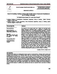
associated polytrauma) and out of the 1090 patients who had trauma to head region, 188 (17.25%) were between 1 month and 18 years of age. 80 (42.55%) children out of the 188 were asymptomatic and had a normal neurologic examination and normal CT brain (done in 48 children) and were discharged from the ER itself. These children were followed up after 7 days and were not found to be symptomatic or having any focal deficit. Out of 188 children, 57.45% (mean age 5.41 ± 4.20 years, n = 108) required admission out of which 22% (n = 24) were admitted in PICU because of severe TBI and all of them required advanced airway management, including ventilation. Age and sex distribution of children admitted with TBI are shown in Table 1. Mean age of males was 6.08 ± 4.77 years and mean age of females was 4.53 ± 3.66 years. Overall two peaks were seen in children admitted with TBI, first in the age group of 1–3 years (38/108, 35%) and second between 6 and 14 years (36/108, 33%). Mode of injury and time of presentation of admitted TBI patients are shown in Table 1. Fifty percent children arrived within 6 h of trauma, 35.11% of children arrived between 6 and 24 h of trauma, and 14.89% arrived between 24 and 72 h. In fact, 83% of children who arrived after 6 h had already sought consultation in another ER or hospital and were referred to our trauma center. Grading of TBI is shown in Figure 1. Table 1: General characteristics of the patients
Characteristics n/mean±SD Case number 108 Age (year) 5.41±4.20 1-11 months 12 (11) 1-3 years 38 (35) 3-6 years 20 (19) 6-14 years 36 (33) 15-18 years 2 (2) Gender Male 60 (55.56) Female 48 (44.44) Prepuberty 70 (64.82) 36 (33.33) Puberty (n) Postpuberty 2 (1.85) GCS >13 54 (50) 9-12 30 (27.78) ≤8 24 (22.22) CT pathology 66 (61.11) Yes 42 (38.89) No 20 (18.52) Time after TBI 15.02±20.35 h GCS: Glasgow coma scale, CT: Computed tomography, TBI: Traumatic Brain Injury, SD: Standard deviation
Journal of Neurosciences in Rural Practice ¦ Volume 9 ¦ Issue 2 ¦ April-June 2018
227
Dara, et al.: Traumatic brain injury in pediatric population
Deficit at presentation and imaging findings of admitted TBI patients are shown in Table 2. Nearly 16.67% of patients presented with bleeding from ear immediately or within few hours of trauma and 11.11% had associated vomiting and 3.7% also had focal seizures. Severity of TBI Fall from Height Animal hit
RTA
Fall from stairs
Obejct fall over head
0 4 4
46
Mild (13-15)
0 4 2 14
4 0 10
10
10
Moderate (9-12)
Severe (3-8)
Figure 1: Severity of traumatic brain injury
Table 2: Mode of injury and time of presentation to the hospital