source (for example a hospital) but there is a system that allows to find out which .... these profiles, a monitor calibration tool and a post-acquisition ... GOCR, a freeware OCR software. .... IHE Stress Testing Workflow Integration Profile. 2.
Int J CARS (2011) 6 (Suppl 1):S74–S78 DOI 10.1007/s11548-011-0592-2
WORKFLOW AND STANDARDS
Experience with IHE profiles in a national project for medical data sharing E. Bellon1, P. Neyens1, M. Vinkx1, M. Feron1, M. Sweertvaegher1, B. Van den Bosch1 1 University Hospitals Leuven, Dept. of Information Technology, Leuven, Belgium Keywords IHE � Telemedicine � PACS � eHealth Purpose With this presentation we want to share initial experiences in deploying IHE integration profiles, primarily XDS (Cross-Enterprise Document Sharing) and XCA (Cross Community Access). This is part of a national project in Belgium that aims at making medical history of a patient available to any physician who is currently in contact with that patient, regardless of which institution that information was collected in. In this project the medical documents typically remain at the source (for example a hospital) but there is a system that allows to find out which documents are available and to retrieve them. That system is implemented on two levels: regional partnerships between health care providers maintain an index of documents available within that hub, while a national metahub manages information about which hubs have any data on a specific patient. Our hub will group around 20 hospitals throughout the Flanders region. That national system is being developed around a Belgian standard for medical communication, as that standard was already used in a number of local initiatives. In our hub, in contrast, we have decided to use IHE profiles. We therefore must build bridges between the local standard and IHE and match the concepts in IHE to the policies in the national project. More in general, we study possibilities to advance the use of IHE in a gradual fashion. Methods Document sharing within the hub is in principle based on the XDS profile. In practice, however, current information systems in the connecting hospitals do not provide an XDS interface. At the same time we believe that the largest bottleneck in this project is exactly the effort required to connect such local information systems. Commercial software has been identified that enables those systems to connect using HL7 feeds. This solves some of the connectivity problems but not all. Already in the local setting, connecting medical information systems is more involved than just sending HL7; in an organization that extends beyond the own institution, complexity is even much higher. For example, at the time at which the information is generated the global (national) patient ID may not yet be known, or the patient may not yet have provided consent for data sharing. Handling such exceptions could be rather difficult (e.g., putting data aside and implementing an explicit trigger to transmit it later). Patient merges, which occur frequently in medical practice, can now be at the local as well as the global level. From experience in technical pilots we are now finalizing adaptations to the system that enable to concentrate most, if not all, of the complexity within the central software. The HL7 feeds from the local systems can take abstraction from these aspects, and local workflows need only be adapted in minimal ways.
123
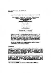
We did not limit ourselves to the basic XDS profile. For example, we want to provide access to previous data right after consent has been provided, but in the absence of such consent the XDS index may not yet have been populated. A lot of consideration must go to protection of personal data anyhow. We use a combination of XDS and XCA. The XCA profile was designed to exchange information between loosely coupled domains. In our project we deploy XCA for its possibilities to query information dynamically (whereas XDS assumes that the index has been populated before). This makes is possible to define a separate XDS domain for each hospital (in which even the index data is contained) while from the outside this looks as a single domain that always contains up to date information. Most importantly, this gives us more implementation options within the hub while still sticking to IHE profiles. By deploying the IHE PIX profile (Patient Identifier Cross Referencing) the local systems can keep using local patient IDs. This is particularly important for information feeds that the hospital does not want to interfere with, such as DICOM communication with a PACS. Results The system is still in technical pilot, internally as well as in connectivity to other (non IHE) hubs and the national metahub. As illustrated in Fig. 1, in the technical implementation we try to rigorously stay with IHE profiles internally, even if the environment in which this system must operate is not particularly IHE friendly. The bridge between this IHE hub and the other hubs in the Belgian project is implemented as an XCA responding and initiating gateway. For the connecting systems, and for the interactive viewing software provided over the Internet, the other hubs therefore look like IHE hubs. Current work includes integrating the national concepts for user authentication (in part based on the electronic ID card) and for secure communication, and this in such a way that we can keep using commercial components that were developed in international competition.
Fig. 1 The eHealth hub rigorously adheres to IHE profiles internally. Conversions to and from the national standard are concentrated in a gateway that implements the IHE XCA profile. The information systems within the hospitals typically use HL7 feeds to provide documents to the local repository that acts as an XDS results server
Int J CARS (2011) 6 (Suppl 1):S74–S78 Conclusion As expected, IHE profiles cannot be used right out of the box to implement a large scale data sharing project. However, our experience so far suggests that adaptation to local policies and requirements does not require fundamental changes in these profiles but rather a more creative combination of profiles. We still must experience, however, to what extent we will be able to use existing or developing IHE profiles in implementing the specific rules for fine-grained access control.
Clinical implementation of the IHE presentation of grouped procedures integration profile-implementation details and workflow benefits in a neuroradiology section G. Wendt1 1 University of Wisconsin - Madison, Radiology, Middleton, USA Keywords PACS � PGP � IHE Purpose The implementation of Presentation of Grouped Procedures (PGP) integration profile, as described in the IHE Technical Framework, on CT and MR scanners as well as the PACS addresses the problem faced by many institutions where there are multiple requested procedures performed in one acquisition. This paper will discuss the problems solved by PGP, the steps necessary to implement such a solution and the benefits to subspecialty groups such as neuroradiology. Materials and Methods The University of Wisconsin - Madison is a level one trauma center and a tertiary care hospital. As a result many exams (CT and MR) consist of several thousand images and cover several body parts. This leads to several problems in a subspeciality softcopy environment: there is only one study listed while several reports must be generated; when one radiologist marks a study as read, those reading the other procedures won’t see them in their in-box; and the radiologist and referring clinician are required to search for relevant images within a large data set. This is a particular problem in a neuroradiology section where an individual patient may have several neuroradiology exams in a single visit to the scanner suite. These problems are magnified in a trauma setting where there are often many other subspecialty exams done, sometimes adding hundreds or thousands of images that are not part on the neuroradiology exams. These and other problems with potential solutions will be discussed. Results Vendor requirements include modification of the modality hardware and software. PGP support also requires support of Scheduled Workflow and Consistent Display of Images integration profiles. The modality vendor developed two new software applications to support these profiles, a monitor calibration tool and a post-acquisition application designed to sub-group images and create grayscale presentation states. The PACS required software modification to support the PGP information passed from the modalities. The system implements virtual study splits by receiving PGP information as Grey Scale Presentation State (GSPS) objects from the scanner. As a result PACS users are presented with only the relevant images for that body part when they select an exam and the PACS only needs to maintain one data set with all the acquired images. Whenever an exam is viewed the GSPS objects are consulted and only images applicable to the selected study are displayed. Conclusion Implementation of PGP solves several problems faced on a daily basis is a softcopy environment. The most important on these are faster access to relevant images and the elimination of duplicate data. Implementation of PGP in an academic setting offers many benefits to
S75 the neuroradiology section (as well as other subspecialty divisions). It is however, not a trivial implementation at the current time as it requires vendors to cooperate and implement lab to lab testing to ensure proper functionality prior to clinical implementation.
RADIANCE - a dose information consumer and reporter within the IHE REM profile T. S. Cook1, W. W. Boonn1, W. Kim1 1 Hospital of the University of Pennsylvania, Department of Radiology, Philadelphia, PA, USA Keywords CT dose monitoring � IHE � RADIANCE � DICOM RDSR Purpose There is growing interest worldwide in being able to archive, monitor and report radiation dose to patients from modalities that use ionizing radiation, particularly computed tomography (CT). A number of initiatives are underway to facilitate dose information reporting. First, the DICOM Standards Committee has developed a radiation dose structured report (RDSR) that stores dose-related parameters in the DICOM header of a study, rather than on image-based dose sheets or screen captures as historically done [1]. In addition, the American College of Radiology (ACR) is piloting the Dose Index Registry, to which imaging centers across the United States can transmit RDSRs from their studies for storage and aggregate analysis within a national, anonymized database [2]. Finally, the Integrating the Healthcare Enterprise (IHE) initiative has developed the Radiation Exposure Monitoring (REM) Profile, an extensive set of guidelines for vendors and imaging centers seeking to monitor dose indices [3]. Methods Together, these new measures provide a robust toolkit for dose monitoring with exams acquired on newer CT scanners that produce RDSRs. However, there is a vast repository of data worldwide that
Fig. 1 Modification of the IHE REM Profile diagram to demonstrate its application at our institution and to indicate the actors whose roles RADIANCE is able to perform
123
S76 has been and continues to be acquired on legacy CT scanners that produce image-based dose sheets. To assist those imaging centers in implementing dose monitoring systems, we have designed RADIANCE–Radiation Dose Intelligent Analytics for CT Examinations. RADIANCE is a free, IHE-compliant, open-source software package that parses image-based dose sheets as well as the DICOM header for a study and stores relevant data in a relational database for subsequent analysis [4]. However, it is also capable of reading RDSRs generated by newer scanners or writing RDSR representations of legacy dose sheets. As a result, it acts as both a dose information consumer as well as a dose information reporter within the IHE REM Profile (Fig. 1). Results As a dose information consumer, RADIANCE can be configured to query an image archive/manager, such as a PACS, for CT dose sheets from multiple vendors, including Siemens, GE, Toshiba, Philips and NeuroLogica. Standard DICOM query/retrieve operations are performed to obtain only the image-based dose sheet; the diagnostic images from the study are not required. RADIANCE begins by performing optical character recognition (OCR) using GOCR, a freeware OCR software. It then parses the text representation of the dose sheet and validates the extracted dose indices, including CTDI (CT dose index), DLP (dose-length product), x-ray tube voltage and tube current. Additional information about the patient, scanner, institution and examination is parsed from the DICOM study header, and the data is stored in a relational database. Using the anatomy-specific conversion factors (‘‘k’’ factors) set forth by the European Commission, RADIANCE estimates whole-body effective dose for each study from its total DLP and stores this value in the database. RADIANCE is also able to read RDSRs produced by newer scanner models and import those data into the database. Since the database contains protected health information, it is password-protected and intended to reside behind an imaging center’s firewall, rather than on a public network. As a dose information reporter, the analytics dashboard built on the RADIANCE database schema allows users to scrutinize their dose data more carefully, and analyze dose estimates by departmental section, individual scanner, involved personnel (technologist, radiologist or referring physician) or individual patient. Outlier identification is also possible, i.e., detecting studies whose dose estimates exceed a prescribed threshold. With the exception of the patient dashboard, which requires input of the patient’s medical record number, all data presented by the dashboard is completely de-identified in keeping with the REM Profile. One of the actions of a dose information reporter in the REM Profile is to transmit dose information to a registry. Hence, RADIANCE is able to generate de-identified RDSR representations of legacy CT dose sheets, to enable users to participate in dose registries. This was tested in the recent IHE Dose Demonstration, in which RADIANCE fulfilled the roles of both dose information consumer as well as dose information reporter. Using secure FTP, RDSR representations of legacy CT dose sheets were produced by RADIANCE and transmitted to the ACR’s Dose Index Registry to demonstrate communication between a dose information reporter and a dose registry. Conclusion As imaging centers worldwide strive to develop effective measures for archiving, monitoring and reporting radiation dose estimates, the IHE REM Profile provides valuable guidelines for implementation of dose monitoring and reporting. RADIANCE represents an IHEcompliant dose information consumer and reporter that can parse legacy CT dose sheets as well as import new structured radiation dose reports into a central database for subsequent analysis. It provides users with a set of standard dose analytics dashboards as well as the ability to customize new analytics modules for their imaging centers. Using RADIANCE and other actors within the IHE REM Profile increases awareness of the issues surrounding radiation exposure from CT and ultimately improves patient care.
123
Int J CARS (2011) 6 (Suppl 1):S74–S78 References [1] DICOM Standards Committee. DICOM Standard Supplement 127: CT Radiation Dose Reporting. 2007. [2] National Radiology Data Registry. http://www.acr.org/ SecondaryMainMenuCategories/quality_safety/NRDR.aspx. Accessed March 11, 2011. [3] Radiation Exposure Monitoring. http://wiki.ihe.net/index.php? title=Radiation_Exposure_Management. Accessed March 11, 2011. [4] TS Cook, SL Zimmerman, ADA Maidment, W Kim, WW Boonn. Automated Extraction of Radiation Dose Information for CT Examinations. JACR 11(7): 871-77.
Multi-source data integration for semantic annotation of medical imaging reports I. Lassoued1, W. Barhoumi1, E.Zagrouba1 1 ISI, SIIVA, Ariana, Tunisia Keywords Medical report � Ontology � Semantic annotation Purpose Annotation of medical images and reports is an increasingly challenging issue for practicians in their daily activity. Indeed, images contain implicit knowledge, about anatomy and abnormal structures, which is usually deduced manually by the viewers. However, in addition to the high cost, manual annotations suffers from human mistakes. Besides, the extracted knowledge is generally not recorded in a structured manner nor directly linked to the image. Thus, images cannot be easily indexed and retrieved given their semantic content (e.g., find all images containing particular anatomy or representing particular abnormalities). In the last decade, DICOM Structured Reporting (SR)[1] specification improves the documentation of diagnosis images and waveforms. The specification supports the interchange of expressive compound reports in which the critical features shown by images and waveforms can be denoted unambiguously by the observer, and thus indexed and retrieved selectively by subsequent reviewers. Findings may be expressed by the observer as: 1) text, codes, numeric measurements; 2) computer-generated coordinates of specific regions of interest within images or waveforms; or 3) references to comparison images, sound, waveforms, curves, and previous report information. DICOMSR-IR contributes to the establishment of indexing and retrieval systems using the semantic content of patient reports while combining the annotations produced by the expert [2] with the elements of the ontology. These systems do not consider rich low-level image features in annotation process. In [3], authors proposed a multi-cue approach for automatic medical image annotation using global and local features. However, the expert cannot intervene in the automatic annotation process and does not carry out the textual comments linked to images. Moreover, the observational and historical findings recorded by the observer may include any evidence referenced as part of an interpretation procedure of image content. Indeed, we assist and guide the choice of experts during the annotation process of its medical imaging reports. Mainly, these annotation techniques are based on the integration of multisource data which takes into account many aspects of data ontology presentation mechanism, old report annotations and low level images information. Methods The proposed semi-automatic annotation technique is composed of two components (Fig. 1). First, we exploit elements of the modular ontology to annotate text and image in the input report. The proposed system contains a management module as well as a visualization tool for the studied ontology. It allows to load modular ontology and to present it in the same annotation session. In fact, the ontology used in the proposed system is a modular ontology. All elements of a module
Int J CARS (2011) 6 (Suppl 1):S74–S78
S77
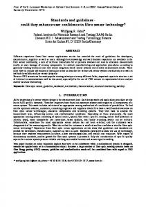
Fig. 2 Recall/precision curves Fig. 1 Proposed annotation procedure for medical reports (concepts, instances, relations, properties) are viewed as a tree. This presentation facilitates the management, operation and reuse of large ontology in the annotation process. The expert has the possibility to search an element (concept, property and relation) of the ontology from the search box provided by the system. Moreover, the system uses the already recorded annotation for old reports to annotate different part (images and text) of the studied report by combining mutually text annotation and image annotation. For text annotation, the expert can choose a report to annotate. Then, the system proceeds to retrieve similar reports in the database, in order to provide a set of possible annotations from the annotations already made on old reports. At the first step, the annotated text is split into a set of words. Then, the system compares words of text to annotate with annotations already existing in the database. The result of the algorithm is annotation suggestions for the input text. The annotator uses this result and can modify it by adding or removing items. The annotator can also select some text and annotate it directly from the used ontology. For image annotation, the annotator specifies firstly what he wants to annotate: region, part of the image or the full image. If the selected element is a feature region, the expert must choose a threshold for the gray level and a germ pixel inside the concerned region for a region growing process. The result of this step is a feature region. Then, the photometric properties of the selected element are computed and used to retrieve similar cases in the database using a similarity measure to suggest most relevant annotations to the annotator. Indeed, Euclidean distances of all photometric properties are combined to form a single weighted measure, which is used as a measure of similarity between images and regions. Thus, the annotator can use the annotations suggested by the proposed system or modify them by using the elements of the ontology. Our system allows annotations update already made to the text, image or regions by adding or taking away elements of this ontology in the annotation session. Results To evaluate objectively the proposed system, three Recall/Precision curves illustrating the reliability of the approach are presented (Fig. 2). A set of five DICOM SR reports are decoded and presented by the suggested system to validate it and to evaluate its performances. Each report contains n images with their comments and observations. We seek the annotation of image report by the existing annotations in the database. The first curve illustrates the recall rate according to the precision of the suggestions given by the annotation system based on the exploitation of annotations associated to images text comments. The second and the third curve show the recall rate according to the precision of the suggestions given by the annotation
system based respectively on the exploitation of low-level descriptors of the images, and on joint exploitation of low-level information of images and comments. Indeed, these results show that the combination of low-level descriptors of images and comments produces accurate results comparatively to the use of suggested annotations using only the textual comments or only low level descriptors. Conclusion In this paper, a semi-automatic tool for semantic annotation of medical reports is proposed. The proposed solution allows annotating text and images which present different parts of a medical report. This is done by using an integration of multiple data sources: the annotations of old reports, low-level information of images and different elements of ontology. We propose to improve our approach by adapting our semantic annotation technique to the remaining elements of modern medical records. References [1] W. Dean Bidgoog, ‘‘Clinical importance of the DICOM structured reporting standard’’, The International Journal of Cardiac Imaging, pp. 307-315, 1998. [2] S. Mhiri1,S.Despre´s, E.Zagrouba, « Unification d’ontologies he´te´roge`nes pour l’indexation se´mantique du compte-rendu standardise´ des examens du patient » , Atelier The´matique GDR I3.3,Grenoble, pp. 31-36, 2007. [3] T.Tommasi, F.Orabona, B.Caputo, ‘‘Discriminative cue integration for medical image annotation’’, Pattern Recognition Letters, pp. 1996-2002, 2008.
DICOM based digital electrocardiography T. Salas1, C. Ru´bies1, J. Guanyabens1 1 Age`ncia d’Avaluacio´, Informacio´ i Qualitat en Salut, Pla Imatge Me`dica, Barcelona, Spain Keywords Digital � Electrocardiography � DICOM � PACS Purpose Interoperability ECG standards are not generally implemented by manufacturers. Enable information exchange between different systems for acquisition, visualization and post-processing of signals, and integration with electronic health records, independently from manufacturers and technological platforms, would help to improve quality of care, research and teaching activities. Additionally it also can reduce acquisition and maintenance costs and foster innovation. Methods General requirements of the system are: 1. Based on existing standards
123
S78
Int J CARS (2011) 6 (Suppl 1):S74–S78
2. Obtain from the device the original signal and clinical parameters 3. Information must consist of structured and coded data 4. Grant access to every healthcare professional through Histo`ria Clı´nica Compartida (Catalonia’s shared health record) 5. Grant access to citizens through Carpeta Personal de Salut (Personal Health Folder) 6. Allow advanced post-process capabilities (measure, compare, CAD tools) for specialised diagnose Results Design of the system has been made based on existing standards and IHE TF: 1. 2. 3. 4. 5.
IHE Stress Testing Workflow Integration Profile IHE Resting ECG workflow (draft for trial implementation) IHE Radiology and Cardiology technical frameworks DICOM worklist to manage electrocardiography workflow DICOM waveforms for communication, storage, and retrieval of waveform data
System will be connected to the Catalonian Shared Health Record, which provides the means to make this information available to every healthcare professional in Catalonia The Age`ncia d’Informacio´, Avaluacio´ i Qualitat en Salut del Departament de Salut de Catalunya is responsible for the design of the system and interoperability specifications. When needed, it will also develops SW components to transform proprietary formats into standard ones. Development of the system has being made through collaboration with vendors (PACS, ECGs and viewers), and healthcare providers. Conclusions Deploying of the system is being made through pilot projects, which include sharing of information between Hospitals and Primary Care centres.
123
Currently 15.000 ECgs from 6 pilots projects have been digitised and stored locally in DICOM waveform format. A copy of these ECGs has been sent to the Repositori Central d’imatge Me`dica (Medical Imaging Central Repository) and are available for clinicians and citizens. Healthcare providers and vendors involved in this first phase of the project are: Healthcare providers: Hospital Universitari Joan XXIII, Hospital Sant Jaume de Calella, Hospital de Blanes, Hospital de Campdeva`nol, Hospital de Terrassa, Hospital d’Igualada, Hospital de Berga, Centre Hospitalari Althaia de Manresa Hospital Municipal de Badalona. ECG vendors: Gemmed, Philips, General Electic and Mortara. SW vendors: UDIAT Centre Diagno`stic (ECG viewer) SW integrators: C2C, Costaisa These projects show that: 1. a DICOM based system fully supports healthcare professionals requirements for electrocardiography 2. DICOM WL provides the most effective integration mechanism between information systems and medical devices 3. using a standard format to store, retrieve and display test results allows easy sharing of information through a viewer with basic capabilities In order to accelerate the adoption of this model, Department de Salut de Catalunya (Catalonia’s Health Department) will use public procurement to assure new ECGs are provided with the necessary interoperability capabilities.