Schizophrenia Bulletin vol. 35 no. 1 pp. 19–31, 2009 doi:10.1093/schbul/sbn162 Advance Access publication on November 27, 2008
Working memory and DLPFC inefficiency in schizophrenia: The FBIRN study
S. G. Potkin1,2, J. A. Turner2, G. G. Brown3, G. McCarthy4, D. N. Greve5, G. H. Glover6, D. S. Manoach5, A. Belger7,8, M. Diaz8, C. G. Wible9, J. M. Ford10, D. H. Mathalon10, R. Gollub5, J. Lauriello11,12, D. O’Leary13, T. G. M. van Erp14, A. W. Toga15, A. Preda2, K. O. Lim16, and FBIRN
reaction time with increasing memory load. The mean BOLD signal in the DLPFC was significantly greater in the schizophrenic group than the healthy group, particularly in the intermediate load condition. A secondary analysis matched subjects for mean accuracy and found the same BOLD signal hyperresponse in schizophrenics. Conclusions: The increase in BOLD signal change from minimal to moderate memory loads was greater in the schizophrenic subjects than in controls. This effect remained when age, gender, run, hemisphere, and performance were considered, consistent with inefficient DLPFC function during working memory. These findings from a large multisite sample support the concept not of hyperor hypofrontality in schizophrenia, but rather DLPFC inefficiency that may be manifested in either direction depending on task demands. This redirects the focus of research from direction of difference to neural mechanisms of inefficiency.
2 Department of Psychiatry and Human Behavior, University of California Irvine, Irvine, CA 92697; 3Department of Psychiatry, University of California San Diego, San Diego, CA 92161; 4Department of Psychiatry, Yale University, New Haven, CT 06520; 5 Neuroimaging Division, Department of Psychiatry, Massachusetts General Hospital, Charlestown, MA 02129; 6Lucas Imaging Center, Stanford University, Palo Alto, CA; 7University of North Carolina, Chapel Hill, NC; 8Brain Imaging and Analysis Center, Duke University Medical Center, Durham, NC 27710; 9Department of Psychiatry, Harvard Medical School and Brockton VAMC, Radiology, Brigham Woman’s Hospital, Boston, MA 02115; 10 University of California, San Francisco, CA; 11Department of Psychiatry, University of New Mexico, Albuquerque, NM 87131; 12 The Mind Research Network, Albuquerque, NM 87131; 13Department of Psychiatry, University of Iowa, Iowa City, IA 52242; 14 Department of Psychology, University of California Los Angeles, CA 90095; 15Department of Neurology, University of California Los Angeles, CA 90095; 16Department of Psychiatry, University of Minnesota, Minneapolis, MN
Key words: fMRI/schizophrenia/working memory/ dorsolateral prefrontal cortex/cortical inefficiency
Background: The Functional Imaging Biomedical Informatics Network is a consortium developing methods for multisite functional imaging studies. Both prefrontal hyperor hypoactivity in chronic schizophrenia have been found in previous studies of working memory. Methods: In this functional magnetic resonance imaging (fMRI) study of working memory, 128 subjects with chronic schizophrenia and 128 age- and gender-matched controls were recruited from 10 universities around the United States. Subjects performed the Sternberg Item Recognition Paradigm1,2 with memory loads of 1, 3, or 5 items. A region of interest analysis examined the mean BOLD signal change in an atlas-based demarcation of the dorsolateral prefrontal cortex (DLPFC), in both groups, during both the encoding and retrieval phases of the experiment over the various memory loads. Results: Subjects with schizophrenia performed slightly but significantly worse than the healthy volunteers and showed a greater decrease in accuracy and increase in
Introduction Several lines of evidence suggest working memory, and the dorsolateral prefrontal cortex (DLPFC) component in particular, as a critical domain of dysfunction in the pathophysiology of schizophrenia. In neuropsychological studies, patients with schizophrenia have been found to show performance deficits on nearly all measures of functioning, with working memory among the domains most severely affected.3,4 The working memory deficits are associated with the severity of negative symptoms and impairments in social and occupational functioning.5–9 Structural neuroimaging studies provide convergent evidence indicating that working memory anatomical correlates are affected in schizophrenia. Specifically, a relatively greater degree of reduction in frontal and temporal cortical volumes compared with posterior cortical volumes in patients with this disorder have been reported.10 After accounting for individual differences in gyral patterning and shape, the DLPFC is a key cortical region in which gray matter is reduced in volume in schizophrenic patients compared with their clinically unaffected MZ cotwins, changes that are correlated with the
1 To whom correspondence should be addressed; Department of Psychiatry and Human Behavior, University of California, Irvine, 5251 California Avenue, Suite 240, Irvine, CA 92617; tel: 949-8248040, fax: 949-824-3324, e-mail:
[email protected].
Ó The Author 2008. Published by Oxford University Press on behalf of the Maryland Psychiatric Research Center. All rights reserved. For permissions, please email:
[email protected].
19
S. G. Potkin et al.
degree of cognitive dysfunction and negative symptom severity in the patients.11 Schizophrenia is characterized by longer reaction time and less accurate working memory performance, especially as the memory load increases. The brain activation patterns responsible for the poor performance remain controversial.2,12,13 Manoach et al observed greater activation in the DLPFC in the schizophrenic group compared with controls and a positive correlation between activation and task performance. DLPFC activation appears to be strongly affected by memory load12; therefore, performance changes with increasing memory load require consideration. Performance is a relevant issue as schizophrenic subjects have shown that peak activation of the working memory system may be reached at a lower memory load than in normal controls and that the decline in DLPFC activity observed in some studies of schizophrenia may be related to exceeding the performance capacities of the schizophrenic subjects.13,14 A difference in relative task difficulty between cases and controls may account for whether DLPFC activation is lower or higher than normal in schizophrenic patients in any given paradigm.15,16 In healthy volunteers (HV), BOLD signal in the DLPFC region appears to increase with increasing memory load12,17 but on some tasks activation appears to asymptote (and may decline) at the highest load levels.18 Patients appear to reach peak activation of the working memory system at a lower processing load than do healthy controls.14 Thus, at least on certain tasks, at low levels of difficulty, patients with schizophrenia may use greater prefrontal resources yet achieve lower accuracy compared with healthy subjects (ie, inefficiency), while at higher levels of difficulty, patients may fail to sustain the prefrontal network that processes the information, achieving even lower accuracy as a result.19–24 Kindermann et al,25 however, observed a different pattern of brain response even when schizophrenia subjects performed within the range of their performance capacities. Further evidence for abnormal circuitry has been suggested by the Wolf et al26 finding of reduced functional connectivity between the DLPFC and the temporal lobe structures during the encode process, while demonstrating increased connectivity between the ventral lateral PFC and temporal lobe, that they hypothesized as compensatory activation. Zhou et al27 observed evidence for bilateral DLPFC functional increased connectivity in first-episode patients using resting fMRI BOLD measures. These and other studies13,28 suggest that schizophrenic subjects activate different brain networks in performing memory tasks. Scheuerecker et al29 pointed out that these different networks or patterns of compensatory circuitry are insufficient as memory task demands increase. Additional evidence of abnormal circuitry in the PFC and temporal lobe and in the uncinate and arcuate fasciculi that connect them has been observed in diffusion 20
tensor-imaging studies (reviewed by Kubicki et al30 and Kanaan et al31). While the degree of increased or decreased activity in the DLPFC during the working memory task appears to be a function of task difficulty, most studies average across fMRI runs. Esslinger et al32 in a mental maze task (nonworking memory) observed decreases in activation in patients between early and late trials in contrast to control subjects who showed an increased activation with time. The temporal aspects of brain activation patterns in working memory tasks have not been sufficiently studied. The above findings of both hypo- and hyperactivation in schizophrenic patients compared with normal controls appear related to task, load, performance relative to capacity, ability to compensate, and perhaps temporal variation over time. One important limitation of the above referenced studies is their small sample size, which precludes disentangling these various influences. A metaanalysis by Van Snellenberg et al33 concluded that the magnitude of WM performance differences between schizophrenia subjects and HV was associated with the activation differences in the DLPFC. Meta-analyses address the small sample size by combining studies in the analysis, although they are necessarily limited by the assumptions inherent in equating subjects and task paradigms from different studies. This limitation can be addressed by large multicenter studies. Multicenter studies offer the possibility of developing datasets which are more representative of the population, including differences in health care, comorbidities, racial, and socioeconomic characteristics. To meaningfully combine fMRI data obtained at multiple sites requires methods for reducing intersite variance which, if left unchecked, mitigates the value of the increased sample size. To accomplish this goal, the Functional Imaging Biomedical Informatics Research Network (FBIRN) consortium develops common image acquisition procedures, and assessment tools, calibration, and QA methods to minimize intersite variance, and developed a common data storage and computational environment. Methods The participating institutions in this study were University of California: Irvine (UCI), Los Angeles; University of New Mexico, University of Iowa, University of Minnesota, Duke University/University of North Carolina, Brigham and Women’s Hospital, Massachusetts General Hospital (MGH), and Yale University. Analyses were performed at UCSD, Yale, MGH, and UCI. All data are reported by site code rather than site name. Subjects All sites received local Institutional Review Board approval for this study. Healthy comparison subjects (HV) and schizophrenic/schizoaffective (SZ) male and
DLPFC Activation During Working Memory in Schizophrenia
female adults between the ages of 18 and 70 were recruited for this study. All subjects had regular hearing levels (no more than a 25-db loss in either ear), had sufficient eyesight or were correctable to be able to see the visual display, were fluent in English, and were able to perform the cognitive tasks in this study. No female subjects were pregnant; all subjects were screened for contraindications to MRI. Subjects were excluded if they had a current or past history of a major medical illness; previous head injury or prolonged unconsciousness; substance and/or alcohol dependence; IQ less than 75 (as measured by the North American Adult Reading Test [NAART]); or if they were using migraine treatments. Control volunteers were excluded if a first-degree family member had a diagnosis of a psychotic illness. Subjects with schizophrenia or schizoaffective disorder meeting Diagnostic Standard Manual-IV criteria were allowed in the study; schizophreniform subjects were excluded. Patients were also excluded if they currently had significant extrapyramidal symptom or tardive dyskinesia. Subjects were required to be clinically stable with no significant changes in their psychotropic medications in the previous 2 months. Clinical measures Prior to participating in scanning procedures, all subjects received extensive diagnostic evaluations by experienced raters. Subjects were diagnosed using the Structured Clinical Interview for Diagnosis (November 2002 NonPatient34 and Patient35 version); demographic and other socioeconomic information was collected by interview. Other measures collected on all subjects included the Edinburgh Handedness Inventory,36 Fagerstom Test for Nicotine Dependence,37 and the NAART.38 In addition, all patients received the Scales for the Assessment of Positive39 and Negative Symptoms,40 the Calgary Depression Scale,41 Schedule for the Deficit Syndrome,42 and the InterSePT Scale for Suicidal Thinking.43 Movement disorders were measured with the Abnormal Involuntary Movement Scale,44 Barnes Akathisia Rating Scale,45 and the Simpson-Angus Scale.46 Rating methods for the symptom scales were standardized across sites through cross-site group training sessions by experienced clinical raters and by having the raters at each site rate videotapes of several subjects for comparison with expert assessments. Imaging methods There were 6 3T scanners, 1 4T scanner, and 2 1.5T scanners used in data collection. Both Siemens and GE scanners were included and 1 Marconi (Picker) scanner. Scanning protocols The scanning session consisted of a localizer scan as needed to identify the AC-PC axis; any shimming that
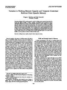
a site used (higher order when possible); a 3D T1weighted scan, (FSPGR on GE, MP-RAGE on Siemens scanners, 24 cm FOV, 1.2–1.5 mm slice thickness, 160– 170 slices as needed to cover the entire head, sagittal orientation); a T2 scan which set the slice prescription for the remaining EPI scans (FOV 22 cm, 27 slices if possible, 4 mm thickness with a 1 mm gap, 256 3 192 matrix). B0 field mapping scans were acquired on Siemens sites only. The functional scans were T2*-weighted gradient echo EPI sequences, with TR = 2, TE = 30 ms, flip angle 90°, acquisition matrix 64 3 64, 22 cm FOV, 27 slices when possible, 4 mm thick with 1 mm gap, and oblique axial AC-PC aligned. Each scan session consisted of a brief training session to familiarize the subject with the paradigms and placement in the scanner for about 1.5 h during which structural and functional images were collected. At least 24 h later and no more than 3 weeks later, the subject repeated the entire session. Only the first visit is reported here. Subjects were to have a normal night’s sleep the night before each scan, no more than one alcoholic drink the night before, and abstain from drinking coffee within 2 h prior to lying down in the scanner. Subjects who smoked refrained from smoking starting 40 min before lying down in the scanner. Cognitive paradigm methods The stimuli and responses were presented and collected using E-prime software, using an SRBox response device (Psychology Software Tools, Inc.). These E-Prime programs are now available at http://www.nbirn.net/. ‘‘Visual stimuli’’ were delivered using various methods. Several sites in the consortium used an LCD projector, with the presentation focused onto a back-projection screen mounted in the magnet bore. Several sites were using projectors onto head coil–mounted mirrors; others used MRI-compatible goggles. Prior to scanning, subjects completed at least one practice run without coaching; the behavioral analysis was run immediately to determine that the subjects were performing at greater than 75% correct. At each site, subjects were scanned according to the same protocol. The Sternberg Item Recognition Paradigm (SIRP) was collected as part of the larger protocol. The order of the tasks was constant, insofar as possible. The stimulus generation computer and scanner were linked by a trigger signal. All scanning paradigms began with a 6-s (3 TR) countdown to allow for dummy acquisitions. To maintain motivation, subjects received an additional 5 cents for every correct response. The SIRP timing and design are as follows (see figure 1, below). During the ‘‘encode’’ condition, subjects memorize a set of target digits. They are then presented with probes (single digits) and respond by indicating whether the probe is a target (a member of the memorized set) or a foil (not a member of the memorized set). 21
S. G. Potkin et al.
Fig. 1. Time Course of the SIRP Design.
This version of the SIRP task consisted of 3 working memory loads, of 1, 3, or 5 target digits (in red), followed by a series of probe digits (in green). Two memory sets for each of the 3 loads were presented in each run of the paradigm. Each condition includes both an encode and probe epoch. Subjects were asked to learn the sets of red digits and instructed to press with their index finger if the green probe digit matches one of the targets and with their middle finger if it does not. The order of the 3 memory load conditions was pseudorandom. In between memory sets, subjects fixated on a flickering cross. The flickering interval was 2 s: 1.85 s on and 0.15 s off (with the exception of the first interval, which lasts 2.8 s). During the encode epochs, red target digits were presented for 6 s. For the 1 and 3 target condition to match for visual stimulation, asterisks (*) were
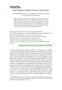
Fig. 2. Image of the DLPFC Mask Shown in Standard MNI Space.
22
used in place of digits so that 5 items were on the screen during every encode epoch. Following the encode epoch, there was a 2.7-s ‘‘delay’’ and a longer ‘‘probe’’ epoch. During the probe epoch, subjects were shown 14 individual probes, serially, in green. Within each condition, half of the probes were targets, half foils, and each member of the target set was presented at least once. Probe digits are random integers between 0 and 9 and are not repeated within a single memory set; no more than 3 consecutive digits were targets within a memory set. The timing of the probe digits was 2.7 s, made up of random jitter around a 1.1 s display. The overall probe epoch lasted 38 s. Analysis methods Behavioral analysis The mean accuracy for each load level was calculated for each subject, averaging over all
DLPFC Activation During Working Memory in Schizophrenia
blocks of the same condition in each run. Mean response times were calculated both for all trials and for correct trials only. A mixed-model design assessed the effects of memory load and run within subject and site and diagnosis across subjects on both measures. Imaging analysis Errors in the imaging data coming from all the sites were identified by examining the content of the XCEDE XML47 files that accompanied the shared NIfTI images, by checking image header information and by visual inspection of the images. Given our a priori hypothesis that activation in the DLPFC during the working memory task would distinguish schizophrenia subjects from control subjects, our primary analysis focused on the DLPFC as the region of interest (ROI). We describe the preprocessing and analysis steps that lead to the summary measure for the ROI. Images were processed with a developmental version of the FBIRN Image Processing System (FIPS), an image analysis pipeline primarily using routines from the FMRIB Software Library (FSL).48 In the version of FIPS used here, preprocessing steps were separated from the remainder of the FIPS pipeline, with XML and related files developed to track provenance information. The data were corrected for head movement using FSL’s MCFLIRT (usually aligning to the middle volume); PRELUDE and FUGUE were used to B0 correct images at sites where field maps were collected and ‘‘slicetimer’’ to correct images for slice-timing differences.49 To equilibrate images for potential site differences in the BOLD signal due to spatial smoothness, we smoothed all 3-D volumes to 8 mm FWHM.50 In the first-level analysis, for each subject’s run, the functional time series was high-pass filtered, intensity normalized to 10 000 and spatially normalized by a 12parameter affine transformation to MNI-152 atlas space.51 A linear model was fit to each subject’s preprocessed functional time series for each SIRP run to estimate regression parameters for encode and probe conditions. The linear model also included temporal derivatives of the gamma function to account for specific temporal shifts of the BOLD response that might vary over load level, event type, and run. Both linear and quadratic terms were included in the model to account for baseline drift. Contrasts of parameter estimates— copes—were formed to test for load effects during encode and probe events. The magnitude of each cope, along with an estimate of its variability derived from model residuals, was passed to a second-level analysis to combine copes from separate runs, yielding a composite cope value for each contrast of interest for each subject. These composite cope values were formed by the weighted sum of copes from individual runs with the weights inversely proportional to the run-specific variation in each cope value.52 The composite copes were used to map functional contrast.
Following spatial normalization of the composite cope images to the MNI atlas, we calculated a Jaccard index53 between the base image from each functional time series and the MNI-152 atlas51 in order to identify images with poor geometric properties and assessed each run for temporal outliers using AFNI’s 3dToutcount54 tool. Images with poor spatial geometry included spiral images where poor fat suppression appeared to produce flared image intensities at the edge of the head, images with cysts, images poorly rotated during spatial normalizing, and images with poor head placement within the limited FOV. A run was discarded if at least 34 of the 177 volumes from the functional time series were identified as having an outlier spike, if the base image displayed a visually obvious structural flaw, or if the Jaccard index flagged the fit of the base image to the MNI-152 atlas as being more than 1.5 interquartile ranges above the 75th percentile of the remaining values. Altogether 1.4% of runs were discarded due to poor temporal or geometric properties. The BOLD values for the ROIs for these runs (see below) were replaced with a mean value calculated from data collected for the subject’s diagnostic group at the site where the subject was scanned. ROI analysis The ROI included all voxels within the mask for Brodmann areas 9 and 46 as defined by the WFU PickAtlas,55,56 without the medial wall, which were significantly activated in either the schizophrenic or healthy volunteer group analyses (p < .05, false discovery rate correction). After obtaining masks of the middle and frontal gyri from the PickAtlas that roughly corresponded to Brodmann areas 9 and 46, we formed the intersection of the PickAtlas areas with a gray matter mask derived from the MIN152 Atlas. The medial surface of the resulting mask was then removed. These steps were taken in order to limit the ROI to cortical areas and to limit the mask to lateral cortex. After labeling left and right hemispheres within the mask, we created voxel-wise activation maps for each group identifying voxels where the BOLD response was significantly related to increasing memory load. The false detection rate was set to .05 to correct for the multiple statistical tests performed within the ROI. We then formed the intersection of each within-group activation map with the anatomical DLPFC mask, producing DLPFC empirical masks for each group. Finally, we formed the union of these 2 group masks to produce the final DLPFC masks used in the study. The aim of this method was to restrict the activity of interest to the DLPFC without washing out group effects due to averaging over a large region—while allowing for the possibility of group differences in activation patterns across subregions of the DLPFC. Because the masks were in part determined empirically, they were created for both encode and probe conditions. The resulting ROIs for the encode and probe conditions are shown in Figure 2.WithintheROI,eachsubject’smeanBOLDsignal 23
S. G. Potkin et al.
diagnosis was significant (HV = 96.1 6 .64, SZ = 92.1 6 .66; F(1,208) = 19.3, p < .0001) and interacted with memory load (F(2, 416) = 5.4, p < .005) as shown in the figure. An ANOVA also showed the effect of memory load on reaction time as expected, with increasing memory load leading to increased reaction time for all trials and correct trials (F(2,2.1) = 741, p < .0001 for the overall RT, similar for RT on correct trials only). Reaction time was approximately 100 ms slower in the SZ subjects at each memory load (F(1,20) = 24, p < .0001); the load by diagnosis interaction (F(2,2.1) = 9.1, p < .0001) showed the difference between SZ and HV increased slightly with increasing memory load (73 ms at 1 item, 109 ms at 5 items).
change was computed for encode and probe conditions separately, for each load. These mean BOLD signal changes were the input to the final analyses. In the final analyses, the BOLD signal from the encode and probe conditions were analyzed in separate ANOVAs contrasting each memory load with fixation. Site and diagnosis were between-subject factors and hemisphere and run were within-subject factors. Significance thresholds were set at p < .05. Results Subjects The clinical and demographic data for the 256 subjects are shown in tables 1 and 2. The distribution of male and female subjects, or right and left handed subjects, was not different by disease status; mean age was similar between the 2 groups. The Full-Scale IQ (FSIQ) estimate from the NAART was significantly higher in HV, as were educational levels. Of these 256 subjects, imaging data were missing from 24 subjects. A further 13 subjects were removed because they responded to fewer than 80% of the trials or they responded at lower than chance levels on any of the 3 runs. The final sample of 217 subjects consisted of 111 HV (43 female) and 106 subjects (40 female) with SZ. This group did not differ from the overall group in mean age, handedness, gender or racial distribution, NAART, clinical symptom ratings, or education levels. All behavioral and imaging results presented below are from these 217 subjects.
Imaging results by ROI In figure 4, the average BOLD signal changes over time from the DLPFC region are shown by memory load and diagnostic group. Relative to fixation, the BOLD signal increases at the onset of the items to be remembered and is maintained (particularly in the higher load conditions) during retrieval. The major difference between the schizophrenic subjects and the controls is the degree of activation at memory load of 3 items relative to memory load of 1. Encode results There were significant effects of site, load, run, and hemisphere on the mean DLPFC BOLD signal change in the encode condition, as well as interactions between run and hemisphere and hemisphere and load. However, there was no significant effect of diagnosis and no interactions with diagnosis. The results are summarized in table 3. The effect of load by hemisphere for the 2 diagnostic groups can be seen in figure 4 and is summarized in figure 5a. BOLD signal response during encoding increased for both groups with increasing memory load. The hemispheric differences are minimal, only significant in the 3-item condition (left > right), and there is no interaction with diagnosis. The effect of run within the scanning ses-
Behavioral data The mean accuracy by memory load and diagnostic group are shown in figure 3. An ANOVA with diagnosis as a between-subject variable and load level as withinsubject variables showed the effect of memory load was as expected ((F(2, 416) = 19.8, p < .0001), with increased load leading to reduced accuracy. The effect of Table 1. Demographic Information Demographic Characteristics
Patients
Controls
Number of subjects
128
Race (% Caucasian) Gender (% male) Handedness (% right) Mean age (SD)
Statistical Significance
% Reporting
128
—
100
70.3
77.3
ns
99
71.9
62.5
ns
100
89.8 Range: 18–65
38.0 (11.6)
89.8 36.2 (11.9)
ns
100
ns
100
Subject’s mean years of education (SD)
Range: 5–24
13.3 (1.9)
15.9 (2.3)